- 91 -
Morphologic Analysis of
Crista Galli Using Computed Tomography
Jong Jun Kim, MD1, Jae Hyeong Cho, MD1, Jae Won Choi, MD1, Hyun Woo Lim, MD1, Yong Jin Song, MD1, Soo-Jung Choi, MD2 and Nam-Kyung Yeo, MD1
1Department of Otolaryngology, Gangneung Asan Hospital, University of Ulsan College of Medicine, Gangneung, Korea
2Department of Radiology, Gangneung Asan Hospital, University of Ulsan College of Medicine, Gangneung, Korea
ABSTRACT
Objective : We often observe the variation of Crista galli (CG) which lies in the midline above the cribriform plate on computed tomography (CT) scans. We investigated the variations in CG and the factors which affect its pneumatization. Materials and Methods : We analyzed the CT images of 818 chronic rhinosinusitis patients between July 2003 and July 2011. We investigated height, position relative to the cribriform plate, degree of pneu- matization, and cell origin for the pneumatization in CG. We analyzed the relationship between several factors (age, sex, and position of CG) and pneumatization of CG. Results : The average height of CG was 17.98 mm. In 13.9% of subjects, the base of CG did not extend below the level of the cribriform plate. In 84.2%, CG extended less than 50% of its height below the cribriform plate. In 1.8%, CG extended more than 50% of its height below the cribriform plate. Pneumatization of CG was found in 12.2%. Except one, every pneumatization was connected with the frontal sinus. The rate of pneumatization was significantly different depending on age. Conclusion : Our study demonstrated that CG showed various morphology and pneumatizaiton. The pneumatization of CG was mainly originated from frontal sinus and related to aging.
KEY WORDS : Crista galli · Computed tomography · Pneumatization · Ethmoid bone · Frontal sinus.
INTRODUCTION
Osteomeatal unit computed tomography (OMU CT) scans frequently allow us not only to detect chronic rhi- nosinusitis (CRS) but also to decide the range of endo- scopic sinus surgery (ESS) through the assessment of the extent and severity of disease. OMU CT scans provide the important information about the anatomic variations of the paranasal sinuses. Several previous studies described marked pneumatization of the ethmoid bone including the Haller’s cells, agger nasi cells, concha bullosa, and Ono- di’s cells.1-3)
Crista galli is a large triangular process at the midline on the superior surface of the cribriform plate anchors a fold(falx cerebri) of dura mater in the cranial cavity. The falx cerebri attaches to its thin and slightly curved pos-
terior border, whereas its shorter thicker anterior border is joined to the frontal bone by 2 small alae, completing the margins of the foramen caecum.4) In the embryo, the ethmoidal cartilage consists of both a mesial mass(the me- sethmoid), which extends from the sphenoid to the tip of the nasal process, and a pair of lateral masses developed from the lateral nasal processes(the ectethmoid), lateral to the olfactory sacs. The terminal portion of the mesial mass persists as the cartilaginous nasal septum, whereas ossi- fication of the upper portion becomes the perpendicular plate and Crista galli.
The morphologic variation of Crista galli is often ob- served on the preoperative OMU CT scans. Pneumatiza- tion of the Crista galli is one of the recognized incidental findings on CT scans. Even though the pneumatization of Crista galli is not common, obstruction of the pneumatized Crista galli ostium may lead to chronic sinusitis and mu- cocele formation within this structure.5) However, there is a paucity of information concerning the anatomic charac- teristics of the Crista galli. The purpose of this study is to examine the morphologic variations of the Crista galli and the factors which affect its pneumatization. In order to ac- complish this, we investigated axial and coronal CT scans Address correspondence and reprint requests to Nam-Kyung Yeo, De-
partment of Otorhinolaryngology, Gangneung Asan Hospital, University of Ulsan College of Medicine, 415, Bangdonglee, Sachunmyun, Gangne- ung, 210-711, South Korea
Tel: +82-33-610-3308, Fax: +82-33-610-4960 E-mail: [email protected]
Received for publication on April 4, 2012 Accepted for publicatoin on July 18, 2012
OMU CT scans were obtained from 1059 patients, who were diagnosed as CRS preoperatively at our institution from July 1, 2003 through July 31, 2011. Patients with previous histories of facial trauma, nasal surgery or sinus carcinoma were excluded from the investigation in order to minimize the chance of acquired anatomic defects. Fi- nally, 818 consecutive axial and coronal CT scans of the paranasal sinuses were made to assess the height, the posi- tion, the existence of pneumatization, and the connection site of the pneumatization in the Crista galli.
CT acquisition and images Analysis
The OMU CT scans were retrospectively reviewed with the Picture Archiving and Communication System (PACS, Peta Vision, Asan Medical Center, Korea). CT examinations were performed with a 64 channel MDCT (Lightspeed VCT, GE Healthcare, Milwaukee, USA; scan parameter, 120kVp, 250mAs; scan time, 1000msec; ma- trix size, 512x512) without contrast enhancement. The patients were scanned in the supine position (gantry tilt, about 21 degrees through anterior margin of frontal sinus and nostril), and coronary editing of 1-mm thick slices from the front of the frontal sinus to the end of the sphe- noid sinus was done. The CT scans were routinely evalu- ated with a window width/level of 2000/265 HU for axial scans and 2000/350 HU for coronal scans.
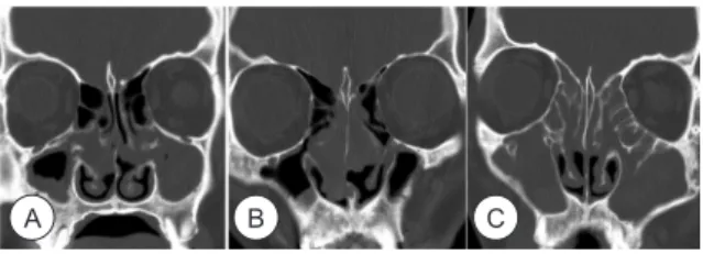
The position of the Crista galli was evaluated according to Hajiioannou’ method6) The position of the Crista galli was categorized into three grades according to the loca- tion related to the cribriform plate (Fig. 1).
Type I—base of the Crista galli is located at the level of the cribriform plate.
Type II—less than 50% of the height of the Crista galli
Crista galli were completely intact other than its caudal margin or there was an extension of either frontal sinus or ethmoid complex into the Crista galli. In addition, we evaluated the existence of mucosal thickening extending from adjacent frontal or ethmoid sinus into the pneuma- tized Crista galli. All scans were reviewed separately by one radiologist and one otolaryngologist. Any different opinions were resolved by consensus.
Lastly, we also analyzed the relationship between other factors (age, sex, and type of Crista galli) and the pneu- matization of Crista galli. The study was performed with the approval of the internal review board (number 2010- 051).
Statistical Analysis
Results were statistically analyzed using SPSS software (version 12; SPSS Inc, Chicago, Illinois). Differences in the rate of pneumatization of Crista galli depending on age, sex, and type of Crista galli were analyzed by using Chi square test. P values < 0.05 were considered statisti- cally significant.
RESULTS
Eight hundred eighteen sets of OMU CT scans were evaluated. The patient population was comprised of 512 men and 306 women, with a median age of 44.51 (range, 7-91). Six hundred thirty-three patients (77.4%) had bilat- eral CRS and 185 patients (22.6%) had unilateral CRS.
The average height of Crista galli was 17.98 ±3.7 mm.
Crista galli type I, in which the base of the Crista galli did not extend below the level of the cribriform plate, was found in 13.9% of subjects (114/818). Type II, the Crista galli extended less than 50% of its height below the cribri- form plate, was the most common position of Crista galli, which accounted for 84.2% of subjects (689/818). Type III, in which the Crista galli extended more than 50% of its height below the cribriform plate, was found in 1.8% of the CT scans (15/818).
Pneumatization in Crista galli was found in 100 patients (12.2%). There were 19 cases with only minimal pneuma- tization and 81 cases with extensive pneumatization (Fig.
2). Every pneumatization was connected with adjacent paranasal sinuses. Except one case, every pneumatization was connected with the right (46 cases), left (38 cases) or interseptal frontal sinus (15 cases) (Figs. 2, 3). Only one
Fig. 1. Position of the Crista galli noted by relation to the cribri- form plate. Type I was regarded as when base of the Crista galli was at the level of the cribriform plate (A). Type II was less than 50% of the height of the Crista galli below the level of the cribri- form plate (B). Type III was greater than 50% of the height of the Crista galli below the level of the cribriform plate (C).
A B C
patient, who had bilateral undeveloped frontal sinuses, showed pneumatization of Crista galli connected to the right ethmoid sinus (Fig. 4). In 44 cases, inflammatory disease in a frontal sinus extended directly into the pneu- matized Crista galli (Fig. 5).
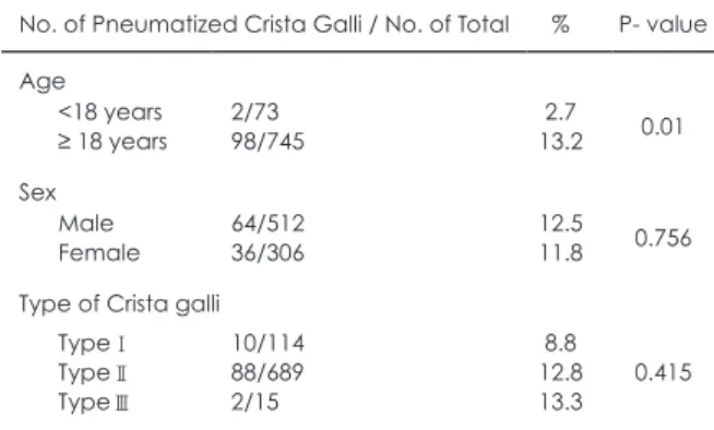
We analyzed the relationship between variable factors and pneumatization of Crista galli (Table 1). The age was found to have an effect on the rate of pneumatiza- tion of Crista galli (P=0.01). Among the 73 CT scans of patients under the age of 18, there were 2 cases of Crista galli pneumatization (2.7%). Among the 745 CT scans of patients with 18 and over the age of 18, there were 98 cases with pneumatization of the Crista galli (13.2%). In contrast, the rate of pneumatization was not affected by sex, or type of Crista galli.
DISCUSSION
Embryologically, the Crista galli is derived from the ethmoid bone. In the second fetal month, it is formed together with the central structures of the anterior skull base and the perpendicular plate of the ethmoid bone by the mesethmoidal cartilage which is derived from the pre- sphenoidal cartilage.4) 6) Then, most ossification of the cartilaginous Crista galli starts at approximately 2 months of postnatal life, steadily increases in ossification to 14
Fig. 2. Pneumatization in Crista galli from frontal sinus. Coronal (A) and axial CT scans (B) of the paranasal sinuses in a 37-year- old man show the extensive pneumatization (arrow) of the Crista galli from the left frontal sinus. The other coronal (C) and axial CT scans (D) of the paranasal sinuses in a 54-year-old woman show minimal pneumatization (arrow) of the Crista galli from the right frontal sinus.
Fig. 3. Pneumatization in Crista galli from interseptal frontal sinus.
Coronal (A, B, and C) and axial CT scans (D, E, and F) of the paranasal sinuses in a 45-year-old man show the typical pneu- matization (arrow) of the Crista galli from the interseptal frontal sinus (star).
Fig. 4. Pneumatization in Crista galli from ethmoid sinus. Coronal (A, B, and C) and axial CT scans (D, E, and F) of the paranasal sinuses in a 30-year-old woman show the pneumatization (white arrow) of the Crista galli from the ethmoid air cell (star). Bilateral frontal sinuses were undeveloped (black arrow).
Fig. 5. Extension of the inflammatory disease into the pneuma- tized Crista galli. Axial (A) and coronal CT scans (B) of the para- nasal sinuses in a 39-year-old man show thick mucosal thickening extending from the right frontal sinus into a well-pneumatized Crista galli (arrow).
Table 1. Rate of Pneumatization of Crista Galli.
No. of Pneumatized Crista Galli / No. of Total % P- value Age
<18 years
≥ 18 years
2/73 98/745
2.7 13.2 0.01 Sex
Male Female
64/512 36/306
12.5 11.8 0.756 Type of Crista galli
TypeⅠ TypeⅡ TypeⅢ
10/114 88/689 2/15
8.8 12.8 13.3
0.415 A
A
A D
D
B
B E
E
C
C F
F
A
C
B
B
D
However, recent studies demonstrated that pneumatiza- tion of the Crista galli came primarily from the adjacent frontal sinuses by radiologic analysis.2) 4) Especially, Som et al. showed that there was 13% of Crista galli pneuma- tization from either the left or right frontal sinuses in 200 adult CT scans, and there were no cases of crista pneuma- tization in the 132 CT scans of children 0–7 years of age.4) According to their suggestion, some Crista galli pneu- matization would be present in the children if the pneu- matizing cells were from the ethmoid complex, because the Crista galli is from the ethmoid bone and the ethmoid complexes are pneumatized at birth.
In the present study, we also demonstrated that pneu- matization of the Crista galli is generally from the frontal sinus. On the 818 OMU CT sca ns, all of crista pneumati- zation except one was connected with frontal sinus. Only one patient, who had bilateral undeveloped frontal sinus- es, showed the pneumatization of Crista galli connected to the right ethmoid sinus. Additionally, we showed that there was statistically significant difference in the rate of Crista galli pneumatization between children and adults groups. On the CT scans of the children and puberty pe- riods adolescents (<18 years) whose frontal sinuses have not reached full size,8) we found 2.7% of Crista galli pneu- matization. On the other hand, we had 13.2% of adults (≥
18 years) with Crista galli pneumatization. This finding supports the concept that the Crista galli pneumatization is almost always from the frontal sinus and age related de- velopment of frontal sinus is an important factor to affect the pneumatization of Crista galli.
In the present study, we showed that the Crista galli was most commonly extended less than 50% of its height be- low the cribriform plate like Hajiioannou’s study.6) They suggested that the pneumatization of Crista galli might be related to the position of Crista galli, because pneumati- zation was present in 75% of type III Crista galli which descends markedly towards the nasal cavity. However, we didn’t find statistically significant difference of the rate in crista pneumatization depending on the position of Crista galli. Hajiioannou et al. did not explain how the position of Crista galli affected the pneumatization and there was a limitation in this study since the number of cases of type III Crista galli was too small (n=8).
Even though the pneumatization of Crista galli is not common, obstruction of the pneumatized Crista galli os- tium may lead to chronic sinusitis and mucocele formation within this structure.5) In the present study, nearly half of
that the mucocele arising from the Crista galli was further complicated by its intracranial extension.9) Therefore, it is important to recognize this entity preoperatively and to differentiate it from an adjacent air cell to avoid extension of surgery into the cranial vault.
To the best of our knowledge, this study is the first re- port to analyze the morphologic characteristics of Crista galli using computed tomography in Asian patients. Our findings are not surprising, because we showed the similar position or pneumatization of Crista galli when compared to the previous reports. However, we suggested another evidence that the primary origin of pneumatization of the Crista galli is from the frontal sinus according to the anal- ysis of 818 CT scans.
In conclusion, our study demonstrated that the Crista galli showed various morphology and pneumatizaiton.
The pneumatization of Crista galli was mainly extended from frontal sinus and related with aging. This finding may have surgical implications when disease is present in the Crista galli.
저자역할(Author Contributions)
김종준, 여남경 은 본 연구에서 모든 자료에 접근할 수 있으며 자료의 완전성과 자료 분석의 정확성에 책임을 지고 있습니다. 연구 기획 : 여 남경. 자료 해석 및 분석 : 김종준, 최재원, 조재형, 임현우, 송용진, 최 수정, 여남경. 논문초안 : 김종준, 여남경. 논문수정 : 김종준, 최재원, 조재형, 임현우, 송용진, 최수정, 여남경. 연구 총괄 : 여남경.
REFERENCES
1) Basić N, Basić V, Jukić T, Basić M, Jelić M, Hat J. Computed tomographic imaging to determine the frequency of anatomical variations in pneumatization of the ethmoid bone. Eur Arch Oto- rhinolaryngol 1999;256:69-71.
2) Robinson M, Donlon D, Harrison H, Houang M, Stammberger H, Wolf G. Variations of the paranasal sinuses in Melanesians as ob- served by CT. Rhinology 2010;48:11-17.
3) Braun H, Stammberger H. Pneumatization of turbinates. Laryngo- scope 2003;113:668-672.
4) Som PM, Park EE, Naidich TP, Lawson W. Crista galli pneumati- zation is an extension of the adjacent frontal sinuses. Am J Neuro- radiol 2009;30:31-33.
5) McLaughlin RB Jr, Rehl RM, Lanza DC. Clinically relevant fron- tal sinus anatomy and physiology. Otolaryngol Clin North Am 2001;34:1-22.
6) Hajiioannou J, Owens D, Whittet HB. Evaluation of anatomical variation of the Crista galli using computed tomography. Clin Anat 2010;23:370-373.
7) Belden CJ, Mancuso AA, Kotzur IM. The developing anterior skull base: CT appearance from birth to 2 years of age. Am J Neuroradiol 1997;18:811-818.
8) Frederick KK, Juan CO. Characteristics of normal and abnormal postnatal craniofacial growth and development. In: Paul WF, Bruce HH, eds. Cummings Otolaryngology Head & Neck Surgery, 5th ed. Philadelphia, PA: Mosby; 2010:2613-2637.
9) Wingate J, Rechtweg JS, Grand W, Jouandet M, Balos L, Wax MK.
Mucocele of the Crista galli. J Otolaryngol 2001;30:43-46.