Kor. J. Fertil. Steril., Vol. 31, No. 4, 2004, 12
정자형성 과정에서 Vascular Endothelial Growth Factor 및 Endothelin-1 발현의 면역조직화학적 연구
부산대학교 의과대학 비뇨기과학교실
박성우・박현준・박남철
The Influences of Vascular Endothlelial Growth Factor and Endothelin-1 on Speramtogenesis in Testis
Sung Woo Park, Hyun Jun Park, Nam Cheol Park
Department of Urology, Pusan National University Hospital, Pusan, Korea
Objective: The effects on spermatogenesis by expression of vascular endothelial growth factor (VEGF) and endothelin-1 (ET-1) were investigated.
Materials and Methods: Testicular specimens were obtained from 40 infertile males due to primary testicular failure and from 10 fertile males with other urologic problems. The specimens of infertile males were devided into 4 groups according to histologic findings; Sertoli cell only syndrome (A), maturation arrest (B), hypospermatogenesis (C) and sloughing and disorganization (D). VEGF and ET-1 expression were detected with immunohistochemical stain.
Results: VEGF expression on Leydig cell was detected in all cases. But, VEGF expression rates on germ cell were significantly higher in infertile group B, C, D compared to that of the control group (p<0.05). ET-1 expression rates on Leydig cell was significantly lower in all infertile group compared to that of the control group (p<0.05). But, ET-1 expression rates on Sertoli cell was significantly higher in all infertile group compared to that of the control group (p>0.05). In germ cell of infertile group, LH, FSH and prolactin were significantly decreased, and estradiol is increased in positive stain group on ET-1 immunohistochemical stain (p<0.05). VEGF and ET-1 expression were not correlated mean seminiferous tubule diameter (p>0.05).
Conclusions: Abnormal spermatogenesis would be reflected in VEGF expression in germ cell.
Key Words: Vascular endothelial growth factor, Endothelin-1, Spermatogenesis, Testis
남성불임은 다양한 원인에 의해 유발되지만 그 중 정자형성 (spermatogenesis)장애에 의한 일차성 고환부전이 약 50~75%를 차지한다.1 일차성 고환부 전의 치료는 과거 다양한 경험적 약물요법이 이용 되어 왔으며, 최근에는 보조생식술의 발달로 인해 미 성숙정자를 이용한 난자세포질내 정자주입술 (intra-
cytoplasmic sperm injection, ICSI)이 시도되고 있으나 두 방법 모두 치료 성공률에서 한계를 드러내고 있 다. 따라서 고환에서의 정자형성 장애의 원인을 분 자생물학적 수준에서 규명하여 이를 근거로 정자형 성 기능의 개선을 유발할 수 있는 치료법의 개발이 필요하다. 이러한 목적으로 고환내 정자형성 기능
주관책임자: 박남철, 우) 602-739 부산광역시 서구 아미동 1가 10번지 부산대학교병원 비뇨기과
Tel: (051) 240-7349, Fax: (051) 247-5443, e-mail: [email protected]
이 장애되는 원인을 규명하고자 많은 노력이 시도 되어 왔으며 여기에는 정자형성의 각 단계에서 정 세포 (germ cell)의 분화를 조절하는 효소, 성장인자 또는 이들과 관련된 항원항체 혹은 유전자에 대한 연구가 주류를 이루고 있다.2~4 이들 인자 중 종양 세포에서 처음 발견되어지고 혈관신생 및 유지에 관 련되는 것으로 알려진 vascular endothelial growth fa- ctor (VEGF) 및 endothelin-1 (ET-1)이 고환내 혈관 뿐 만 아니라 Sertoli 세포, Leydig 세포 및 정세포의 생 리적 기능 유지에 관여하는 것으로 보고된 바 있다.5~9 저자는 특발성 남성불임에서 정자형성장애의 원 인을 알아보기 위해 정상 임신능을 가지는 남성과 남성 불임환자의 고환생검조직에서 VEGF 및 ET-1 의 발현 정도를 면역조직화학염색방법으로 관찰하 여 정자형성기능에 미치는 영향을 분석하였다.
연구 대상 및 방법
1. 연구 대상
불임을 주소로 내원한 비폐색성 무정자증 환자 40 례 (불임군)와 전립선암, 외상 혹은 기타 원인으로 고 환적출술 혹은 생검을 통해 병리조직학적으로 정상 정자형성이 확인된 환자 10례 (대조군)를 대상으로 하였다. 불임군은 정자형성장애 정도에 따라 Sertoli cell only syndrome, maturation arrest, hypospermatogen- esis 및 sloughing and disorganization으로 분류하고 이 들을 각각 불임 A, B, C 및 D군으로 하였다. 불임 A, B, C, D 및 대조군을 각각 10례 총 50례를 대상으로 하였다.
2. 연구 방법 1) 혈청 호르몬 검사
불임군 전례에서 혈청 황체호르몬 (luteinizing ho- rmone, LH), 난포자극호르몬 (follicular stimulating hormone, FSH), 테스토스테론 (testosterone), 에스트 라디올 (estradiol), 프로락틴 (prolactin) 등을 첫 내원 시 측정하였다.
2) 고환생검 및 병리조직학적 검사
정색마취와 음낭 피부에 국소마취를 시행한 후 wi- ndow법으로 채취된 고환조직을 파라핀 포매시킨 뒤 hematoxylin-eosin (H-E) 염색을 시행하였다.
3) 면역조직화학검사 (1) VEGF 발현
파라핀 포매된 고환 조직을 4~5 µm 두께로 잘라 서 1시간 가량 가열한 후 xylene으로 5분간 4회, alcohol로 2분간 네 번 처리 후 흐르는 물에 3분간 세척한 뒤, citrate buffer를 이용하여 중화시키고 au- toclave로 15분간 건조시킨 뒤 H2O2로 10분간 처리 하였다. 이어서 PBS buffer를 이용하여 2분간 3회 중 화시킨 뒤 1:250으로 희석된 항 VEGF 항체를 1차 항체 (mouse monoclonal antibody, Santa Cruz Biote- chnol, USA)로 사용하여 4℃에서 하룻밤을 반응시킨 후 biotin이 부착된 이차항체에 streptavidin을 30분 간 반응시키고 PBS에 5분간 3번씩 세척 후, AEC 로 발색하였다. Hematoxylene으로 대조염색을 시행 한 뒤 mounting solution을 도포하였다.
(2) ET-1 발현
상기 방법과 동일하게 시행하였으며 1:250으로 희석된 항 ET-1 항체를 1차 항체 (mouse monoclonal antibody, Abcam Biotechnol, USA)로 사용하였다.
4) 면역조직화학염색의 판정
대상 환자의 고환조직에서 VEGF 및 ET-1의 발현 양 상은 한명의 숙련된 비뇨병리 전문의에 의해 무작위로 선정된 400배 확대 시야 열군데에서 분석되었다. 발현 정도는 세포막과 세포질 염색이 안 되는 경우를 음성 (-), 세포막과 세포질 염색이 25% 이내로 염색될 때 약 양성 (1+), 25~75%로 염색될 때 중등도 양성 (2+) 및 75% 이상 염색될 때 강양성 (3+)으로 분류하였다.10,11
3. 통계학적 분석
각 군간의 비교는 Fisher's exact test 및 Mann Whi- tney U test를 이용하였고, VEGF와 ET-1 발현간의 상호연관성을 조사하기 위해서는 Chi-square test를 이용하여 분석하였다. p값이 0.05 미만인 경우 통 계학적으로 유의 하다고 판정하였다.
결 과
1. 면역조직화학염색 소견 1) VEGF 발현
(1) 정세포
정세포에서 VEGF의 발현은 불임 A군 및 대조군
의 모든 세포에서 음성이었다. 그러나 불임 B, C 및 D군의 정조세포 (spermatogonia)에서 +1은 각각 2례
(20%), 2례 (20%) 및 1례 (10%)였고, +2는 불임 C 군에서만 1례 (10%)로 각군별 양성률은 각각 20%,
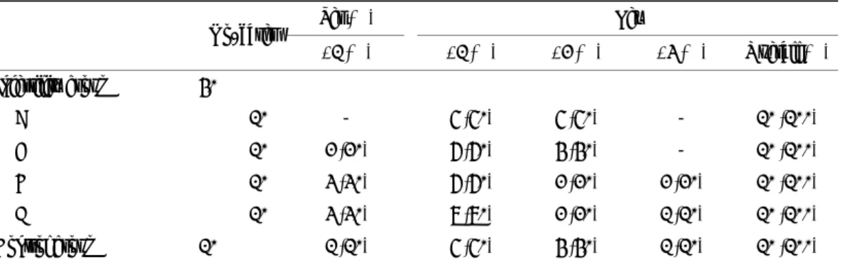
Table 1. VEGF expression rate on germ cell according to the grade of maturation
SG SC ST
No. cases
+1 (%) +2 (%) +1 (%) +2 (%) +1 (%)
Overall (%) Infertility group 40
A 10 - - - - - -
B 10 2 (20) - 3* (30) - - 3* (30)
C 10 2 (20) 1 (10) 5* (50) 2 (20) 2 (20) 7* (70)
D 10 1 (10) - 3* (30) - - 3* (30)
Control group 10 - - - - - -
A; Sertoli cell only syndrome, B; maturation arrest, C; hypospermatogenesis, D; sloughing and disorganization, SG; spe- rmatogonia, SC; spermatocyte, ST; spermatid *; p<0.05 vs control group. calculated by Mann-Whitney U test
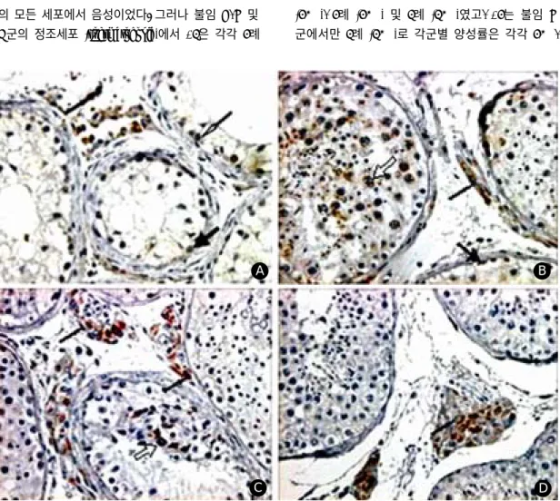
Figure 1. Immunohistochemical stains for VEGF (×400). A; Sertoli cell only syndrome B; maturation arrest C; hypo-
spermatogenesis D; control group. Sertoli cell; thick black arrow, Leydig cell; thin black arrow, Spermatogonia; thin white arrow, Spermatocyte; thick white arrow.A B
C D
30% 및 10%였다. 정모세포 (spermatocyte)에서 +1은 각각 3례 (30%), 5례 (50%) 및 3례 (30%)였고, +2는 불임 C군에서만 2례 (20%)로 각군별 양성률은 30%, 70% 및 30%였다. 그리고 정자세포 (spermatid)에서 는 불임 C군에서만 +1이 2례 (20%)에서 관찰되었 다. 따라서 정세포에서 VEGF의 발현율은 대조군 및 불임 A군에서 음성이었으나, 불임 B, C 및 D군에 서 평균 42.5% (30%~70%)로 유의한 차이를 나타 내었으며 정자형성장애 정도에 따른 유의한 차이는 없었다 (Table 1) (Figure 1).
(2) Sertoli 세포
Sertoli 세포에서 VEGF의 발현은 불임 A군을 제외 한 불임 B, C, D 및 대조군에서 각각 2례 (20%), 3례 (30%), 3례 (30%) 및 1례 (10%)였으며 전례에서 염색 정도는 +1이었다. 따라서 Sertoli 세포에서 VEGF의
양성률은 불임군 및 대조군에서 평균 20% (0%~30%) 및 10%로 유의한 차이가 없었으며, 정자형성장애 정 도에 따른 유의한 차이도 없었다 (Table 2) (Figure 1).
(3) Leydig 세포
Leydig 세포에서 VEGF 발현은 불임군 및 대조군 모두에서 양성 소견을 나타내었다. 불임 A, B, C, D 및 대조군에서 +1은 각각 5례 (50%), 6례 (60%), 6례 (60%), 7례 (70%) 및 5례 (50%), +2는 각각 5례 (50%), 4례 (40%), 2례 (20%), 2례 (20%) 및 4례 (40%), +3 은 불임 C, D 및 대조군에서만 각각 2례 (20%), 1례 (10%) 및 1례 (10%)였다 (p>0.05). 따라서 Leydig 세 포에서 VEGF의 전체적인 양성률은 불임군 및 대조 군에서 모두 100%로 유의한 차이가 없었으며, 정세 포나 Sertoli 세포에 비하여 높은 양성률을 나타내 었다 (Table 2) (Figure 1).
Table 2. VEGF expression rate on Sertoli cell and Leydig cell according to the grade of maturation
Ser (%) Ley
No. cases
+1 (%) +1 (%) +2 (%) +3 (%) Overall (%)
Infertility group 40
A 10 - 5 (50) 5 (50) - 10 (100)
B 10 2 (20) 6 (60) 4 (40) - 10 (100)
C 10 3 (30) 6 (60) 2 (20) 2 (20) 10 (100)
D 10 3 (30) 7 (70) 2 (20) 1 (10) 10 (100)
Control group 10 1 (10) 5 (50) 4 (40) 1 (10) 10 (100) A; Sertoli cell only syndrome, B; maturation arrest, C; hypospermatogenesis, D; sloughing and disorganization, Ser;
Sertoli cell, Ley; Leydig cell, In all, p>0.05 vs control group. calculated by Mann-Whitney U test
Table 3. ET-1 expression rate on germ cell according to the grade of maturation
SG (%) SC (%) ST (%) Overall (%)
No. cases
+1 +1 +1 +1
Infertility group 40
A 10 - - - -
B 10 1 (10) 1 (10) - 2 (20)
C 10 - 1 (10) - 1 (10)
D 10 1 (10) 2 (20) 1 (10) 3* (30)
Control group 10 - - - -
A; Sertoli cell only syndrome, B; maturation arrest, C; hypospermatogenesis, D; sloughing and disorganization, SG; spe- rmatogonia, SC; spermatocyte, ST; spermatid *; p<0.05 vs control group. calculated by Mann-Whitney U test
2) ET-1 발현 (1) 정세포
정세포에서 ET-1의 발현은 불임 B 및 D군의 정 조세포에서 +1이 각각 1례 (10%)로 관찰되었으며, 불 임 B, C 및 D군의 정모세포에서 +1은 각각 1례 (10%), 1례 (10%) 및 2례 (20%)였다. 그리고 정자 세포는 불임 D군의 1례 (10%)에서만 +1로 관찰되 었다. 따라서 정세포에서 ET-1은 대조군 및 불임 A군에서 음성이었으며 불임 B, C 및 D군에서는 평균 20.0% (10%~30%)의 양성률을 나타내어 대조 군 및 불임군 사이, 그리고 불임군간에 정자형성장 애 정도에 따라 유의한 차이는 없었다 (Table 3) (Fi- gure 2). 다만 불임 D군과 대조군의 정세포 간에는 ET-1의 발현이 유의한 차이를 보였다 (p<0.05).
(2) Sertoli 세포
Sertoli 세포에서 ET-1의 발현은 대조군에서 음성 이었으나 불임 A, B, C 및 D군에서는 +1이 각각 3 례 (30%), 6례 (60%), 3례 (30%) 및 3례 (30%)였다.
따라서 Sertoli 세포에서 ET-1의 양성률은 불임군 및 대조군에서 평균 37.5% (30%~60%) 및 음성으로 유 의한 차이를 보였으나 정자형성장애 정도에 따른 유의한 차이는 없었다 (Table 4) (Figure 2).
(3) Leydig 세포
Leydig 세포에서 ET-1의 발현은 불임 A, B, C, D 및 대조군에서 +1은 각각 7례 (70%), 6례 (60%), 7 례 (70%), 4례 (40%) 및 7례 (70%)로 나타났고, +2는 대조군에서만 3례 (30%)로 유의한 차이를 보였다. 따 라서 Leydig 세포에서 전체적인 양성률은 불임군 및 대조군에서 평균 60.0% (40%~70%) 및 100%로 유
A
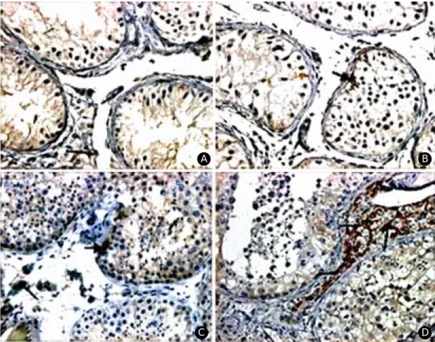
Figure 2. Immunohistochemical stains for VEGF (×400). A; Sertoli cell only syndrome B; maturation arrest C; hypo-
spermatogenesis D; control group Sertoli cell; thick black arrow Leydig cell; thin black arrow.B
D
C
의한 차이를 나타내었으나 정자형성장애 정도에 따 른 유의한 차이는 없었다 (Table 4) (Figure 2).
3) 정세포에서 VEGF와 ET-1 발현의 상호관계 불임군의 정세포에서 VEGF 및 ET-1의 양성률은 각각 32.5% 및 15.0%였으며 대조군에서는 모두 음 성이었다. 따라서 정세포에서 VEGF 및 ET-1의 발 현간에 의미있는 관련성은 없었다 (p>0.05) (Table 5).
3. 호르몬치와 VEGF 및 ET-1의 발현 간의 상 호관계
불임군에서 정세포의 VEGF 양성 및 음성군의 LH, FSH, testosterone, estradiol 및 prolactin 치는 각각 6.40±5.85 mIU/ml 및 5.58±4.20 mIU/ml, 11.16±7.13 mIU/ml 및 13.72±11.62 mIU/ml, 4.69±1.39 ng/ml 및 4.51±1.30 ng/ml, 22.85±12.04 pg/ml 및 31.99±16.71 pg/
Table 5. Correlation with expression of VEGF and
ET-1 on germ cell in infertility groupET-1 Positive
rate (%) Negative rate (%) Positive rate (%) 2 (5.0) 11 (27.5) VEGF
Negative rate (%) 4 (10.0) 23 (57.5) Calculated by Chi-square test
Table 4. ET-1 expression rate on Sertoli cell, Leydig cell and germ cell according to the grade of maturation
Ser (%) Ley (%)
No. cases
+1 +1 +2 Overall
Infertility group 40
A 10 3* (30) 7 (70) -* 7* (70)
B 10 6* (60) 6 (60) -* 6* (60)
C 10 3* (30) 7 (70) -* 7* (70)
D 10 3* (30) 4 (40) -* 4* (40)
Control group 10 - 7 (70) 3 (30) 10 (100)
A; Sertoli cell only syndrome, B; maturation arrest, C; hypospermatogenesis, D; sloughing and disorganization, Ser; Se- rtoli cell, Ley; Leydig cell, Germ; germ cell, *; p<0.05 vs control group. calculated by Mann-Whitney U test
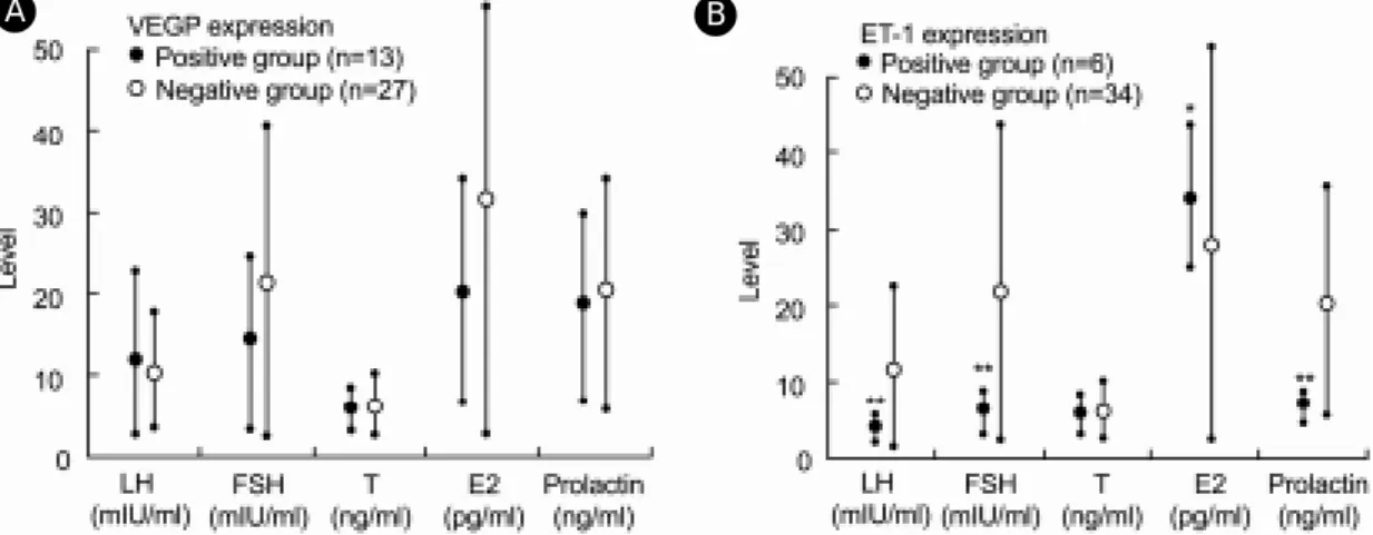
Figure 3. The relationship between serum hormonal level and VEGF, ET-1 expression on germ cell in infertility
group. *; p<0.05, **; p<0.001.A B
ml, 그리고 15.10±9.44 ng/ml 및 11.51±7.39 ng/ml로 양군간에 유의한 차이는 없었다 (p>0.05, 모든 검사 치에서). 그러나 정세포의 ET-1 양성 및 음성군에서 LH, FSH, testosterone, estradiol 및 prolactin 치는 각 각 2.92±1.36 mIU/ml 및 6.36±4.94 mIU/ml, 4.38±
1.74 mIU/ml 및 14.38±10.52 mIU/ml, 4.51±1.65 ng/
ml 및 4.58±1.28 ng/ml, 37.23±6.09 pg/ml 및 27.99±
15.43 pg/ml, 그리고 5.36±0.92 ng/ml 및 13.41±7.85 ng/ml로 testosterone을 제외한 나머지 호르몬은 양군 간에 유의한 차이를 나타내었으나 모두 정상 범위 내에 있었다 (p=0.001, <0.001, >0.05, 0.01, <0.001, 순 서대로) (Figure 3).
고 찰
원시정조세포에서 성숙정자까지 정자형성의 각 단 계를 조절하는 인자를 찾을 수 있다면, 고환의 정자 형성기능 장애의 치료뿐만 아니라 체외에서 미성숙 정자의 성숙을 조장할 수도 있을 것으로 생각된다.
현재 보고된 정자형성 과정에 영향을 미치는 인자 로는 호르몬, 외상, 감염, 성장인자의 소실, 독성물질, 고열 및 방사능 외에 세포고사를 일으키는 유발유 전자로 c-myc, p53, Bax 및 Fas, 억제유전자로 Bcl-2, c-kit 등이 있으며 다른 여러 물질들도 연구 중에 있다.12~16
VEGF는 종양세포에서 가장 먼저 검출되었으며 그 기능으로는 혈관투과성을 증가시키고 발생 초 기에 혈관신생 (vasculogenesis), 성숙 혈관계에서는 내피세포의 유지 및 성장 촉진과 혈관 형성 (angio- genesis)이며, 그 외에 배발생 (embryogenesis), 상처 치유과정 (wound healing), 종양 성장 (tumor growth), 심근 허혈 (myocardial ischemia), 안구내 신생혈관질 환 (ocular neovascular disease) 및 만성 염증질환 등 과 관련이 있는 것으로 알려져 있다.17
VEGF의 작용은 VEGF 수용체와의 결합에 의해 효과를 나타나게 되므로 현재까지는 고환내 세포에 서 이들의 검출 유무로 작용을 추측하고 있는 실정 이다. Ergϋn 등5에 의하면 VEGF는 Leydig 세포와 Sertoli 세포에 분포하며 고환내 혈관에는 검출되지 않았으나 VEGF 수용체는 Leydig 세포, Sertoli 세포 및 고환내 미세혈관에도 존재하여 자가분비 (auto-
crine effect)와 주변분비효과 (paracrine effect)에 의해 작용한다고 하였다. 그러나 이와같은 정세관 주변 세포들에서 VEGF 수용체의 결합 후 나타나는 정자 형성기능 등의 구체적인 이차 효과에 대해서는 아직 정확히 밝혀지지 않았다.
Huminiecki 등6에 의하면 유전자조작으로 VEGF 발현을 증가시킨 쥐에서 고환내 VEGF가 정세포에 직접 작용하여 정자형성기능을 억제하거나 혈관 내 피세포에 작용하여 간접적으로 불임을 일으킬 수 있 다고 하였으며, 부고환에서도 수축력 증가와 수분흡 수의 장애를 일으켜 정자의 성숙 및 배출을 억제한 다고 하였다. Korpelainen 등7은 VEGF가 혈관 내피 세포의 성장 및 투과성을 조절하는 주요 조절인자 이나 비내피세포에도 작용을 한다고 하였으며, 유전 자조작으로 VEGF를 과발현시킨 쥐에서 불임이 유 발된다고 하였는데, 이는 고환내에서 정세포를 통해 조정과정을 직접 억제하거나 미세혈관의 수와 크기 가 증가되어 전체적인 고환의 온도가 상승하여 유 발된다고 하였다. 그리고 부고환에서도 섬유소원 (fi- brinogen)의 누출에 의해 강내에 섬유소 (fibrin)가 축 적되거나 재흡수가 원활히 되지 않아 부고환의 부 종을 일으킨다고 하였다.
또한 Obermair 등18에 의하면 체외수정 (in vitro fer- tilization, IVF)을 시행한 사람의 정액내 VEGF가 정 상 수준 이상으로 증가할 경우나 정상 수준 이하로 감소할 경우 임신률이 의미 있게 감소한다고 하였는 데, 이는 VEGF 증가시 불임을 야기시킬 수도 있지 만 정상 고환기능 및 조정기능을 위해서는 적정 수준 의 고환내 농도가 유지되어야 한다는 것을 시사한다.
본 연구에서도 Leydig 세포에서는 VEGF의 발현 이 양군의 모든 조직에서 관찰되었으나 정세포와 Se- rtoli 세포에서는 불임군에서 대조군에 비하여 VEGF 의 발현이 증가하였다. 불임 A군의 경우 정세포가 존재하지 않기 때문에 모두 음성이었으며 대조군에 서도 정세포에 염색이 되는 경우는 없었으나 불임 B, C 및 D군의 정모세포에서는 대조군에 비해 높 은 양성률을 나타냈으며 그 중에서도 불임 C군이 가 장 높은 양성률을 나타내었다. 즉 고환내에서 VEGF 는 비정상적 정자형성 단계에서 특히 정세포내 발현 이 증가하였다.
Endothelin 중 ET-1은 1988년 Yanagisawa에 의해
처음 발견된 가장 강력한 혈관수축물질로, norepin- ephrine보다 100배, angiotensin II보다 10배 강력한 작용을 한다.20,21 ET-1은 고환내 Sertoli 세포와 Leydig 세포에서 합성되며 고환내 농도는 혈중 농도의 100 배 가량 된다.21,22 ET-1은 Leydig 세포, Sertoli 세포 에 주로 분포하며 그 외에 정자세포에 분포하며 정 세포 및 정조세포에 존재한다는 보고도 있었다.8,9
수용체는 ETA와 ETB가 있으며 ETA는 주로 ET-1 과 결합을 하나 ETB는 다른 모든 아형과 비슷한 친 화도를 가지고 결합한다.9,23 혈관 평활근내 ETA는 혈관수축과 평활근세포의 성장을 조절하고 ETB 수 용체를 통하여 혈관확장을 일으킨다.24,25 ETA와 ETB
는 Leydig 세포, Sertoli 세포 외에 ETA는 정자세포 에 ETB는 미세혈관과 고유층 (lamina propria)에 존 재한다고 하였다.9
고환내 ET-1의 기능을 요약하면 혈관수축 및 확 장, 스테로이드 합성 자극, 정자의 유출, 정자형성기 능의 조절 등이라 할 수 있다.8,9,26,27 혈관수축 및 확 장에 대한 효과는 Collin 등27의 Sprague-Dawley rat 을 이용한 실험에서 잘 나타나는데, 고환내 ET-1을 직접 주사하였을 때 용량에 따른 혈류의 감소 정 도와 지속시간이 상관관계를 보였다. ET-1의 ETA를 매개로 한 혈관수축은 비교적 서서히 이루어지며 오 래 지속되는 특징이 있으므로 어떤 상황에 대처하 는 급성 반응 시 나타나기보다는 정상적인 혈관 긴 장상태를 유지하는데 중요역할을 하나 ETB는 ET- 1과 ET-3에 비슷하게 반응하여 혈관확장을 일으키 며 nitric oxide (NO)나 prostacyclin 등에 의해서도 유 사반응을 보였다.20
그리고 근육양 세포 (myoid cell)에 작용하여 세정 관 수축력을 조절하는데 Tripiciano 등28에 의하면 이 는 Sertoli 세포에서 ECE-1 (endothelin converting en- zyme-1)의 주기적인 발현에 의해 이루어진다고 하였 다. 그밖에 세정관의 수축을 조절하는 것은 arginine vasopressin, TGF-β, PDGF, oxytocin, prostaglandin 등 이 있으며 정세관의 확장을 일으키는 것은 adreno- medullin이 밝혀져있다.29 본 연구에서는 Sertoli 세포 에서 ET-1이 발현될 경우, 정세관의 수축에 의해 직 경이 감소되지는 않았다. 그 이유는 ET-1 단독으로 정세관의 수축을 조절하는 것이 아니라 앞에서 언급 한 여러 조절인자들이 상호연관 되어지기 때문일
것이다.
본 연구에서는 Leydig 세포에서의 ET-1의 발현은 불임군에서 대조군에 비하여 의미 있게 감소되었으 나 오히려 Sertoli 세포에서는 증가되는 양상을 나타 내었고 정세포에서는 통계학적으로 유의한 변화는 없었다. 즉, 정량적 분석이 미흡하지만 ET-1의 발현 양상이 대조군에서는 Leydig 세포가 주를 이루었으 나 불임군에서는 전체적으로 발현이 감소되면서 Se- rtoli 세포에서는 증가되는 양상을 나타내어 정자형성 기능을 위해선 Leydig 세포에서 ET-1이 필요할 것 이라고 추측할 수 있었다.
Fantoni 등30은 난포자극호르몬을 쥐의 체내 투여 시 용량에 비례하여 ET-1이 감소되는 것을 확인하 였고 Romanelli 등31은 황체호르몬방출호르몬 (leu- tenizing hormone releasing hormone, LHRH)와 ET-1을 동시 투여 시 상승효과를 내어 테스토스테론 분비를 더욱 촉진시킴을 증명하였으며, Ergul 등32은 ET-1 이 정상 Leydig 세포에서 테스토스테론 외에 여러 호르몬 합성에 중요 역할을 한다고 보고하였다.
본 연구에서는 불임군에서 정세포의 VEGF 양성 및 음성군 간에 유의한 차이를 보이가 양성일 경우 에스트라디올만 의미 있게 감소하였으며 나머지 호 르몬은 통계학적 유의성이 없었다. 그러나 정세포내 ET-1이 양성일 경우 황체호르몬, 난포자극호르몬 및 프로락틴은 의미 있게 감소하였으나 에스타라디올 은 증가하였으며 테스토스테론의 변화는 통계학적 유의성이 없었다. 즉, 유사한 혈관영향인자인 VEGF 와 ET-1의 고환내 발현 정도와 혈중 호르몬과의 관 계는 상이하게 나타났다.
이상의 연구 결과 원발성 고환부전에 의한 남성불 임환자에서 여러 가지 원인이나 전신 상태에 의해 고환내 정자형성기능이 영향을 받을 수 있지만, 원 인에 관계없이 VEGF 및 ET-1의 고환내 과분비나 부족 시 정자형성장애의 원인임을 확인하였다. 따라 서 정자형성에 관여하는 고환내 분비물질의 규명은 불임의 원인에 관계없이 고환내 환경을 정자형성에 적합하게 유지하여 원인불명의 불임을 치료할 수 있을 뿐만 아니라 보조생식술을 위한 미성숙 정세포 의 체외 배양 및 성숙에도 이용될 수 있을 것으로 생 각된다.
참 고 문 헌
1. Hargreave TB. Introduction. In: Hargreave TB, Soon TE, editors. The management of the male infertility.
Singapore: PG Publishing 1990; 2-21.
2. Hedger MP, Meinhardt A. Cytokines and the imm- une-testicular axis. J Reprod Immunol 2003; 58: 1- 26.
3. Holstein AF, Schulze W, Davidoff M. Understanding spermatogenesis is a prerequisite for treatment. Re- prod Biol Endocrinol 2003; 1: 107.
4. Huleihel M, Lunenfeld E. Regulation of spermatoge- nesis by paracrine/autocrine testicular factors. Asian J Androl 2004; 6: 259-68.
5. Ergϋn S, Kilic N, Harneit S, Paust HJ, Ungefroren H, Mukhopadhyay A, et al. Microcirculation and the va- scular control of the testis. Adv in Experim Med and Biol 1997; 424: 163-80.
6. Huminiecki L, Chan HY, Lui S Poulsom R, Stamp G, Harris AL, Bicknell R. Vascular endothelial growth factor transgenic mice exhibit reduced male fertility and placental rejection. Mol Hum Reprod 2001; 7:
255-64.
7. Korpelainen EI, Karkkainen MJ, Tenhunen A, La- kso M, Rauvala H, Vierula M, et al. Overexpression of VEGF in testis and epididymis causes infertility in transgenic mice: Evidence for nonendothelial tar- gets for VEGF. J Cell Biol 1998; 143: 1705-12.
8. Maggi M, Barni T, Orlando C, Fantoni G, Finetti G, Vannelli GB et al. Endothelin-1 and its receptors in human testis. J Androl 1995; 16: 213-24.
9. Ergϋn S, Harneit, Paust HJ, Mukhopadhyay AK, Ho- lstein AF. Endothelin and endothelin receptor A and B in the human testis. Anat Embryol 1999; 199:
207-14.
10. Nalbandian A Dettin L, Dym M, Ravindranath N.
Expression of vascular endothelial growth factor re- ceptors during male germ cell differentiation in the mouse. Biol of Reprod 2003; 69: 985-94.
11. Senger DR, Connelly DT, Van De Water L, Feder J,
Dvorak HF. Purification and NH2-terminal amino se- quence of guinea pig tumor secreted vascular perm- eability factor. Cancer Res 1990; 50: 1774-8.
12. Blanchard KT, Allard EK, Boekelheide K. Fate of germ cells in 2, 5-hexanedione-induced testicular in- jury. Toxicol Appl Pharmacol 1996; 136: 5-12.
13. Richburg JH, Boekelheide K. Mono- (2-ethyhexyl) phtalate rapidly alters both Sertoli cell vimentin fila- ments and germ cell apoptosis in young rat testes.
Toxicol Appl Pharmacol 1996; 137: 42-50.
14. Kim ED, Barqawi AZ, Seo JT, Meacham RB. Apo- ptosis: its importance in spermatogenic dysfunction.
Urol Clin North Am 2002; 29: 755-65.
15. Zhang XW, Xu B. Differential regulation of p53, c- myc, Bcl-2, Bax and AFP protein expression, and caspase activity during 10-hydroxycamptochecin- induced apoptosis in Hep G2 cells. Anticancer Drugs 2000; 11: 747-56.
16. 양주헌, 박현준, 박남철. 정자형성시 세포고사에 대한 c-kit 유전자의 면역조직화학적 연구. 대한 비뇨회지 2003; 44: 1124-30.
17. Zachary I. Vascular endothelial growth factor. Int J Bio & Cell Biol 1998; 30: 1169-74.
18. Obermair A, Obruca A, Phl M, Kaider A, Vales A, Leodolter S, et al. Vascular endothelial growth factor and its receptor in male fertility. Fertil Steril 1999;
72: 269-75.
19. Yanagisawa M, Kurihara H, Kimura S, Tomobe Y, Kobayashi M, Mitsui Y et al. A novel vasoconstr- ictor poptide produced by vascular endothelial cells.
Nature 1988; 332: 411-5.
20. Ellahham SH, Charlon V, Abassi Z, Calis KA, Cho- ucair WK. Bosentan and the endothelin system in co- ngestive heart failure. Clin Cardiol 2000; 23: 803-7.
21. Fantoni G, Morris PL, Forti G, Vanelli GB, Orlando C, Barni T et al. Endothelin-1: A new autocrine/
paracrine factor in rat testis. Am J Physiol 1993; 265:
E267-74.
22. Matsumoto H, Suzuki N, Onda H, Fujino M. Abun- dance of endothelin-3 in rat intestine, pituitary gland and brain. Biochem Biophys Res Commun 1989;
164: 74-80.
23. Sakaguchi H, Kozuka M, Hirose S, Ito T, Hagiwara H. Properties and localization of endothelin-1-spe- cific receptors in rat testicles. Am J Physiol 1992;
263: R15-8.
24. Ohlstein EH, Douglas SA. Endothelin-1 modulates vascular smooth muscle structure and vasomotion:
Implications in cardiovascular pathology. Drug De- velop Res 1993; 29: 108-28.
25. Takuwa Y. Endothelin in vascular and endocrine system: Biological activities and its mechanism of action. Endocr J 1993; 40: 489-506.
26. Conte D, Morris PL, Forti G, Vanelli GB, Orlando C, Barni T et al. Endothelin stimulates testosterone se- cretion by rat Leydig cells. J Endocrinol 1993; 136:
R1-4.
27. Collin O, Damber JE, Bergh A. Effects of endothelin- 1 on the rat testicular vasculature. J Androl 1996;
17: 360-6.
28. Tripiciano A, Peluso C, Morena AR, Palombi F, Ste- fanini M, Ziparo E et al. Cyclic expression of endo- thelin-converting enzyme-1 mediates the functional regulation of seminiferous tubule contraction. J Cell Biol 1999; 145: 1027-38.
29. Rossi F, Ferraresi A, Romagni P, Silvestroni L, San- tiemma V. Angiotensin II stimulates contraction and
growth of testicular peritubular myoid cells in vitro.
Endocrinol 2002; 143: 3096-104.
30. Fantoni G, Morris PL, Forti G, Vannelli GB, Orlando C, Barni T et al. Endothelin-1: A new autocrine/
paracrine factor in rat testis. Am J Physiol 1993;
265: E267-74.
31. Romanelli F, Fillo S, Isidori A, Conte D. Stimulatory action of endothelin-1 on rat Leydig cells: Involve- ment of endothelin-A subtype receptor and phospho- lipase A2-arachidonate metabolism system. Life Sci- ences 1997; 61: 557-66.
32. Ergul A, Glassberg MK, Majercik MH, Puett D.
Endothlin-1 promotes steroidogenesis and stimulates protooncogene expression in transformed murine Le- ydig cells. Endocrinology 1993; 132: 598-603.