Research Article Open Access
The effects of the angles of the knee and heel-off on the muscle activity during a bridge exercise
Byeong-jo Kim, PT, PhD⋅Su-kyoung Lee, PT, PhD
†⋅Jung-hoon Lee, PT, PhD⋅Hae-yeon Kwon, PT, MD Department of Physical Therapy, College of Nersing and Healthcare Sciences, Dong-Eui University
Received: July 27, 2015 / Revised: August 3, 2015 / Accepted: August 17, 2015
ⓒ 2015 J Korean Soc Phys Med
| Abstract |
1)PURPOSE: The purpose of this study is to investigate the effects of the angles of the knee and heel-off on the muscle activity during a bridge exercise.
METHODS: 15 healthy adult men and women with the balance ability and joint working range required for performing a bridge exercise participated in this study, in which 120°, 90° and 60° angles of the knee-flexion and heel-off were applied during the bridge exercise.
RESULTS: Our data showed that there were significant differences in muscle activities of elector spinae and rectus abdominis when 120°and 60°angles of the knee were applied, of internal oblique when 120°and 60°were applied, and external oblique when 90°and 60°were applied. When heel-off was applied, there were significant differences in muscle activities of elector spinae and rectus abdominis when 120°and 60°were applied, of internal oblique when 120°and 60°were applied, and external oblique when 90°and 60°were applied.
CONCLUSION: In this study on an application of heel-off to the bridge exercise, we showed that the effect of
†Corresponding Author: [email protected]
This is an Open Access article distributed under the terms of the Creative Commons Attribution Non-Commercial License (http://creativecommons.org/licenses/by-nc/3.0) which permits unrestricted non-commercial use, distribution, and reproduction in any medium, provided the original work is properly cited.
the angles of the knee on the muscle activities of elector spinae, rectus abdominis, internal oblique and external oblique were all similar to the regular bridge exercise, but overall muscle activities were increased with heel-off when compared with the regular bridge exercise.
Key Words: Bridging exercise, Heel off, Knee angle, Muscle activity
Ⅰ. Introduction
The bridge exercise is a trunk stabilization exercise that prevents the damages that result from the repetitive stimulations of various muscles, ligament tissues, and joints around the spine during daily life (Jeon, 2010). Stevens et al (2007) argue that the bridge exercise is a posture that makes patients with back pain comfortable, reducing their pain. It also allows patients to retrain large local muscles to coordinate at the proper ratio. Furthermore, this exercise is frequently performed in clinical settings and prescribed to patients because it is easy for therapists to teach.
An investigation into the effect the bridge exercise has
on muscular activation in normal people revealed that
performing this exercise on an unstable surface increased
the activation of the trunk muscles. Furthermore, starting
Table 1. muscle attachment sit
Muscle Attachment sites
Internal oblique Umbilicus and ASIS in middle region
External oblique 15cm outside from the umbilicus
Rectus abdominis 3cm upperfrom theumbilicus
Erector spine 2cm later from L3 spinal proces
this exercise on a stable floor and changing to an unstable floor improved stability and balance in the lumbar muscles in patients with back pain (Park, 2012). Hong et al (2010) find that setting different support surface stability levels during the bridge exercise contributes to greater muscular activation and endurance. Bjerkerfors et al(2010) report that deep muscle activity may increase when deep abdominal muscle training is applied to the bridge exercise.
The multifidus and transverse abdominis contract earlier than the other muscles involved in stabilization during every body movement to maintain the body’s balance (Hodges and Gandevia, 2000). Furthermore, the internal oblique, external oblique, and quadratus lumborum muscles perform both stabilization and motility functions (Lee et al, 2013).
In this study, the bridge exercise was altered. The heel was raised and the knee angle was changed during the bridge exercise to provide gradual resistance using body weight while simultaneously applying the optimum knee angle at which trunk muscle stabilization increased. Raising the heel and changing the knee angle may be an efficient exercise method. Therefore, the change in muscular activation after the heel was raised and the knee angle was changed during the bridge exercise was examined.
Ⅱ. Method
1. Subjects
The study included 15 healthy male and female adults with the ability to balance and the range of joint motion required to complete the bridge exercise. The participants
also understood the details of study and chose to participate.
The selection criteria were as follows.
- Individuals who had not experienced back pain during the previous six months
- Individuals who had no problem walking independently and had not experienced any muscle strength weakening in their lower limbs and trunk
- Individuals who had no complaints about pain during the motion and no contracture of the hip joints that limited the necessary motion
- Individuals who had no surgical or neurological history related to the musculoskeletal system of the body and lower limbs; and
- Individuals who agreed to participate in the study.
2. Electromyogram and Data Processing
1) Electromyogram (EMG)
In this study, EMG electrodes were attached to the internal oblique, external oblique, rectus abdominis, and erector spine muscles to measure their activation during the bridge exercise. The ground electrode was attached to the dominant anterior superior iliac spine (ASIS) (Arokoski et al, 2001; Cram et al, 1998). To prepare for the EMG measurement, adhesive Ag/AgCl surface electrodes were attached after alcohol disinfection to minimize potential skin resistance at the sites to which the electrodes were attached (Table 1).
2) EMG Data Processing
The EMG was measured using a surface electromyograph
Table 2. General characteristics of subjects (N=15)
Variables Mean±SD Range
Age (yrs) 22.84±2.19 20~28
Height (cm) 164.15±8.30 150~178
Weight (kg) 57.84±11.80 42~80
(Biopac Student Lab MP 36, Biopac Systems, Inc., USA) with a band pass filter that removed the noises from the surrounding environment. Furthermore, the root mean square data of each muscle was measured for five seconds in the anatomical position. The muscle activation was measured three times for five seconds in each posture, and the %RVC value was calculated after the exclusion of one second before and after this measurement. This calculation standardized the data from each participant so it could be comparatively analyzed.
3. Experimental Procedure
1) Bridge Exercise
In this study, based on the bridge exercise that is generally used in clinical settings, the subjects lied down in the supine position with their feet parallel to each other, their soles in full contact with the floor, and both knees bent at 60°. They kept both hands comfortably spread on the floor with their palms facing downward. In this position, the subjects raised their pelvis based on the rater’s signal and aligned their trunk with their pelvis. They maintained this posture for five seconds (Jeon, 2010; Stevens et al, 2006). Before this experiment, each subject practiced the bridging position with their spine and pelvis maintained in the neutral position for 10 minutes and both their arms were positioned at specific angles so their lower limbs stayed parallel and at an appropriate angle. The subjects received continuous feedback to adjust their specified posture.
2) Heel Raising using the Ankle Joint During the Bridge Exercise
The participants’ knees were bent at 120°, 90°, and 60°, and then, their heels were raised during the bridge exercise.
When their heels were raised, the participants were instructed to maintain an angle of maximum contraction for five seconds. The order in which the participants were asked to position their knees in the three knee angles was determined randomly. A five-minute resting period was given after each exercise to avoid muscle fatigue, which can significantly affect the EMG measurements.
4. Statistical Processing
The collected data were analyzed using SPSS 18.0 for Windows. The activation of each muscle measured during the bridge exercise was analyzed using one-way ANOVA.
Each significant difference was tested post-hoc using Bonferroni's correction. The statistical significance level was set to a=0.05.
Ⅲ. Results
1. General Characteristics of the Subjects
The study consisted of 15 normal male and female adult
participants. Their ages ranged from 20 to 28, with a mean
age of 22.84±2.19 years. Their mean height was
164.15±8.30 cm, and their mean weight was 57.84±11.80
kg (Table 2).
Table 3. Comparison of trunk muscle activation among the various bridging exercise position (unit: %RVC)
Muscle 120 90 60 F P
ES
*1171.5±82.54 1374.11±105.64 1614.81±154.28
‡3.53 .038
EO
*185.51±15.65 202.01±18.88 254.38±18.96
‡£4.03 .025
RA
*170.07±11.59 184.32±12.30 220.00±15.48
‡3.77 .031
IO
*250.33±33.78 287.01±34.09 392.61±40.01
‡£4.19 .022
Mean±SE,
*p<.05
ES: elector spinae, EO: external oblique, RA: rectus abdominis, IO: internal oblique
‡
significant difference between 120° and 60° (P<0.05).
£
significant difference between 90° and 60° (P<0.05).
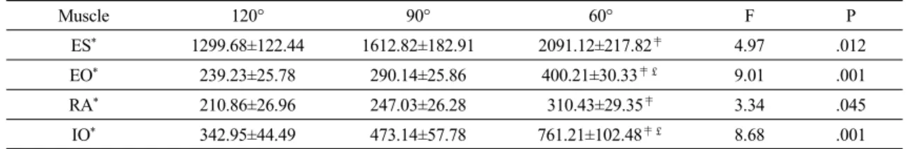
Table 4. Comparison of trunk muscle activation among the various bridging exercise position with heel off
Muscle 120° 90° 60° F P
ES
*1299.68±122.44 1612.82±182.91 2091.12±217.82
‡4.97 .012
EO
*239.23±25.78 290.14±25.86 400.21±30.33
‡£9.01 .001
RA
*210.86±26.96 247.03±26.28 310.43±29.35
‡3.34 .045
IO
*342.95±44.49 473.14±57.78 761.21±102.48
‡£8.68 .001
Mean±SE,
*p<.05
ES: elector spinae, EO: external oblique, RA: rectus abdominis, IO: internal oblique
‡
significant difference between 120° and 60° (P<0.05).
£