I. Introduction
The filling of bone defects resulting from trauma or periodontal disease requires bone grafts1). Autogenous bone grafts are some disadvatages relat- ed to this modality, especially lack of sufficient avail- able bone and the need for a second intraoral or extraoral surgical site, which increases patient incon- venience and morbidity2). Therefore, there have led to the development of new synthetic or natural bone substitutes2).
Bovine derived xenograft (BDX) is a natural bone substitute made from bovine bone via a proprietary extraction procedure. Osteoblasts form a layer on the BDX mesh and, osteoid and finally lamellar bone covers BDX trabecullae. BDX scaffold persists for some time which permits the correction of alveo- lar defects with permanent results 4).
Biomaterials, such as calcium phosphate ceramics, appear to be suitable alternatives to bone grafts.
Biphasic calcium phosphate(BCP) ceramics prepared from a close association of hydroxyapatite (HA) and
β-tricalcium phosphate(β-TCP) were developed in the 1980s5). Calcium phosphate ceramics have been used mostly because of their close chemical and crystal resemblance to bone mineral. BCP is biocom- patible and osteoconductive; it is able to promote new bone formation on contact, but it seems to have no intrinsic osteoinductive properties6). Macroporous type of BCP have been used for bone substitution for orthopedic surgery, and was carried out lots of experiments in vivoand in vitrofor a long time5).
Recently, The microstructure in the macropore surface enlarged greatly the surface area for protein adsorption, more proteins could be absorbed on the surface; the larger surface area could also facilitate ion exchanges and bone-like apatite surface forma- tion by the process of dissolution and re-precipita- tion7). Therfore, the micro- and macro-porous BCP may be more favorable for bone ingrowth. But, micro- and macro- porous BCP has not been stud- ied sufficiently yet. The purpose of this study was to evaluate the potential of micro- and macro porous BCP compared with BDX to stimulate bone regener-
Effects of biphasic calcium phosphate on bone formation in human fetal osteoblasts
Kye-Chul Shin, Kil-Young Jang, Myoung-Ku Lee, Ho-Sang Yoon, Jae-Bong Song, Hyun-A Kim, Sung-Hee Pi, Hyung-Shik Shin, Hyung-Keun You
Department of Periodontology, School of Dentistry, Wonkwang University
대한치주과학회지 : Vol. 35, No. 1, 2005
Corresponding author : You Hyung-Keun, Department of periodontology, School of Dentistry, Wonkwang University, 344-2 Shinyong-dong, Iksan, Chonbuk, 570-749 Korea
ation in periodontal and implant dentistry.
II. Materials and methods
1. Culture of human fetal osteoblastsHuman fetal osteoblastic cell line (hFOB 1.19 ; American Type Culture Collection, Manassas, VA, USA) that have the ability of production and calci- fication of bone matrix protein22)were plated at 5×
104 cells/well of 6-well plate containing 2 ml of DMEM/F-12 HAM (Sigma, St. Louis, MO, USA), with 10% FBS (Gibco BRL, Grand island, NY, USA) and 0.03mg/ml of G-418 (Duchefa, Netherlands). Cells were cultured at 34℃ in an atmosphere of 5% CO2, 95% air and 100% humidity. The four groups of dif- ferent concentrations within 1㎍/m~1ng/ml in both BDX (Bio-Oss , Geistlich-Pharma, Wolhusen, Switzerland) and BCP (MBCP , Biomatlante, France) were added in each experiment.
2. Cell number counting
hFOB 1.19 were plated in 6-well culture dishes at 1×10⁴cells/well. After 24 h, cells were exposed to BDX and BCP. The number of viable cells after try- pan blue exclusion was counted under microscope at 3 and 5 days of incubation. There were four cul- tures in each group at each time.
3. Collagen synthesis analysis
To measure the total collagen synthesis of hFOB 1.19 indirectly, hydroxyproline contents were mea- sured (Rojkind et al.1979). Briefly, hFOB 1.19 were plated in 60-mm plates at 3×10⁵ cells with 50 ㎍/ml ascorbic acid and 10mM sodium β-glycerophos- phate. After cells were reached a confluence, BDX and BCP were added and cultured 3 days more.
After hydrolyzing at 100℃ for 24 hours, samples were filtered, collected, and completely dried at 60℃
and 50㎕ methanol was added for removing of HCl.
Remaining precipitates were dissolved with 1.2 ml of 50% isopropanol, and placed at room temperature for 10 minutes after mixing with 200㎕ chloramin-T solution (Sigma). 1.0 ml of Ehrlich reaction reagent (Sigma) was mixed and cultured at 50℃ for 90 min- utes, and chilled at room temperature. Collagen syn- thesis was measured at 557 nm wave length using spectrophotometer (Beckman, Fullerton, CA, USA).
And the standard concentrations of protein were cal- culated using BCA protein assay reagent (Pierce, Rockford, IL, USA). Results were expressed as colla- gen/total protein(㎍/mg/ml).
4. Alkaline Phosphatase (ALP) activity
After hFOB 1.19 were plated in 6-well plates at 1
×105cells/well, it was cultured until a confluence in 50 μg/ml ascorbic acid, 10 mM sodium β-glyc- erophosphate. After BDX and BCP were added, each group was incubated for 3 days more. Each 0.1 ml of suspension was mixed with 0.1 M glycine NaOH buffer (pH 10.4) 0.2 ml, 15 mM pNPP (para- nitrophenyl phosphate, Sigma) 0.1 ml, 0.1% Triton X-100/saline 0.1 ml and 0.1 ml of sterilized distilled water, and the reaction was stopped by 0.1 N NaOH 0.6 ml. The cultured cells were transported on 96- well plate and the absorbance was measured at 410 nm in ELISA reader. And the standard concentrations of protein were calculated using BCA protein assay reagent (Pierce). Results were expressed as nmol of para-nitrophenol released per min/mg protein.
5. Western blot analysis
hFOB 1.19 plated in 100-mm dishes at 5×10⁵ cells/well were reached a confluence, 1ng/ml of
BDX and BCP were added in experimental group.
And then each group was incubated for 21 days more. Protein was isolated from cells using lysis buffer under the conditions recommended by the manufacturer. Protein concentration was deter- mined by BCA solution. The denatured supernatant containing 100 ㎍ of protein was electrophoresed in a 15% SDS-polyacrylamide gel and transferred onto a PVDF (Immobilon-P membrane, Milipore, Bedford, MA, USA). To reduce nonspecific antibody binding, the membrane was incubated in a blocking solution (Zymed, San Francisco, CA, U.S.A.) for 1 hours at room temperature. And then osteocalcin(OC, Biogenesis, Kingston, NH, USA) and bone sialoprotein(BSP, Chemicon, Temecula, CA, USA) as first antibody activated for 90 minutes.
After washing with PBS, the membrane was treated with anti-mouse IgG-alkaline phosphatase conjugat- ed secondary antibody for 1 hours, and washed again with PBS. The membrane was then incubated in ECL Western Star Substrate reagent (Amersham, Buckinghamshire, UK), and exposed to Hyperfilm- MP (Amersham) for a few minutes.
6. Statistical analysis
Statistical significance was evaluated for by one way analysis of variance (ANOVA) using SPSS (v10.0, Chicago, IL, USA) program of computer. The mean, and the standard deviation was calculated for each variable. Differences were considered statisti- cally significant at P<0.05.
III. Results
1. Cell number counting of BDX and BCP on hFOB 1.19
Cell proliferation by BDX and BCP was deter- mined by cell number counting (Zhang et al., 2000;
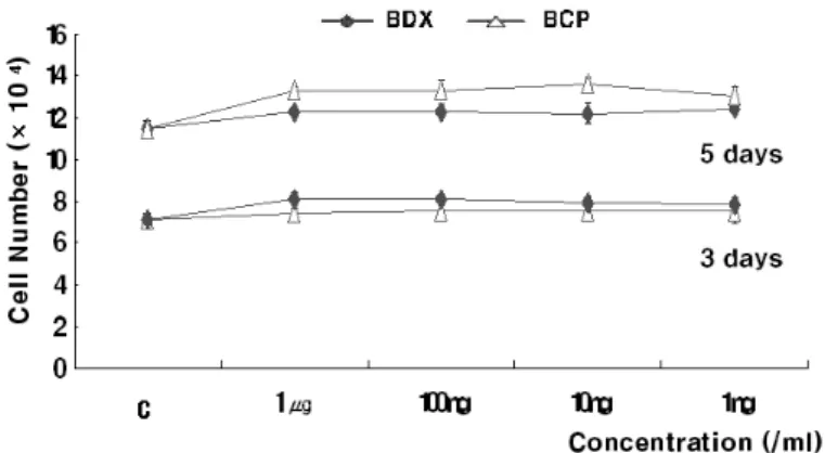
Watson et al., 1998). The proliferation results of the cultures on control and experimental groups are summarized in Figure 1. BDX and BCP treated did not show a significant increase than control at 3 and 5 days, indicating that BDX and BCP have no effect on cell proliferation. And, there were no significant differences between BDX and BCP, and between each concentration of BDX and BCP (P>0.05).
Figure 1. Effect of BDX and BCP on cell proliferation of hFOB 1.19.
hFOB 1.19 were plated in a 6-well plate at 1×10⁴cells/well and cultured in EMDM/F-12 HAM containing BDX and BCP for 3 and 5 days. Values represent averages from four independent experiments and standard devi- ation. C : control
2. Collagen synthesis of BDX and BCP on hFOB 1.19
The measurement of collagen synthesis is demon- strated as Figure 2. Collagen synthesis increased in all experimental groups. 1ng/ml(5.79±0.31) and 10ng/ml(5.64±0.29) of BDX and 1ng/ml(5.96±0.09), 10ng/ml(6.19±0.28) and 100ng/ml(5.89±0.29) of BCP were significantly increased compare with the
control group. Especially, 10ng/ml(5.64±0.29) of BDX and 10ng/ml(6.19±0.28) of BCP showed the highest collagen synthesis than other groups. No sig- nificant difference was observed between BDX and BCP , and between each concentration of BDX and BCP in the quantity of collagen synthesis (P<0.05).
3. ALP activity of BDX and BCP on hFOB 1.19
Figure 2. Collagen synthesis of hFOB 1.19 treated with BDX and BCP(㎍/mg/ml). Cells were plated in 100- mm plates at 5×10⁴ cells/well and cultured in EMDM/F-12 HAM until cells reached a confluence.
After that, BDX and BCP were added and cultured for another 3 days more. Values represent aver- ages from each independent experiments and standard deviation. C : control
*and†: Statistically significant difference compared with control (p<0.05)
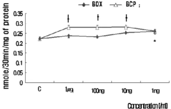
Figure 3. Effect of BDX and BCP on ALP activity of hFOB 1.19.
hFOB 1.19 were plated in 6-well plates at 1×10⁵ cells/well and cultured in EMDM/F-12 HAM until cells reached a confluence. After that, BDX and BCP were added and cultured for another 3 days more. Values represent averages from each independent experiments and standard deviation. C : control
*and†: Statistically significant difference compared with control (p<0.05)
Increased ALP activity is associated with an increase in osteoblastic differentiation. The measure- ment of ALP activity is demonstrated as Figure 3.
Generally, ALP activity is increased with the reduc- tion of concentrations. 1ng/ml(0.26±0.01) of BDX and 10ng/ml(0.28±0.02), 100ng/ml(0.28±0.01) and 1㎍/ml(0.28±0.02) of BCP were significantly increased compare with control group. Especially, 1ng/ml(0.26±0.01) of BDX and 10ng/ml(0.28±
0.02) of BCP showed the highest ALP activity than other groups. No significant difference was observed between BDX and BCP, and between each concentration of BDX and BCP in ALP activity (P<0.05).
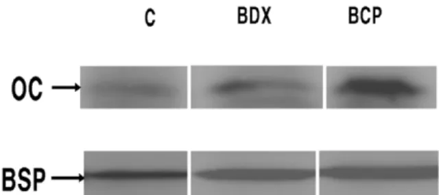
4. Expression of OC and BSP in hFOB 1.19
Because OC and BSP, non-collagenous matrix proteins, is one of the major markers of bone forma- tion in vitro, the expression level of OC and BSP was investigated using western blot analysis. As shown in Figure 4, OC and BSP in hFOB1.19 were increased to 1ng/ml of BDX and BCP. Western blot analysis indicated that the expression of OC in BCP were increased than that of BDX. But, there is no
difference between BDX and BCP in the expression of BSP.
IV. Discussion
The aim of the present study was to evaluate that BCP will be a usable bone grafting material in den- tistry by comparing it with commonly used xenogenic bone BDX.
The present study has performed a preliminary research (data not shown) on collagen synthesis and ALP activity at concentrations of 100mg/ml~1ng/ml, and we obtained proper results at 1㎍/ml~1ng/ml.
Thus, four groups of different concentration within 1
㎍/m~1ng/ml were used in this experiment method.
The osteoblastic cell proliferation indicates that cells were responsive to mitogenic factors present in the graft material6). In previous studies, Stephan et al. reported that they cultured rat calvarial osteoblasts on BDX and they found no significant differences in cell proliferation8). Aybar B. found no significant differences in various calcium phosphate materials on cell proliferation using neonatal rat cal- varial osteoblasts9). Toquet J. reported that the cell number was not different on BCP and control, indi- cating that BCP did not modify human mesenchy-
Figure 4. Western blot analysis for intracellular levels of OC and BSP in hFOB 1.19. First lane is control group and the second is BDX group, and the last one is BCP group. Cell extract equivalent to 100μg/ml of total cellular protein of hFOB 1.19 was electrophoresed by 15% SDS-PAGE and transferred to a PVDF membrane. The intracellular protein levels of OC and BSP in hFOB 1.19 was probed with anti- bodies diluted by 1:1000
mal stem cell proliferation10). These results support- ed that no statistically significant increase in the osteoblastic cell proliferation were detected for BDX and BCP compare with the control in the present study.
Type I collagen is important protein secreted from osteoblasts and exists inside the bone11). Collagen have the largest part of bone tissue among organic matters. If it is not produced properly, the activity of ALP and the production of osteocalcin appear to be quite low12). According to the result of analyzing the ability of collagen synthesis, the BDX and BCP showed a significant increase in our study(figure 2), which is similar to the results of research by the other studies that both BDX and HA showed an increase compared to control group13),14),15). Although the previous report in which BDX enhanced collagen synthesis to much more than that of HA14), this study dose not show significant differences between BDX and BCP. This difference seems that cell type used (human fetal osteoblast cells vs MC3T3-E1 cells) and, tricalcium phosphate in BCP which, accelerated cell differentiation16).
ALP activity is associated with primary calcification in a number of tissues including bone10). Although the function of skeletal ALP in vivois unclear yet, the enzyme is thought to be involved in bone for- mation and calcification. In our work, progressive elevation of ALP activity in both BDX and BCP sug- gests that cells are entering the osteogenesis cascade prior to mineralization, because the elevation of ALP activity reflects an earlier stage of osteoblast differen- tiation17). In previous studies, Hofman et al. report- ed that ALP activity was significantly increased in two different BDX materials in cultured rat calvarial osteoblasts17). And, Sun et al. reported that β-TCP and HA in various calcium phosphate materials is shown a significant increase of ALP activity in rat calvarial osteoblasts. These results supported the
cell differentiation capability of BCP used in our study18). In addition to, Alarm et al. have investigat- ed the pellet-shaped implants prepared from BCP with different ratios of HA and TCP as a carrier of rhBMP-2. It was found that 100% HA and 75%
HA/25% TCP showed higher ALP activity than 25%
HA/75% TCP6). But, 100% HA is very slowly resorbed in clinic and hampers rapid bone turnover in the affected area. Therefore, using a combination of HA and TCP, bone replacement can be enhanced since TCP displays better bioresorption. In the pre- sent study, BCP is composed of 60% HA/40% TCP, and ALP activity is increased significantly similar to results of Alarm et al.6).
BSP and OC are referred to as a marker of the late stage of osteoblastic differentiation in the process of bone mineralization and maturation19). BSP can serve as a hydroxyapatite nucleation center and is essential for matrix mineralization19). BSP has a spe- cific role during the initial phases of bone formation at the cartilage and bone interface19). OC is one of important noncollagenous proteins in the connec- tive tissue resulting from the mineralization of verte- brates20). Also, mRNA of BSP is expressed by osteoblasts earlier than mRNA of OC21). We attempt- ed to detect BSP and OC by Western blotting using a monospecific antibody. The present study used 1ng/ml of BDX and BCP because it is shown a dis- tinct increase in ALP activity and collagen synthesis assay. It was resulted in an incresed expression of BSP and OC compare with control. These mean that BDX and BCP accelerated the mineralization of matrix and the late stage of osteoblast differentia- tion.
These findings suggest that BCP and BDX may have a differentiating effect on osteoblasts with an improvement of bone matrix synthesis, and without effects of cellular proliferation, which is typical in differentiated cells. The in vitro approach with
human osteoblasts may be a useful model for the assessment of cellular response to new biomaterials prior to their applicationin vivo.
V. Acknowledgement
This paper was supported by Wonkwang University in 2004.
VI. References
1. Artzi Z, Nemcovsky CE, Tal H. Efficacy of porous bovine bone mineral in various types of osseous deficiencies: clinical observations and literature review. Int J Periodontics Restorative Dent 2001;21:395-405.
2. Becker W, Urist M, Becker BE, Jackson W, Parry DA, Bartold M, Vincenzzi G, De Georges D, Niederwanger M. Clinical and histologic obser- vations of sites implanted with intraoral autolo- gous bone grafts or allografts. 15 human case reports. J Periodontol 1996;67:1025-33.
3. Simion M, Dahlin C, Trisi P, Piattelli A.
Qualitative and quantitative comparative study on different filling materials used in bone tissue regeneration: a controlled clinical study. Int J Periodontics Restorative Dent 1994;14:198-215.
4. Benke D, Olah A, Mohler H. Protein-chemical analysis of Bio-Oss bone substitute and evidence on its carbonate content. Biomaterials 2001;22:1005-1012.
5. Gauthier O, Bouler JM, Aguado E, Pilet P, Daculsi G. Macroporous biphasic calcium phos- phate ceramics: influence of macropore diame- ter and macroporosity percentage on bone ingrowth. Biomaterials 1998;19:133-139.
6. Alam MI, Asahina I, Ohmamiuda K, Takahashi K, Yokota S, Enomoto S. Evaluation of ceramics composed of different hydroxyapatite to tricalci-
um phosphate ratios as carriers for rhBMP-2.
Biomaterials 2001;22:1643-51.
7. Yuan H, Kurashina K, de Bruijn JD, Li Y, de Groot K, Zhang X. A preliminary study on osteoinduction of two kinds of calcium phos- phate ceramics. Biomaterials 1999;20:1799-806.
8. Stephan EB, Jiang D, Lynch S, Bush P, Dziak R.
Anorganic bovine bone supports osteoblastic cell attachment and proliferation. J Periodontol 1999;70:364-9.
9. Aybar B, Bilir A, Akcakaya H, Ceyhan T. Effects of tricalcium phosphate bone graft materials on primary cultures of osteoblast cells in vitro. Clin Oral Implants Res 2004;15:119-125.
10. Toquet J, Rohanizadeh R, Guicheux J, Couillaud S, Passuti N, Daculsi G, Heymann D. Osteogenic potential in vitro of human bone marrow cells cultured on macroporous biphasic calcium phosphate ceramic. J Biomed Mater Res 1999;44:98-108.
11. Mizuno M, Kitafima T, Tomita M, Kuboki Y. The osteoblastic MC3T3-E1 cells synthesized C-termi- nal propeptide of type I collagen, which pro- moted cell-attachment of osteoblasts. Biochim Biophys Acta 1996;1310:97-102.
12. Kim, SY. et. al. Effects of orthovanadate on col- lagen and fibronectin synthesis, and alkaline phosphatase activity in MC3E3-E1 osteoblast cells. J of Korean Orthop Assoc 2003;38:133-41.
13. Zambonin G, Grano M. Biomaterials in orthopaedic surgery: effects of different hydrox- yapatites and demineralized bone matrix on pro- liferation rate and bone matrix synthesis by human osteoblasts. Biomaterials 1995;16:397- 402.
14. Matsumoto T, Kawakami M, Kuribayashi K, Takenaka T, Minamide A, Tamaki T. Effects of sintered bovine bone on cell proliferation, colla- gen synthesis, and osteoblastic expression in
MC3T3-E1 osteoblast-like cells. J Orthop Res 1999;17:586-92.
15. Zambonin G, Camerino C, Greco G, Patella V, Moretti B, Grano M. Hydroxyapatite coated with heaptocyte growth factor (HGF) stimulates human osteoblasts in vitro. J Bone Joint Surg Br 2000;82:457-60.
16. Ehara A, Ogata K, Imazato S, Ebisu S, NakanoT, Umakoshi Y. Effects of alpha-TCP and TetCP on MC3T3-E1 proliferation, differentiation and min- eralization. Biomaterials 2003;24:831-6.
17. Hofman S, Sidqui M, Abensur D, Valentini P, Missika P. Effects of Laddec on the formation of calcified bone matrix in rat calvariae cells cul- ture. Biomaterials 1999;20:1155-66.
18. Sun JS, Tsuang YH, Liao CJ, Liu HC, HangYS, Lin FH. The effects of calcium phosphate particles on the growth of osteoblasts. J Biomed Mater
Res 1997;37:324-34.
19. Mizuno M, Imai T, Fujisawa R, Tani H, Kuboki Y. Bone sialoprotein (BSP) is a crucial factor for the expression of osteoblastic phenotypes of bone marrow cells cultured on type I collagen matrix. Calcif Tissue Int 2000;66:388-396.
20. Ducy P, Schinke T, Karsenty G. The osteoblast:
a sophisticated fibroblast under central surveil- lance. Science 2000;289:1501-4.
21. Duarte WR, Shibata T, Takenaga K, Takahashi E, Kubota K, Ohya K, Ishikawa I, Yamauchi M, Kasugai S. S100A4: a novel negative regulator of mineralization and osteoblast differentiation. J Bone Miner Res 2003 ;18:493-501.
22. Harris SA, Enger RJ, Riggs BL, Spelsberg TC.
Development and characterization of a condi- tionally immortalized human fetal osteoblastic cell line. J Bone Miner Res 1995;10:178-86.
- 국문초록 -
Biphasic Calcium Phosphate가 태아골모세포의 골 형성에 미치는 영향
신계철, 장길용, 이명구, 윤호상, 송제봉, 김현아, 피성희, 신형식, 유형근 원광대학교 치과대학 치주과학 교실
목
목적적 :: 이 연구의 목적은 치과 영역에서 골 재생을 촉진하기 위해, 현재 많이 사용하고 있는 BDX(bovine-
derived xenograft)와 비교하여 BCP(biphasic calcium phosphate)의 효과를 알아보기 위함이다.
실
실험험 재재료료 및및 방방법법:: 본 연구는 태아골모세포주(hFOB 1.19)를 사용하였으며, 사용된 골 이식재에 따라 2개의 실험군으로 구분하였고, 각 실험에 적절한 농도의 BDX와 BCP를 첨가하였다. 그리고, 세포 증식도 검사, 교원 질 합성량 분석, 염기성 인산분해효소 활성도 측정, Western blot 분석을 통한 OC과 BSP의 발현 정도등의 실험 을 진행하였다.
결
결과과 :: BDX와 BCP는 대조군과 비교하여 세포 증식에서 유의한 차이가 없었지만, 교원질 합성량, 염기성 인
산분해효소의 활성, 그리고 OC과 BSP의 발현에 있어 대조군과 비교하여 유의하게 증가를 보였다. 그러나, 두 이식재간의 유의한 차이는 보이지 않았다.
결
결론론 :: 본 실험실적 연구에서 BCP는 골모세포분화에 긍정적인 영향을 미침으로써 효과적인 이식재로 사용할
수 있음을 가늠할수 있었다.
주요어 : In vitro; bone regeneration; biphasic calcium phosphate; Xenografts