Odontogenic infection involving the secondary fascial space in diabetic and non-diabetic patients: a clinical comparative study
Je-Shin Chang, Kil-Hwa Yoo, Sung Hwan Yoon, Jiwon Ha, Seunggon Jung, Min-Suk Kook, Hong-Ju Park, Sun-Youl Ryu, Hee-Kyun Oh
Department of Oral and Maxillofacial Surgery, School of Dentistry, Chonnam National University, Gwangju, Korea
Abstract(J Korean Assoc Oral Maxillofac Surg 2013;39:175-181)
Objectives: This retrospective study was performed to evaluate the clinical impact of diabetes mellitus on the prognosis in secondary space infection.
Materials and Methods: Medical records, radiographic images, computed tomography, and microbial studies of 51 patients (25 diabetic patients and 26 non-diabetic patients) were reviewed. Patients were diagnosed as secondary fascial space infections with odontogenic origin and underwent treatment at Chonnam National University Hospital, in Department of Oral and Maxillofacial Surgery, from January 2007 to February 2009.
Results: Compared to patients without diabetes, patients with diabetes were presented with the following characteristics: older age (diabetic patients:
62.9 years, non-diabetic patients, 47.8 years), more spaces involved (diabetic patients, 60%; non-diabetic patients, 27.3%), more intense treatment, longer hospitalization (diabetic patients, 28.9 days; non-diabetic patients, 15.4 days), higher white blood cell and C-reactive protein values, higher incidence of complication (diabetic patients, 40%; non-diabetic patients, 7.7%), and distinctive main causative microorganisms.
Conclusion: These results suggest that the prognosis of diabetic patients is poorer than that of non-diabetic patients in secondary space infections since they had greater incidence rates of involved spaces, abnormal hematologic findings, more complications, and additional procedures, such as tracheostomy.
Key words: Diabetes mellitus, Diabetes complications, Bacterial infections, Abscess, Cellulitis
[paper submitted 2013. 6. 17 / revised 2013. 7. 31 / accepted 2013. 8. 5]
susceptible to complications or infections of the blood vessels because of the functional alteration of the polymorphonuclear leukocytes in diabetic patients. Moreover, diabetic patients are reported to have problems with chemotaxis, phagocytosis, and bactericidal function, with gram-negative bacilli species as the main causative infectious bacteria in the oral cavity6.
There are two routes of infection in the oral and maxillofacial area: one is the route via the root apex, and the other is the route via the deep periodontal pocket. The fascial spaces that could be directly affected by odontogenic infections are called
‘primary spaces’, and they include canine, infratemporal, buccal, submental, submandibular, and sublingual spaces.
Failure to control the infections may cause them to spread to secondary spaces including temporal, masseteric, pterygo- mandibular, lateral pharyngeal, retropharyngeal, and pre- vertebral spaces7. It is difficult to treat patients who have these space infections without drainage of purulent exudates, since those are connected to primary spaces and are surrounded by connective tissues that have poor blood supply. If the infections of diabetic patients spread to secondary spaces,
I. Introduction
Diabetic mellitus (DM) can cause diabetic coma, hypogly- cemia shock, retinopathy of the eye, macular edema, coronary disease, peripheral vascular disease, and cerebrovascular disease1. Patients with DM have higher risk of infection2,3 due to their abnormal phagocytosis, persistent reduction of blood flow, and cell-mediated immune abnormalities typical of diabetic patients4. The World Health Organization classified diabetes as a cause of secondary immune deficiency5.
According to Delamaire et al.4, diabetic patients were more
Hee-Kyun Oh
Department of Oral and Maxillofacial Surgery, Dental Science Research Institute, School of Dentistry, Chonnam National University, 33 Yongbong-ro, Buk-gu, Gwangju 500-757, Korea
TEL: +82-62-220-5436 FAX: +82-62-220-5437 E-mail: cnuh.oms@gmail.com
This is an open-access article distributed under the terms of the Creative Commons Attribution Non-Commercial License (http://creativecommons.org/licenses/by-nc/3.0/), which permits unrestricted non-commercial use, distribution, and reproduction in any medium, provided the original work is properly cited.
CC
Copyright Ⓒ 2013 The Korean Association of Oral and Maxillofacial Surgeons. All rights reserved.
2. Diagnosis of secondary fascial space infection
1) Hematologic examination
Indicators such as white blood cell (WBC), C-reactive protein (CRP), and body temperature were chosen to evaluate the extent of inflammation on the relevant patients.
2) Radiographic examination
Iopromide (Ultravist; Bayer Korea Ltd., Seoul, Korea) was used as contrast media for facial CT with enhancement. The same examiner read the CT images and performed diagnosis.
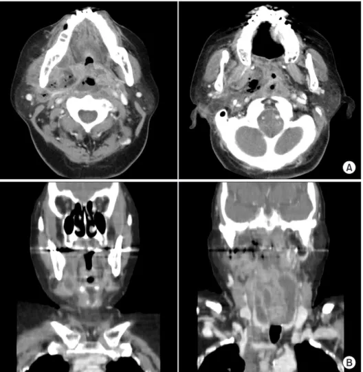
(Fig. 1, Table 1)
3. Surgical procedures
We performed tracheotomy or intubation for emergency patients with dysphagia, dyspnea, and reduced oxygen saturation to secure the patients’ airway. For patients who are not in emergency state, we did antibiotic and fluid therapy first, and then took CT to confirm the localization of the abscess. After that, we performed incision and drainage (I&D) with the insertion of thoracic catheters. During the procedure, we took pus cultures and selected the most specific antibiotics based on the result. The inserted tubes were replaced every 2-3 days. The silastic catheter was replaced with thoracic catheter in accordance with the extent of pus drainage. As radical therapy, root canal therapy or tooth extraction was carried out in case of tooth-origin infections.
We regarded the termination of treatment or time of dis- charge as the moment patients’ clinical symptoms improved, with normal inflammation level in the serum laboratory test and complete treatment of involved microor ganisms.
4. Bacterial culture test
Amoxicillin/clavulanic acid (Augmentin; Ilsung Pharma- ceuticals Co. Ltd., Seoul, Korea) was the drug of choice as empirical antibiotics. Clindamycin (Fullgram; Samjin Pharmaceuticals Co. Ltd., Seoul, Korea) or astromicin sulfate (Fortimicin; Yungjin Pharmaceuticals Co. Ltd., Seoul, Korea) was subsequently used according to the result of a serum laboratory test. During I&D, we took the drained pus using sterile agar gel transport swabs without charcoal (Copan Italia S.p.A., Brescia, Italy). Susceptibility to various antibiotics (β-lactam, aminogylcoside, lincosamide, cephalosporin, and quinolone) was evaluated by measuring the “minimal inhibitory concentration (MIC)” of bacteria cultured for 4-5 they would be more severe, with the patients suffering from
more complications; hence the difficulty of treatment and the need for much longer treatment period compared to patients without systemic diseases6.
Beck et al.8 reported that secondary fascial space infections can cause life-threatening complications in patients with weakened immunological functions such as diabetic patients.
On the other hand, Chen et al.9 noted how diabetic patients normally have unique clinical features in the oral and maxillofacial areas compared to non-diabetic patients. Note, however, that there are not enough clinical studies on the difference between DM patients and non-DM patients in the treatment of fascial infection with odontogenic origin in the maxillofacial area. Therefore, this study was conducted to evaluate clinically the impact of DM on the prognosis of secondary fascial space infection.
II. Materials and Methods
1. Patients
The medical records of 51 patients diagnosed with secon- dary fascial space infections and treated at Department of Oral and Maxillofacial Surgery in Chonnam National University Hospital (Gwangju, Korea) from January 2007 to February 2009 were reviewed. The patients were divided into two groups: DM group and non-DM group. Patients with pre vious history of DM and without previous history of DM but had fasting plasma glucose level of over 126 mg/dL were classified under the DM group. Other patients who had no previous medical history and whose fasting plasma glucose level was under 126 mg/dL were classified under the non- DM group. Note, however, that we reclassified the suspected diabetic patients with an additional glucose tolerance test. At least 25 of the patients had DM, and 26 had no DM. In the DM group, 10 were male and 15 were female. In the non-DM group, 10 were male and 16 were female. All of the patients received treatment while they were hospitalized.
Investigation was done on the age of the patients, type of diffused space, type of antibiotics, duration of drug admini stration, number of surgical operations and tracheos- tomy, clinicopathological test, complications, period of hospitalization, and microorganism involved using the patients’ medical records, radiography, and computed tomography (CT) between the two groups. When more than 2 spaces were involved, the case was classified as multiple secondary space infection.
III. Results
1. Age distribution
The patients’ average age was 62.9 years (range, 23-79 years) in the diabetic group and 47.8 years (range, 13-72 years) in the non-DM group. This suggests that the DM group was much older than the non-DM group (P>0.05).
2. Involved fascial spaces
The masseteric, pterygomandibular, and temporal spaces were the secondary spaces that were mainly involved. Note that 15 patients in the DM group (60.0%) had multiple secon- dary space infections spreading to more than 2 secondary spaces, which was double that observed in the non-DM group (30.0%).(Table 2, P<0.05) We could observe more than 2 involved spaces - sometimes up to 5 spaces - in the DM group, but mostly 2 spaces in the non-DM group, with 2 of them having 3 space infections.(Table 3)
days, after which the specific antibiotics were selected.
5. Statistical analysis
Statistical analysis of each numeral value of the clinical simi- larity in diabetes and non-diabetes patients was performed using t-test (SPSS program ver. 12.0; SPSS Inc., Chicago, IL, USA).
Table 1. Classification of primary and secondary spaces
Primary space Secondary space
Canine space Buccal space Infratemporal space Buccal space Submental space Sublingual space Submandibular space
Parapharyngeal space Temporal space Submasseteric space Infratemporal space Pterygomandibular space
Je-Shin Chang et al: Odontogenic infection involving the secondary fascial space in diabetic and non-diabetic patients: a clinical comparative study. J Korean Assoc Oral Maxillofac Surg 2013
Fig. 1. A. Axial view of computed tomography (CT) scans disclose the fascial space of ptery gomandibular, parapharyngeal, retropharyngeal, and submandibular space. B. Coronal view of CT scans show widened spaces.
Je-Shin Chang et al: Odontogenic infection involv- ing the secondary fascial space in diabetic and non- diabetic patients: a clinical comparative study. J Korean Assoc Oral Maxillofac Surg 2013
performed in the DM and non-DM groups was 1.6 (range, 0-6) and 0.9 (range, 0-1), respectively.(Table 4, P<0.05)
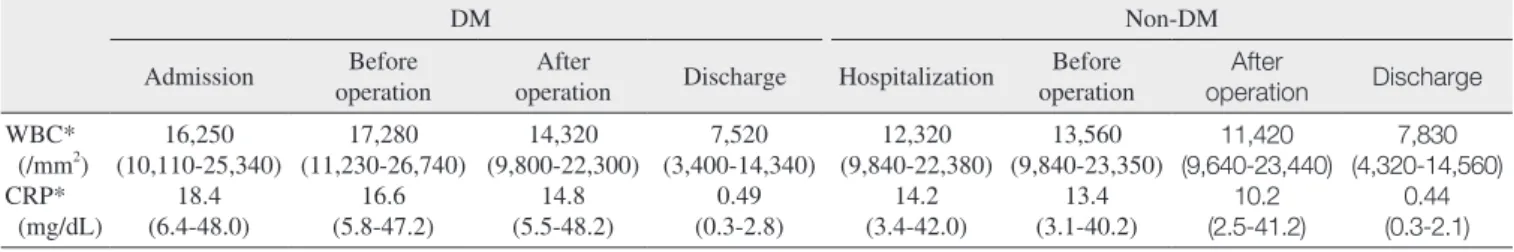
4. Hematologic examination
During the treatment period, the WBC and CRP levels increased at the time of admission and preoperative and postoperative states of the patients but became almost normal at the time of completion of treatment in both DM and non- DM groups. All of those levels were higher in the DM group compared to the non-DM group.(Table 5, P>0.05)
5. Tracheotomy and complication
The tracheotomy ratio was higher in the DM group (20%, 5 people) compared to the non-DM group (3.8%, 1 person) (P<0.05). The complication ratio was 40.0% (10 people) and 7.7% (2 people) in the DM and non-DM groups, respectively (P<0.05). The major complications were airway obstruction, sepsis, cavernous sinus thrombosis, trismus, skin defect, scar contraction, and hypoesthesia.(Table 6)
6. Duration of hospitalization
The mean duration of hospitalization was 28.9 days in the 3. Types of antibiotics, duration of drug administration,
and number of operations
The mean number of antibiotics prescribed was 3.8 in the DM group (range, 2-5) and 3.1 in the non-DM group (range, 2-4). The mean duration of intravenous administration of antibiotics in the DM and non-DM groups was 23.8 days (range, 7-62 days) and 13.8 days (range, 4-28 days), respectively. The mean duration of oral administration after intravenous administration in the DM and non-DM groups was 17.8 days (range, 7-8 days) and 9.4 days (range, 7-17 days), respectively. The mean number of surgical procedures
Table 2. Distribution of secondary spaces
Fascial space DM Non-DM Total
Parapharyngeal space Temporal space Submasseteric space Pterygomandibular space Extended space*
Total
0 (0) 2 (8.0) 4 (16.0) 4 (16.0) 15 (60.0) 25 (100)
4 (15.4) 4 (11.5) 6 (23.1) 4 (15.4) 8 (30.0) 26 (100)
4 6 10 8 23 51 (DM: diabetes mellitus)
*Two or more involved spaces, P<0.05.
Values are presented as number (%) or number only.
Je-Shin Chang et al: Odontogenic infection involving the secondary fascial space in diabetic and non-diabetic patients: a clinical comparative study. J Korean Assoc Oral Maxillofac Surg 2013
Table 3. Number of extended spaces
Number of extended space DM Non-DM Total
2 3 4
≥5 Total
6 5 2 2 15
6 2 0 0 8
12 7 2 2 23 (DM: diabetes mellitus)
Je-Shin Chang et al: Odontogenic infection involving the secondary fascial space in diabetic and non-diabetic patients: a clinical comparative study. J Korean Assoc Oral Maxillofac Surg 2013
Table 4. Count of antibiotics and surgery
Number of treatment DM Non-DM
Antibiotics used
I&D* 3.8 (2-5)
1.6 (0-6) 3.2 (2-4)
0.9 (0-1) (DM: diabetes mellitus, I&D: incision and drainage)
*P<0.05.
Values are presented as mean (range).
Je-Shin Chang et al: Odontogenic infection involving the secondary fascial space in diabetic and non-diabetic patients: a clinical comparative study. J Korean Assoc Oral Maxillofac Surg 2013
Table 5. Comparison of WBC and CRP in the diabetic and non-DM groups
DM Non-DM
Admission Before
operation After
operation Discharge Hospitalization Before operation
After
operation Discharge WBC*
(/mm2) CRP*
(mg/dL)
16,250 (10,110-25,340)
18.4 (6.4-48.0)
17,280 (11,230-26,740)
16.6 (5.8-47.2)
14,320 (9,800-22,300)
14.8 (5.5-48.2)
7,520 (3,400-14,340)
0.49 (0.3-2.8)
12,320 (9,840-22,380)
14.2 (3.4-42.0)
13,560 (9,840-23,350)
13.4 (3.1-40.2)
11,420 (9,640-23,440)
10.2 (2.5-41.2)
7,830 (4,320-14,560)
0.44 (0.3-2.1) (WBC: white blood cell, CRP: C-reactive protein, DM: diabetes mellitus)
*P<0.05, WBC and CRP values were higher in DM except at the discharge.
Each level shows the average of the maximal levels during that period.
Je-Shin Chang et al: Odontogenic infection involving the secondary fascial space in diabetic and non-diabetic patients: a clinical comparative study. J Korean Assoc Oral Maxillofac Surg 2013
patients are vulnerable to infections due to a decrease in the bactericidal effects of neutrophil, cellular immunity, and activity of complement under persistent high blood sugar levels. In addition, chronic diseases such as diabetes occur more often in older patients. In this study, the average age of the DM group was 62.9 years compared to 47.8 years in the non-DM group. These results were similar to other reported studies12,13. Note, however, that aging influences the immune system, making the latter typically less responsive to infection;
vaccination can be more responsive to inflammation14. Lee et al.15 reported that 81.6% of odontogenic infection showed single fascial space involvement. In contrast, 23 patients exhibited more than two fascial space involvements.
(Table 3) In the study of head and neck space infections of odontogenic origin by Rega et al.16, multiple-space infections were more common than single-space infections. Such discord may result from the latency in visiting the hospital.
Note that the DM group had higher incidence of multiple fascial space infections than the non-DM group. This could be related to further progression to deep neck infection as reported by Huang et al.13.
Diabetic patients should be treated based on more stringent criteria due to their decreased immune function. Diabetes is reported to enhance inflammation in general by altering the myeloid and lymphoid functions17,18. Chen et al.12 cited DM group (range, 7-88 days) and 15.4 days (range, 4-35
days) in the non-DM group (P>0.05).
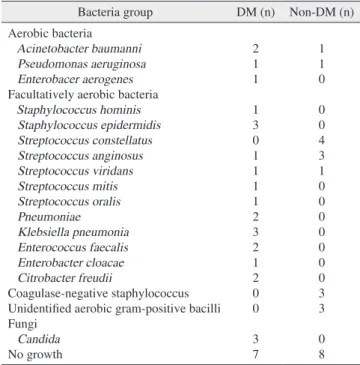
7. Main causative microorganism
Klebsiella pneumoniae, Staphylococcus species, and Candida were the main causative organisms in the DM group.(Table 7)
IV. Discussion
Infections occur when there is a disruption of the balance between the human defense mechanism and bacterial infection mechanism. The more significant factor of the two is human defense mechanism, i.e., local immunity, humoral immunity, and cellular immunity10. Local immunity includes mechanical defense by the surface or mucous membrane of the host itself and defense of normal flora of skin, suppressing the growth of other infectious bacteria. Humoral immunity, a non- cellular immune system, includes the immune system by the complement or immunoglobulin and mainly exists in the serum or exudates. Cellular immunity means the immune system phagocytosis by phagocytes, such as polymorphonuclear lymphocytes, monocytes, and macrophages.
In diabetic patients, among the immunities above are disor- ders of cell-mediated immune system, phagocytosis, and supply of blood, causing increased infection11. There were decreases in the ability of chemotaxis, phagocytosis, and cell destruction, which are factors of innate cellular immunity, and function of diabetic polymorphonuclear lymphocytes and diabetic monocytes in diabetic patients11. Tanaka3 suggested that the function of neutrophil, such as chemotaxis or production of cytokines, is reduced under high blood sugar levels. Similarly, previous studies reported that diabetic
Table 6. Complications of secondary space abscess patients
Complication DM Non-DM
Airway obstruction Sepsis Scar contraction Skin defect Trismus
Cavernous sinus thrombosis Hypoesthesia
Death Total
2 (8.0) 1 (4.0) 2 (8.0) 1 (4.0) 2 (8.0) 1 (4.0) 1 (4.0) 0 10 (40.0)
0 0 0 0 2 (7.7) 0 0 0 2 (7.7) (DM: diabetes mellitus)
Values are presented as number (%).
Je-Shin Chang et al: Odontogenic infection involving the secondary fascial space in diabetic and non-diabetic patients: a clinical comparative study. J Korean Assoc Oral Maxillofac Surg 2013
Table 7. Distribution of microorganisms
Bacteria group DM (n) Non-DM (n)
Aerobic bacteria Acinetobacter baumanni Pseudomonas aeruginosa Enterobacer aerogenes Facultatively aerobic bacteria Staphylococcus hominis Staphylococcus epidermidis Streptococcus constellatus Streptococcus anginosus Streptococcus viridans Streptococcus mitis Streptococcus oralis Pneumoniae Klebsiella pneumonia Enterococcus faecalis Enterobacter cloacae Citrobacter freudii
Coagulase-negative staphylococcus Unidentified aerobic gram-positive bacilli Fungi
Candida No growth
2 1 1 1 3 0 1 1 1 1 2 3 2 1 2 0 0 3 7
1 1 0 0 0 4 3 1 0 0 0 0 0 0 0 3 3 0 8 Je-Shin Chang et al: Odontogenic infection involving the secondary fascial space in diabetic and non-diabetic patients: a clinical comparative study. J Korean Assoc Oral Maxillofac Surg 2013
the more frequently occurring complications were airway obstruction, scar contraction, and trismus. These results were consistent with the findings of previous studies22,23.
Common odontogenic infections are usually associated with five or more types of bacterial strains - including both aerobic and anaerobic bacterial colonies - rather than just a single strain. Aerobic streptococcus is a predominant bacterial strain at the beginning of the infection, and anaerobic bacteria gradually increased as the infection became chronic24. Warnke et al.19 reported that several bacterial strains formed colonies in 98% of patients with abscesses. Viridans streptococcus was the most common strain. According to Gilmer and Moody25, the streptococci or staphylococci were observed in patients with acute dental infections in the early stage of infections.
The proportion of anaerobic bacteria - Klebsiella pneumonia and Streptococcus epidermidis in our study - increased as the infection spread to secondary space or deep neck space. The type of bacterial growth differed between the DM group and non-DM group: Klebsiella pneumoniae in the DM group and Streptococcus constellatus in the non-DM group. We could observe fungal infections, such as Candida infection, in a group of diabetic patients.
Based on the result of this study and literature review, fascial space infections in diabetic patients tend to extend the secondary space and worsen. Korea is already an aging society and is expected to become an aged society in 201726, and almost 20% of the elderly (>60 years old) have DM27. Therefore, oral hygiene care in elderly should be emphasized, especially among diabetic patients. Likewise, early diagnosis, strict control of blood sugar, empirical use of a wide range of antibiotics, and active surgical treatments are important.
V. Conclusion
In this study, we carried out image, physical, and clinical evaluations of 25 diabetic and 26 non-diabetic patients diagnosed with secondary space abscesses and treated at Department of Oral and Maxillofacial Surgery in Chonnam National University Hospital.
1. The average age of the DM group was 62.9 years (range, 23-79 years), which was higher than the non-DM group’s 47.8 years (range, 13-72 years; P>0.05).
2. The rate of multiple secondary spaces infection of the DM group (60%) was higher than that of the non-DM group (27.3%, P<0.05).
3. The mean number of prescribed antibiotics was 3.8 (range, 2-5) in the DM group and 3.1 (range, 2-4) in the non- the need for closer observation and more active surgical
treatments for diabetic patients who had deep neck infections since they have poorer prognosis than non-diabetic patients.
In this study, the difference in the number of antibiotics was insignificant. Augmentin was administered as empirical antibiotic for patients with secondary space infections, with clindamycin and/or aminoglycoside added in severe cases.
Antibiotics were administered every 8 hours to maintain MIC. Although Warnke et al.19 attempted to change the administration method from intravenous administration for 3 days to oral administration to treat abscesses, antibiotics were administered orally after an improvement in the aspects of physical inflammation and clinical features. Note, however, that the period of antibiotics administration and period of hospitalization were longer for diabetic patients, and they underwent more surgical intervention. These results can be said to be a reflection of persistent inflammation and delayed healing, which could be due to prolonged inflammatory response to cytokine dysregulation and enhanced fibroblast apoptosis by diabetes20,21.
In this study, the DM group showed higher WBC level and CRP level in the blood at the time of treatment as well as more post-operative complications compared to the non- DM group. These results were consistent with the findings of previous studies12,13. Thus, it is important to treat those patients in an active manner, such as active surgical incision, pus drainage, and use of empirical antibiotics. The result of our study is similar to that of a previous study of Huang et al.13, i.e., diabetic patients are vulnerable to deep neck infections, with much more complications and increase in the duration of hospitalization. In particular, tracheotomy and emergency I&D were required for 2 patients in the DM group due to the spread of infection to more than 5 spaces concerning airway obstruction. Tracheostomy and higher initial values of WBC and CRP suggest the severity of the infection, and they were significantly higher in the DM group. As complications, scar contraction and skin defect might be related to a wider extent of operation site and the delayed healing in diabetic patients.
Cavernous sinus thrombosis was managed in consultation with the department of internal medicine. Huang et al.22 also reported low recovery rates among diabetic patients, which may have increased the number of complications. Deep neck infections may result in complications such as upper airway obstruction, descending mediastinitis, jugular vein thrombosis, venous septic emboli, carotid artery rupture, adult respiratory distress syndrome, septic shock, and disseminated intravascular coagulopathy23. In our study,
9. Chen MK, Wen YS, Chang CC, Huang MT, Hsiao HC. Predis- posing factors of life-threatening deep neck infection: logistic regression analysis of 214 cases. J Otolaryngol 1998;27:141-4.
10. Plouffe JF, Silva J Jr, Fekety R, Allen JL. Cell-mediated immunity in diabetes mellitus. Infect Immun 1978;21:425-9.
11. Geerlings SE, Hoepelman AI. Immune dysfunction in patients with diabetes mellitus (DM). FEMS Immunol Med Microbiol 1999;
26:259-65.
12. Chen MK, Wen YS, Chang CC, Lee HS, Huang MT, Hsiao HC.
Deep neck infections in diabetic patients. Am J Otolaryngol 2000;
21:169-73.
13. Huang TT, Tseng FY, Yeh TH, Hsu CJ, Chen YS. Factors affecting the bacteriology of deep neck infection: a retrospective study of 128 patients. Acta Otolaryngol 2006;126:396-401.
14. Cavanagh MM, Weyand CM, Goronzy JJ. Chronic inflammation and aging: DNA damage tips the balance. Curr Opin Immunol 2012;24:488-93.
15. Lee WH, Ahn KM, Jang BY, Ahn MR, Lee JY, Sohn DS. Clinico- stastical study of inpatients of abscess in fascial spaces for the last 5 years. J Korean Assoc Oral Maxillofac Surg 2004;30:497-503.
16. Rega AJ, Aziz SR, Ziccardi VB. Microbiology and antibiotic sensitivities of head and neck space infections of odontogenic origin. J Oral Maxillofac Surg 2006;64:1377-80.
17. Graves DT, Kayal RA. Diabetic complications and dysregulated innate immunity. Front Biosci 2008;13:1227-39.
18. Nikolajczyk BS, Jagannathan-Bogdan M, Shin H, Gyurko R. State of the union between metabolism and the immune system in type 2 diabetes. Genes Immun 2011;12:239-50.
19. Warnke PH, Becker ST, Springer IN, Haerle F, Ullmann U, Russo PA, et al. Penicillin compared with other advanced broad spectrum antibiotics regarding antibacterial activity against oral pathogens isolated from odontogenic abscesses. J Craniomaxillofac Surg 2008;
36:462-7.
20. Naguib G, Al-Mashat H, Desta T, Graves DT. Diabetes prolongs the inflammatory response to a bacterial stimulus through cytokine dysregulation. J Invest Dermatol 2004;123:87-92.
21. Liu R, Desta T, He H, Graves DT. Diabetes alters the response to bacteria by enhancing fibroblast apoptosis. Endocrinology 2004;145:2997-3003.
22. Huang TT, Tseng FY, Liu TC, Hsu CJ, Chen YS. Deep neck infection in diabetic patients: comparison of clinical picture and outcomes with nondiabetic patients. Otolaryngol Head Neck Surg 2005;132:943-7.
23. Huang TT, Liu TC, Chen PR, Tseng FY, Yeh TH, Chen YS. Deep neck infection: analysis of 185 cases. Head Neck 2004;26:854-60.
24. Al-Qamachi LH, Aga H, McMahon J, Leanord A, Hammersley N. Microbiology of odontogenic infections in deep neck spaces: a retrospective study. Br J Oral Maxillofac Surg 2010;48:37-9.
25. Gilmer TL, Moody AM. A study of the bacteriology of alveolar abscess and infected root canals. JAMA 1914;LXIII:2023-4.
26. Statistics Korea. Population projection for Korea: 2010-2060.
Daejeon: Statistics Korea; 2012.
27. Korea Centers for Disease Control & Prevention. Chronic disease.
In: Jun BY, ed. Korean National Health Statistics 2011. Daejeon:
Ministry of Health & Welfare; 2012.
DM group (P>0.05). The number of surgical operations performed was 1.6 (range, 0-6) in the DM group and 0.9 (range, 0-1) in the non-DM group (P<0.05).
4. The mean period of hospitalization of the DM group was 28.9 days (range, 7-88 days), which was longer than the non- DM group’s 15.4 days (range, 4-35 days; P>0.05).
5. The values of WBC and CRP of the DM group were higher than those of the non-DM group during the treatment period (P>0.05).
6. The DM group recorded a 40.0% occurrence of compli- ca tions, which was higher than the non-DM group’s 7.7%
(P<0.05). The tracheostomy rate of the DM group (20.0%) was also higher than that of the non-DM group (3.8%, P<0.05).
7. The results of bacteria cultivation indicated that the main causative microorganisms of diabetics were Klebsiella pneumoniae and Staphylococcus species.
These results suggest the need to treat diabetic patients as quickly as possible due to their high risk of infections.
References
1. Carton JA, Maradona JA, Nuño FJ, Fernandez-Alvarez R, Pérez- Gonzalez F, Asensi V. Diabetes mellitus and bacteraemia: a comparative study between diabetic and non-diabetic patients. Eur J Med 1992;1:281-7.
2. Muller LM, Gorter KJ, Hak E, Goudzwaard WL, Schellevis FG, Hoepelman AI, et al. Increased risk of common infections in patients with type 1 and type 2 diabetes mellitus. Clin Infect Dis 2005;41:281-8.
3. Tanaka Y. Immunosuppressive mechanisms in diabetes mellitus.
Nihon Rinsho 2008;66:2233-7.
4. Delamaire M, Maugendre D, Moreno M, Le Goff MC, Allannic H, Genetet B. Impaired leucocyte functions in diabetic patients.
Diabet Med 1997;14:29-34.
5. WHO Scientific Group of Immunodeficiency. Immunodeficiency:
report of a WHO scientific group. World Health Organ Tech Rep Ser 1978;(630):3-80.
6. Chang CM, Lu FH, Guo HR, Ko WC. Klebsiella pneumoniae fascial space infections of the head and neck in Taiwan: emphasis on diabetic patients and repetitive infections. J Infect 2005;50:34- 40.
7. Korean Association of Oral and Maxillofacial Surgeons. Infectious disease. 2nd end. Seoul: Dental and Medical Publishing; 2005.
8. Beck HJ, Salassa JR, McCaffrey TV, Hermans PE. Life-threatening soft-tissue infections of the neck. Laryngoscope 1984;94:354-62.