392 Received:June 29, 2016, Revised:July 28, 2016, Accepted:August 3, 2016
Corresponding to:Kyoung Hwa Choi, Division of Allergy and Pulmonology, Department of Internal Medicine, Presbyterian Medical Center, 365 Seowon-ro, Wansan-gu, Jeonju 54987, Korea. E-mail:[email protected]
pISSN: 2093-940X, eISSN: 2233-4718
Copyright ⓒ 2016 by The Korean College of Rheumatology. All rights reserved.
This is a Free Access article, which permits unrestricted non-commerical use, distribution, and reproduction in any medium, provided the original work is properly cited.
Case Report
Journal of Rheumatic Diseases Vol. 23, No. 6, December, 2016 https://doi.org/10.4078/jrd.2016.23.6.392
Case of Follicular Bronchiolitis in Rheumatoid Arthritis
Jong Hwa Lee1, MyungWoo Choi1, Sung Sik Oh1, Mi Rim Choi1, Hyun Ju Yang1, Kwang Min Lee2, Kyoung Hwa Choi3
Departments of 1Internal Medicine and 2Pathology, 3Division of Allergy and Pulmonology, Department of Internal Medicine, Presbyterian Medical Center, Jeonju, Korea
Follicular bronchiolitis is an uncommon bronchiolar disorder that is characterized by the presence of hyperplastic lymphoid follicles with reactive germinal centers. The condition is associated with connective tissue diseases such as rheumatoid arthritis, Sjögren’s syndrome, and immunodeficiency disorders. A 56-year-old man with rheumatoid arthritis was admitted to hospital with a progressively enlarging pulmonary nodule in the left upper lobe. A follow-up contrast tomography scan showed that the nodule had increased in size from 4.2 mm to 6.3 mm over a 3 month period. An open lung biopsy was performed to establish a definite pathologic diagnosis of the pulmonary nodule, which was suspected to be a lung malignancy. The nodule was diag- nosed as follicular bronchiolitis based on the histopathology findings. We describe a patient with follicular bronchiolitis that was confirmed by an open lung biopsy, and is believed to have had rheumatoid involvement. (J Rheum Dis 2016;23:392-395) Key Words. Bronchiolitis, Solitary pulmonary nodule, Arthritis rheumatoid
INTRODUCTION
Rheumatoid arthritis (RA) is a chronic and systemic in- flammatory arthritis that characteristically features peri- articular pain, joint edema, and a reduced range of motion of the joint. In addition to joint-related symptoms, chron- ic inflammation may cause extra-articular symptoms, in- cluding pericarditis, nerve involvement, interstitial lung disease (ILD), and vasculitis. Although the clinical respi- ratory symptoms could be easily overlooked as they are subtle in the early stages, they have a significant influence on the patient’s prognosis [1]. Follicular bronchiolitis is defined as a proliferation of bronchial lymphoid tissue. It is a type of reactive pulmonary lymphoid disorder and does not show evidence of chronic obstructive lung dis- ease or bronchiectasis. It is characterized by hyperplasia of lymphoid follicles and germinal centers around the bronchiole [2,3]. It is a rare airway disease that has been reported in literature to be associated with autoimmune or connective tissue diseases [3], but there are no reports of it occurring in a patient with rheumatoid arthritis in
Korea.
The authors present the case of a patient with RA-re- lated ILD who presented with a growing solitary pulmo- nary nodule that was histologically diagnosed as follicular bronchiolitis.
CASE REPORT
A 56-year-old male visited our hospital for an incidental abnormal finding on a chest radiograph (Figure 1). He was a patient with seropositive rheumatoid arthritis and had been treated with immunosuppressive agents, such as corticosteroids and methotrexate, for seven years. At the time he presented to us, he was a smoker with mild exertional dyspnea. Based on results from pulmonary function tests, his lung function was not restricted.
Physical examination of the chest revealed inspiratory fine crackles. No palpable subcutaneous node or lympha- denopathy was present. HIV antibodies was negative.
There computed tomography (CT) scan of the chest re- vealed linear and reticular densities in the subpleural area
Follicular Bronchiolitis in Rheumatoid Arthritis
www.jrd.or.kr 393
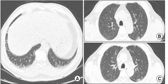
Figure 2. Chest computed to- mography revealed (A) linear, reticular densities and ground glass opacities in the bilateral lower lung fields, as well as (B and C) a slightly increased nod- ule (that had increased in size from 4.2 mm to 6.3 mm) in the apicoposterior segment of the left upper lobe.
Figure 1. The chest radiograph revealed linear densities and small nodules bilaterally in the lower lung fields.
of both lungs, small patchy areas of ground glass opacities (GGOs), as well as a small nodular lesion that was noted in the left upper apicoposterior segment (Figure 2A and 2B). Sputum culture were negative for bacteria, mycobac- teria, fungi. A follow-up chest CT performed three months later revealed that the small nodule near the GGO had become prominent and increased in size (Figure 2C).
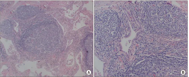
Differential diagnoses included pulmonary involvement of inflammatory or connective tissue disease and ma- lignancy. A lung biopsy was performed. Pathological find- ings from thoracoscopic biopsy revealed peribronchiolar lymphoplasmacytic infiltration with formation of a ger- minal center, suggesting follicular bronchiolitis (Figure 3A and 3B). After the patient underwent a surgical lung biopsy to confirm the diagnosis, the small nodule and
GGO lesion were removed. The patient was treated with immunosuppressants, including prednisolone for rheu- matoid arthritis, and has remained stable at 10 months.
DISCUSSION
Follicular bronchiolitis is a specific respiratory disease that is characterized by hyperplastic lymphoid tissue and follicles near the bronchus and bronchiole, as well as mild lymphocyte and monocyte infiltration in the interstitial area [4]. Yousem et al. [3] reported that follicular bron- chiolitis generally accompanied other diseases and could be classified into three groups. The first type of follicular bronchiolitis accompanies auto-immune or connective tissue diseases, such as rheumatoid arthritis or Sjögren syndrome. The second type occurs in individuals with im- munodeficiencies or a family history of immunodeficiency and may accompany AIDS. The third group is associated with an unknown cause and are predicted to be a result of a hypersensitivity reaction. Follicular bronchiolitis in the present case falls into the first type, as the patient was di- agnosed with rheumatoid arthritis. Major clinical symp- toms include dyspnea, coughing, and fever. Lee et al. [5]
reported coughing and dyspnea with follicular bronchioli- tis while Kim et al. [6] reported coughing and sputum as the major symptoms. The patient in the current case also complained of coughing and sputum. On pulmonary function tests, most patients with follicular bronchiolitis appear normal or show restrictive findings on pulmonary function tests, and rarely show obstructive findings [4].
According to Howling et al. [7], follicular bronchiolitis is primarily observed as a small nodular lesion of less than 3 mm on chest CT; the most common form has a bilateral
Jong Hwa Lee et al.
394 J Rheum Dis Vol. 23, No. 6, December, 2016
Figure 3. Histologic specimen showing (A) hyperplasia of the lymphoid follicle (H&E, ×40) and (B) a benign hyperplastic germinal center (H&E, ×100).
centrilobular distribution. A GGO is also frequently ob- served, along with bronchiectasia, bronchial wall thicken- ing, and emphysema. Rheumatoid arthritis causes varia- ble conditions in the lung, including pleurisy, ILD, small airway disease, and vasculitis. Of these, RA-ILD carries a poor prognosis and has a high mortality rate due to respi- ratory failure that results from disease progression and other complications, including heart disease, lung cancer, and pneumonia [8].
In the present case, the following factors led us to per- form a biopsy in order to differentiate the diagnosis from lung cancer: the patient was a smoker; we observed a nod- ular lesion that was enlarged in size and contour com- pared to a previous chest CT; and RA-ILD was present.
Furthermore, although follicular bronchiolitis frequently accompanies RA, radiological findings of the present study did not show the typical patterns of RA-ILD-asso- ciated follicular bronchiolitis. On a chest CT, follicular bronchiolitis typically presents as small nodules, GGO, and reticulonodular opacities distributed in the cen- trilobular area [7]. However, in the current case, an ipsi- lateral, local nodular lesion was seen, which warranted a histological confirmation to rule out lung cancer. Currently, the role of pulmonary tissue biopsies in patients with connective tissue diseases is unclear [9] and ILD treat- ment guidelines do not recommend pulmonary tissue bi- opsies in patients with connective tissue diseases. However, pathological differentiation via a biopsy may be required to differentiate radiologic atypical finding and accom- panying diseases, such as lung cancer and opportun-
istic/drug-induced pneumonia. Treatment based on an accurate diagnosis plays a critical role in the patient’s prognosis.
Lymphoid interstitial pneumonia is one of the diseases differentiated from follicular bronchiolitis. Both disease are reactive pulmonary lymphoid disorders that present clinically with pulmonary lymphadenopathy, but they are pathologically different. While follicular bronchiolitis mainly shows infiltration of lymphoid follicles and mono- cytes near the bronchus and bronchiole, lymphoid inter- stitial pneumonia involves diffuse infiltration within the interstitium and enlarged alveolar septa [2]. Howling et al. [7] reported that follicular bronchiolitis maybe accom- panied by the above-mentioned histological findings, in which case the pathologist determined the diagnosis based on the main findings. Follicular bronchiolitis is a rarely aggravated and has a relatively good prognosis.
Treatment usually involves steroid therapy, as well as treatment of the underlying diseases. In the current case, the local lesion was surgically removed and we are cur- rently observing the patient since no new lesions were found on the chest CT at the ten-month follow-up visit.
This is the first case in which a progressively enlarging, isolated nodule in a RA-ILD patient was definitively diag- nosed as follicular bronchiolitis via biopsy. While respira- tory infiltrations associated with RA, such as pleural and interstitium infiltration, are well recognized and are quite common, small airway infiltrations, such as follicular bronchiolitis largely go unrecognized. Therefore, it is im- portant to understand various respiratory infiltrations
Follicular Bronchiolitis in Rheumatoid Arthritis
www.jrd.or.kr 395
and accompanying diseases of connective tissue disorders in order to ensure early diagnosis via regular examina- tions and tests.
SUMMARY
Follicular bronchiolitis is a rare small airway disease.
Most of the cases has been reported to be associated with systemic diseases such as connective tissue disease or im- munodeficiency syndromes. This case reports is the first Korean case in which a progressively enlarging, isolated nodule in a RA-ILD patient was diagnosed as follicular bronchiolitis via lung biopsy. Clinicians shoud be aware of various respiratory disorders associated with RA on early stage.
CONFLICT OF INTEREST
No potential conflict of interest relevant to this article was reported.
REFERENCES
1. Archontogeorgis K, Steiropoulos P, Tzouvelekis A, Nena E,
Bouros D. Lung cancer and interstitial lung diseases: a sys- tematic review. Pulm Med 2012;2012:315918.
2. Nicholson AG, Wotherspoon AC, Diss TC, Hansell DM, Du Bois R, Sheppard MN, et al. Reactive pulmonary lymphoid disorders. Histopathology 1995;26:405-12.
3. Yousem SA, Colby TV, Carrington CB. Follicular bronchi- tis/bronchiolitis. Hum Pathol 1985;16:700-6.
4. Aerni MR, Vassallo R, Myers JL, Lindell RM, Ryu JH.
Follicular bronchiolitis in surgical lung biopsies: clinical im- plications in 12 patients. Respir Med 2008;102:307-12.
5. Lee YJ, Park JH, Cho GJ, Lee BC, Kim DS, Suh YL, et al. A case of follicular bronchitis/bronchiolitis. Korean J Med 1993;45:795-801.
6. Kim MS, Lim SC, Kim YH, Na KJ, Kim KS, Kwon KY, et al.
a case report of localized form of follicular bronchitis/bron- chiolitis with fibrosis. Tuberc Respir Dis 1998;45:191-6.
7. Howling SJ, Hansell DM, Wells AU, Nicholson AG, Flint JD, Müller NL. Follicular bronchiolitis: thin-section CT and his- tologic findings. Radiology 1999;212:637-42.
8. Kim EJ, Collard HR, King TE Jr. Rheumatoid arthritis-asso- ciated interstitial lung disease: the relevance of histopatho- logic and radiographic pattern. Chest 2009;136:1397-405.
9. American Thoracic Society; European Respiratory Society.
American Thoracic Society/European Respiratory Society International Multidisciplinary Consensus Classification of the Idiopathic Interstitial Pneumonias. This joint statement of the American Thoracic Society (ATS), and the European Respiratory Society (ERS) was adopted by the ATS board of directors, June 2001 and by the ERS Executive Committee, June 2001. Am J Respir Crit Care Med 2002;165:277-304.