대 한 방사 선 의 학회 지 1996
; 35(3) : 325-333
문맥 고혈합환자에서의 문맥-체측부순환경로:
CT와혈관조영술의 연관1
이경원·정진욱·박재형·연경모·한만청
간문맥 고혈압 환자에서 위장관으로부터 간으로 흐르는 혈류는 간문맥의 혈압이 높아짐에 따라 그 밤향이 바뀌어 간으로부터 매우 다앙한 측부순환경로를 통하여 혈압이 낮은 체순환정 맥으로 흐르게 된다. 측부순환경로는 중재적 시술이나 수술시 심각한 출혈을 발생시킬 수 있으므로 이 측부순환경로 에 대한 사전정보 및 지식은 출혈밤지에 매우 중요하다. 최근 급속조영 역동적 또는 나선식 CT는 측부 순환의 진단에 있어 혈관조영술과 내시경을 상당 부분 대치하고 있다. 하지만 기존의 보고들은 대부분 측부순환경로의 위치 및 빈도에 따른 기술이 많았다. 이 입상화보에서는 CT와 혈관조영술의 연판을 통 해 측부순환경로를 혈류에 따라 보여주고자 하였다.
서 론
간문맥 고혈압 환자에서 위장관으로부터 간으로 흐르는 혈류는 깐문맥의 혈압이 높아점에 따라 그 방향이 바뀌어 간으로부터 매우 다양한 측부순환경로를 통하여 혈압이 낮 은 체순환정맥으로 흐르게 된다. 그중 식도정맥류는 장관 출혈의 흔한 원인이 되므로 임상적으로 중요한 경로라고 할 수 있다. 또한 측부순환은 중재적 시술이나 수술시 심각 한 출혈을 발생시킬 수 있으므로 이 측부순환 경로에 대한 사전정보및 지식은출혈방지에 매우중요한요인이 된다.
예전에는 측부순환의 진단에 있어 혈관조영술이 대표적 인 진단방법으로 이용되었고 현재에도 식도정맥류의 진단 에는 내시경이 가장 정확한 진단방법으로 인정되고 있다.
최근단층영상기술의 발달로다소침습적인 방법언 내시경 이나 혈관조영술 없이도 측부순환의 진단이 가능해졌다.
급속조영 역동적
CT(bolus dynamic CT)
또는 나선식CT(spiral
CT) 는 여러 위치, 다양한 형태의 측부순환의 진단과 간문맥 고혈압의 전체적인 상태를 평가함에 있어 다른 검사방법보다 우월한 것으로 보고되고 있다(1).위장관의 정맥들은 장간막 (mesentery) , 후복강 (retro
peritoneum)
, 대망 (greater omentum) 과 소망(lesseromentum)
등에서 대부분 체순환정맥과 측부순환경로를형성한다. 측부순환 또는 정맥류는 CT에서 주변과 경계가 잘 그려지는 둥글거나 관모양의 균일한 음영의 구조로 나 타나며 인접 혈관과 같은 정도의 조영증강을 보여 럼프절 병증,종양등과의 감별이 용이하다.
1서울대학교 의과대학 진 단방사선과학교실
이 논문은 1996 년 5월 13일 접수하여 1996 년 6월 24일에 채택되었음
325
기존의 보고들은 측부순환경로의 위치 및 빈도에 따른 분류와 진단 기준에 대한 기술이 많았으나 혈류에 따른분 류 및 체순환정맥과 연결된 이후의 혈류방향에 대한 체계 적인 기술은 마약하였다. 이에 저자들은 측부순환 경로를 간문액의 주요분지에 따라 좌위정맥, 버장정맥, 상장간막 정맥, 하장간막정맥, 제대주위정맥 및 간을 경유한 측부순 환경로로 분류하고 체순환정맥과의 연결 및 그 이후의 순 환경로를 알아보았으며 CT에서 나타나는 소견을 혈관조 영술을통하여 이해하고자하였다.
문맥-체측부순환경로
좌위정맥을경유한측부순환
좌위정맥 (coronary vein) 을 경유한 측부순환경로는 문 맥 -체 측부순환경로중 가장 흔히 관찰되는 경로이다. 좌 위정맥은 간문맥의 근위부 또는 비장정맥 (splenic vein) 의 원위부에서 기시하여 주로 식도정맥류 (esophageal
varix)
나 식도주위정맥류 (paraesophageal varix) 로 연결되며(Fig.
1) 위대망정맥 (gastroepiploicvein)
또는 단위정맥 (shortgastric
vein) 과 함께 위정맥류 (gastric varix) 를 형성할 수 있다 (Fig. 1, Fig.3).
식도정맥류, 식도주변정맥 류와 위정맥류는 서로 연결될 수 있으며 혈류는 기정맥(azygos vein)
, 반기정맥(hemiazygos vein)
및 척추주위 정맥총으로 연결되거나, 하횡격막정맥 (inferior phrenic vein) 을 경 유하여 늑간정 맥 (Ïntercostal
vein) 과 심 장횡 격 박정 맥(pericardiacophrenic
vein) 으로 연결될 수 있다(4)(Fig.
2,Fig.
3). 위정맥류는 좌신정맥과 위신창단락(gastrorenal
shunt) 을 형성하기도 한다 (Fig. 1c). 문맥 - 체 측부순환이 좌신청맥으로 단락을 형성한 이후의 혈류대 한 밤사 섭 의 학회 지 1996; 35(3) : 325- 333
경로로서 혈류량이 매우 증가하거나 상장간동맥등의 주변 장기 또는 종괴에 의한 압박의 원인으로 좌신정맥의 혈류 장애가 있을 경우 혈류중 일부는 하대정 맥으로 흐르지 못 하고 다른 측부순환경로를 경유하게 된다. 좌신정맥으로 부터 좌성선정맥(left
gonadal
vein) 으로 역류하여 좌내장골정맥 (left internal iliac vein) 으로 흐르거나 여자의 경
우 골반정맥총(pelvic plexus)을 경유하여 우내장골정맥 또는 우성선정맥을 통해 하대정맥으로 흐를 수 있다.
(Fig.
4). 또한 좌신정맥으로부터 척추주위정맥총을 경유하여 기정맥, 반기정맥으로 연결되기도 한다 (Fig.5).
CT 에서 소망내의 직경 5-6mm의 정맥을 확인하므로 서 좌위정맥을 경유한 측부순환을 진단할 수 있다. 식도정 맥류는 하부 식도벽을 따라 상피하 (subepithelial), 점막하 (submucosal) 에 위치하는 늘어난 정맥드로서 CT에서는 위장관내로 튀어나온 조영증강되는 종괴의 형태나 두꺼위 진 식도벽 자체가 조영증강되는 형태로 나타나게 되며 (2) 혈관조영술에서는 식도를 따라 길게 주행하는 형태를 보 인다. 식도주변정맥류는 식도의 후방으로 후종격동에 위 치하는 굵어진 측부순환 정맥을 의미하며 단순 흉부촬영 상 후종격동 종괴로 오인될 수 있다. 위정맥류 역시 주로
a b
위저부 (gastric fundus) 후내측의 점막하 또는 장막하 (subserosal) 에 위치하므로 (Fig.
la)
내시경이나 바륨위 투시검사에서 점막하종괴 또는점막주름 (rugae) 의 비후로 보이게 되며 식도주위정맥류와 마찬가지로 혈관조영솔 또 는 CT가진단에 매우중요한 역활을한다.비장정맥을 경유한 측부순환경로
비장정맥을 경유한 측부순환로는 비장주위정맥류를 형 성하고 좌하횡격막정맥, 늑간정맥 및 심장횡격막정맥으로 흐르거나 척추주위정맥총을 통하여 기정맥과 반기정맥으 로 흐르게 된다. 또한 좌신정맥과 비장신장단락 (splenore
nal
shunt) 을 형성하기도 하고 (Fig. 6) 부신주위정맥을 경 유하여 좌신정맥으로 연결되기도 한다. 좌신청맥 이후의 혈류는 전술한 바와 같다. 비장주위정맥류는 또한 좌하북 정 맥 (left hypogastric vein) 과 비 장대 정 맥 단락 (splenocaval shunt) 을 형성하기도 하고 단위정맥이나 위대망정
맥을 경유하여 위정 맥류를 형성하기도 한다 (Fig.3). CT에서 비장주위정맥류는 비문 (splenic hilum) 과 비장 주위의 지방조직내에 위치하며 좌측 신장의 외측 및 후측 에서도 발견될 수 있다. 비장신장단락의 경우 CT에서 좌
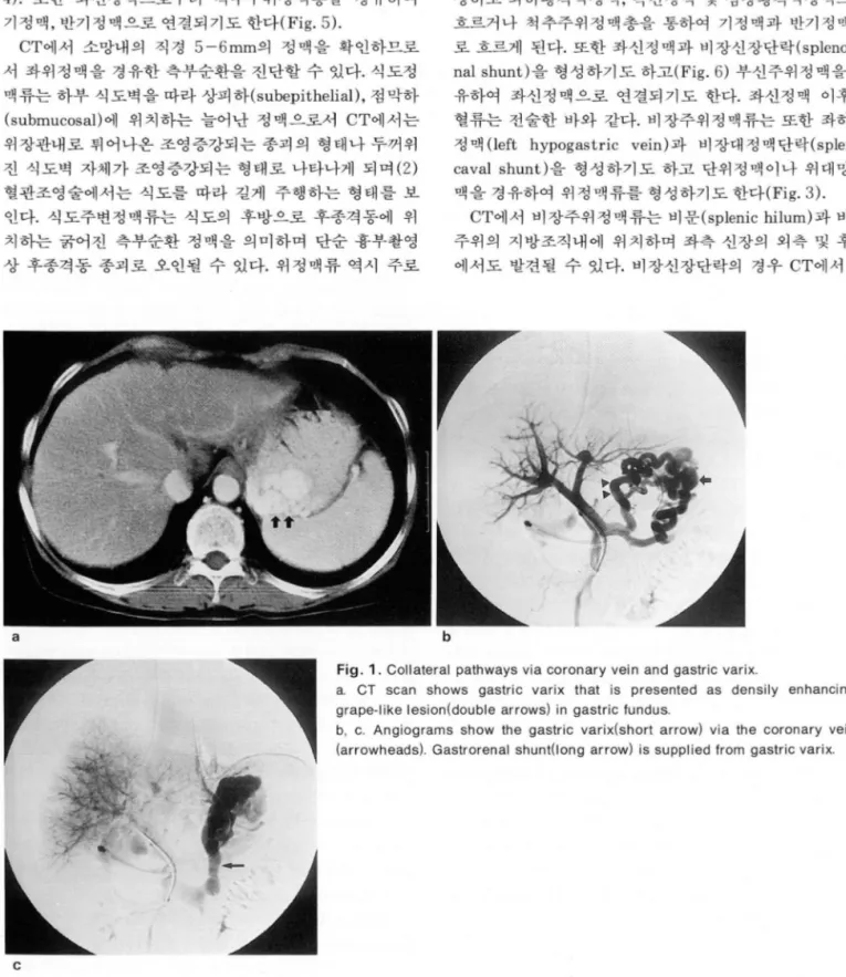
Fig. 1. Collateral pathways via coronary vein and gastric varix
c
a. CT scan shows gastric varix that is presented as densily enhancing grape-like lesion(double arrows)
ingastric fundus.
b
,c. Angiograms show the gastric varix(short arrow) via the coronary
vein(arrowheads). Gastrorenal shunt
(long arrow) is supplied from gastric varix
•
326
이경원 외 ‘ 문맥 고혈압환자에서의 문맥-체측부순환경로
상장간막정 맥을 경유한 측부순환경로
간문맥 으로부터 상장간박정 맥 (superior mesenteric vein)으로 역류된 혈류는 장간막내에서 정맥류 (superior
mesenteric varix)를 형성하고 이로부터 우성선정맥을 경 유하거나 직접 우신정맥과 장간막신단락 (mesorenal sh- unt)을 형성할 수 있다. 장간막정맥류는 후복강정맥류
(retroperi toneal varix) 와 잘 동반되 어 CT에서 장간막과 후복강에 조영증강이 잘 되는 결절 또는 관형 구조가 모여 있는 형태로 나타나며 이로부터 하대정액으로 장간막대정 신문정맥(left renal hilar vein) 과 좌신정맥의 확장을 확
인할 수 있다 (Fig.6a).
a b
i i;
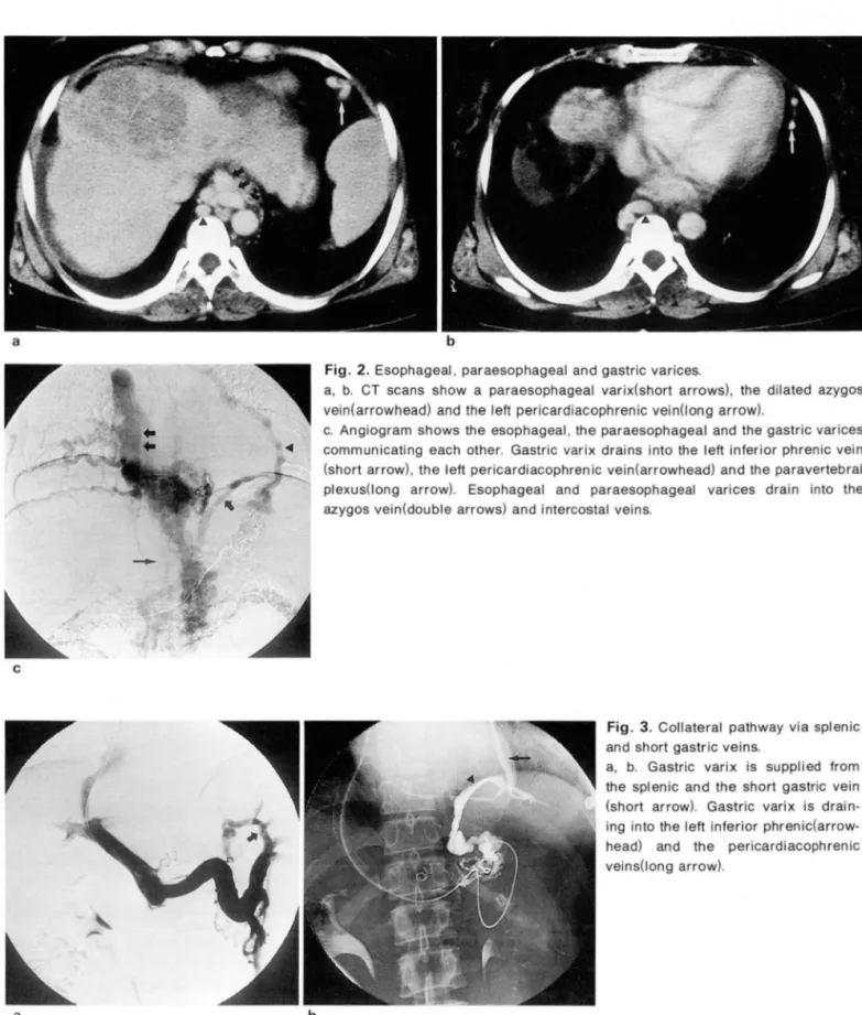
Fig. 2. Esophageal, paraesophageal and gastric varices
a, b. CT scans show a paraesophageal varix(short arrows), the dilated azygos vein(arrowhead) and the left pericardiacophrenic vein(long arrow)
c. Angiogram shows the esophageal, the paraesophageal and the gastric varices communicating each other. Gastric varix drains into the left inferior phrenic vein (short arrow), the left pericardiacophrenic vein(arrowhead) and the paravertebral plexus(l ong arrow). Esophageal and paraesophageal varices drain into the azygos vein(double arrows) and intercostal veins
c
Fig. 3. Collateral pathway via splenic and short gastric veins
a, b. Gastric varix is supplied from the splenic and the short gastric vein (short arrow). Gastric varix is drain- ing into the left inferior phrenic(arrow- head) and the pericardiacophrenic veins(long arrow)
~ -
a b
낀
1 j
대 한 방사 선 의 학회 지 1996: 35(3) : 325- 333
액단락 (mesocaval shunt)을 형성하거나 기정맥, 반기정 맥으로 혈류가 흐르게 된다 (Fig. 7). 또한 상장간막정맥으
로부터 훼십이지장정맥류가 형성되고 기정맥, 반기정맥으 로연결되기도한다.
a b
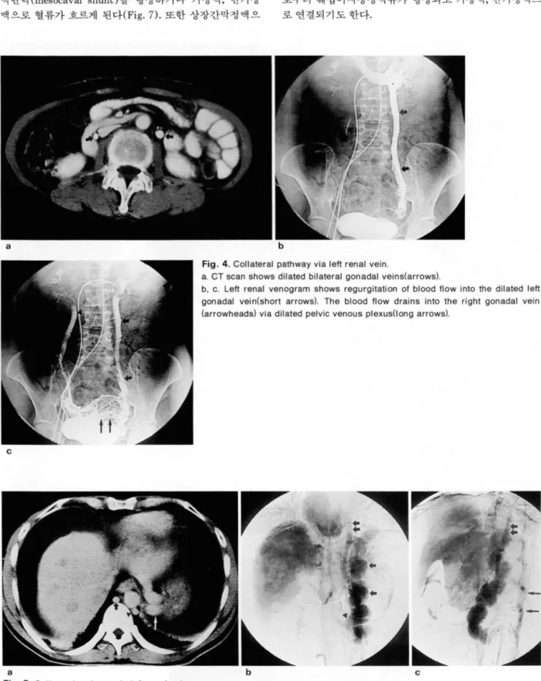
Fig. 4. Collateral pathway via left renal vein.
a. CT scan shows dilated bilateral gonadal veins(arrows)
b, c. Left renal venogram shows regurgitation of blood flow into the dilated left gonadal vein(short arrows). The blood flow drains into the right gonadal vein (arrowheads) via dilated pelvic venous plexus(l ong arrows)
c
i i
a b c
Fig. 5. Collateral pathway via left renal vein
a. CT scan shows dilated azygos(short arrow), hemiazygos vein(arrowhead) and upper portion of gastric varix(long arrow) b, c. Markedly dilated gastric varix(long arrows) and gastrorenal shunt(arrowhead) are seen. Blood flow drains into the paravertebral plexus(long arrows), the azygos and the hemiazygos veins(double arrows)‘
@
잉
이경원 외 ‘ 문맥 고혈압 환자에서의 문맥-체측부순환경로
하잠간막정 맥을 경유한 측부순환경로
상장간막정 맥 과 마찬가지로 하창간막정 맥 (inferior me-
senteric vein) 으로부터 장간막정맥류 (inferior mesente- ric varix) 와 후복강정맥류가 형성되어 하대정맥 (Fig. 8)
a
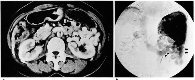
Fig. 6. Collateral pathways via splenic vein and splenorenal shunt
b
a. CT scan shows multiple dilated veins anterior to the left kidney(short arrows). Dilated renal hilar veins(arrowhead) and the left renal vein(long arrow) are also seen
b. Very tortuous and dilated veins(arrows) are seen around and inferior to the spleen. Splenorenal shunt(arrowhead) is formed from perisplenic varix
a b
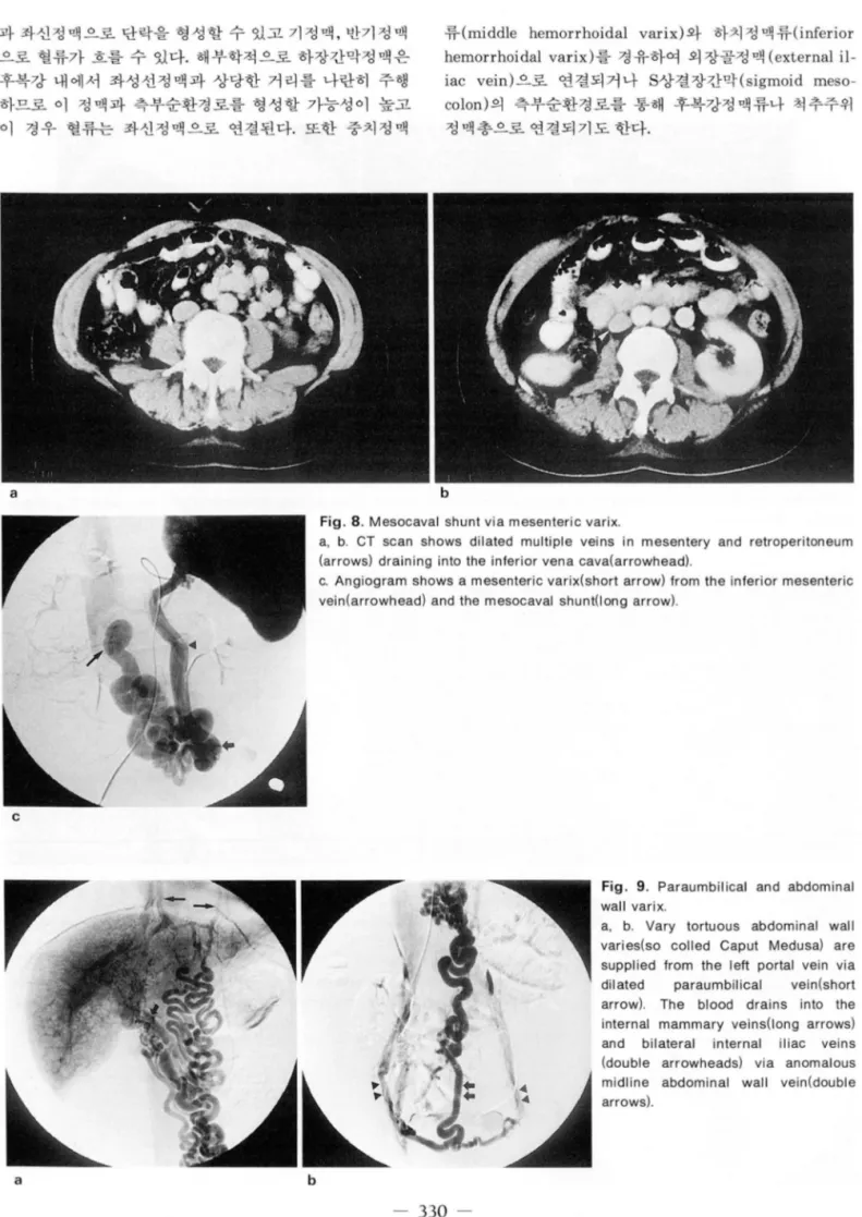
Fig. 7. Mesenteric and retroperitoneal varix
a, b. CT angiogram shows multiple veins in mesentery and retroperitoneum (short arrows) draining into the paravertebral plexus(arrowheads) and the azygos vein(long arrow).
c. SMA portogram shows mesenteric(short arrow) and retroperitoneal varices(ar- rowhead) draining into the paravertebral plexus and the azygos vein(long arrow)
c
왜
대 한 밤 사 선 의 학 회 지 1996 ; 35( 3) : 325-333
과 좌신정맥으로 단락을 형성할 수 있고 기정맥, 반기정맥 으로 혈류가 흐를 수 있다. 해부학적으로 하창간막정액은 후복강 내에서 좌성선정액과 상당한 거리를 나란히 주행 하므로 이 정맥과 측부순환경로를 형성할 가능성이 높고 이 경우 혈류는 좌신정맥으로 연결된다. 또한 중치정맥
류 (middle hemorrhoidal varix) 와 하치 정 맥 류 (inferior hemorrhoidal varix) 를 경유하여 외 장골정 맥 (external il- iac vein) 으로 연결되거나 S상결장간막 (sigmoid meso- colon) 의 측부순환경로를 통해 후복강정맥류나 척추주위 정맥총으로 연결되기도 한다.
a
bFig. 8. Mesocaval shunt via mesenteric varix
a, b. CT scan shows dilated multiple veins in mesentery and retroperitoneum (arrows) draining into the inferior vena cava(arrowhead)‘
c. Angiogram shows a mesenteric varix(short arrow) from the inferior mesenteric vein(arrowhead) and the mesocaval shunt(long arrow).
c
Fig. 9. Paraumbilical and abdominal wall varix
a, b. Vary tortuous abdominal wall varies(so colled Caput Medusa) are supplied from the left portal vein via
‘ 이 lated paraumbilical vein(short arrow
l.
The blood drains into the internal mammary veins(l ong arrows) and bilateral internal iliac veins (double arrowheads) via anomalous midline abdominal wall vein(double arrows)a b
m
씨
이경원 오I
:
문맥 고혈압 환자에서의 문맥-체측부순환경로제대주위정맥을 경유한 측부순환경로 제대주위정맥을 포함하고 있다. 간문맥 고혈압 환자에서 정상 겸상인대 (falciform ligament)는 1-3개의 수축된 제대주위정맥의 수와 직경이 증가하며 좌간문맥으로부터
a b
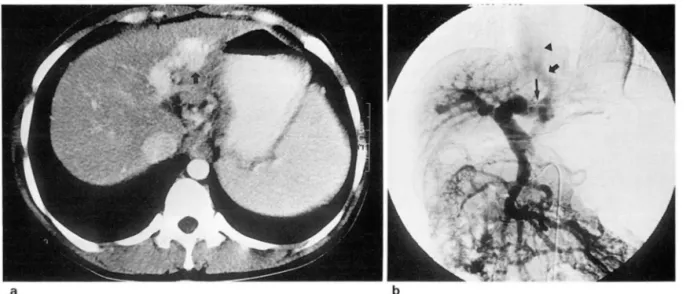
Fig. 10. Intrahepatic collateral pathway‘
a. CT scan shows a shunt Irom the lelt portal vein to the left hepatic vein presented as early enhancing mass-like lesion in left lobe 01 liver(arrow)
b. SMA portogram shows early opaci- lication 01 the left hepatic vein(short arrow) and IVC(arrowhead) via left portal vein(J ong arrow)
a b
Fig. 11. Collateral pathway via colostomy site
a, b. CT scans show displaced IMV(arrow) toward the colostomy site and dillusely enhanced colostomy site(arrowheads)
c. M 비 tiple dilated vascular structures(short arrows) are seen in colostomy site 01 left lower abdomen supplied Irom the inlerior mesenteric vein (arrowheads). Left iliac vein and IVC(long arrows) are early opacilied.
/
c n
μ
대 한 방사 선 의 학회 지 1996: 35(3): 325- 333
역류된 혈류가 이를 통해 전내측으로 제대를 향하게 되고 흔히 1-2개의 확장된 제대정맥을 통해 혈류가 흐르는 경 우가 많다 (5)(Fig. 9). 제대로부터 방사상으로 위치하는 복벽의 정맥류는 Caput Medusa라는 용어로 알려져 있으 며 혈관조영술상 매우 구불구불한 형태를 보인다. 혈류는 양측 하심와부정맥 (inferior epigastric vein) 을 통해 외장 골청맥으로 연결되거나 (Fig. 9b) 상심와부정맥을 경유하 여 내유정맥(internal mammary vein) 으로 연결되기도 하고 (Fig. 9a) 복벽정맥을 통해 흉벽정맥과 연결되기도 한 다.
기타측부순환로
대망정맥류 (omental varix) 는 대망내에서 여러개의 작은 정 맥으로 나타나며 혈관조영술이나 다른 진단방법으로는 쉽게 볼 수 없으나 CT에서는 약 20%에서 발견될 수 있다고 한다(1).
드문 측부순환로로서 간경화 환자에서 식도정맥류 결찰 빛 비장절제술을 시행한 경우 춰l장 의 주변과 내부로 정맥류가 형 성되고 이로부터 좌신정맥과 기정맥, 반기정액으로 혈류가 연 결될수있다
장절제숭 또는 결장루설치술 (colostomy) 등의 수술을 받은 후 장간막정맥과 대망정맥으로부터 결장루주변 또는 복벽 절개 부위로 작고 많은 측부순환경로가 형성되어 체정맥으로 연결될 간내, 경간 측부순환경로 수 있다 (Fig. lla). 이 경우 CT에서는 수술부위에 주변과 경계 간문맥의 측부순환경로는 간내(i ntrahepatic) 및 경간 가 명확히 그려지지 않는 조영층강되는 병소로 관찰된다 (Fig.
(tr anshepatic) 측부순환경로가 있다. 간내 측부순환경로 llb, c).
는 간문맥으로부터 간정맥으로 직접 연결되는 측부순환경 로를의미하며 이 경우 CT에서 확장된 깐정맥이 문맥강조 기 (portal phase)에 조기 조영증캉이 된다(Fig. lOa). 상 창간막동맥에서 조영제를 주사한 후의 문맥조영술에서도 조기에 간정맥과하대정맥이 조영되는것을확인할수있 다 (Fig. 10b).
경 간측부순환로의 예로서 간의 bare area의 정 맥으로부 터 우하횡격막정맥을 경유, 늑간정맥으로 연결되거나 또 는 우심장횡격막정맥으로 혈류가 연결될 수 있다.
결 를응 ‘-
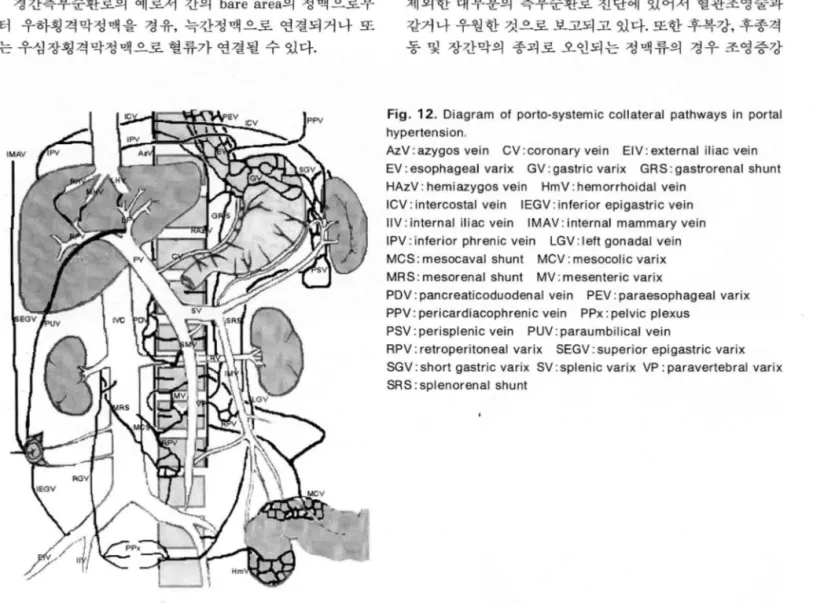
간문맥고혈압 환자에서 문맥 -체측부순환경로는 모식 도 (Fig. 12) 에서 보는 바와 같이 매우 다양한 형태로 나타 난다. 급속조영 역동적 CT나 나선식 CT는 식도정맥류를 제외한 대부분의 측부순환로 진단에 있어서 혈관조영술과 같거나 우월한 것으로 보고되고 있다. 또한 후복강, 후종격 통 및 장간막의 종괴로 오인되는 정맥류의 경우 조영증강
Fig. 12. Diagram 01 porto-systemic collateral pathways in portal hypertension.
AzV: azygos vein CV: coronary vein EIV: external iliac vein
EV: esophageal varix GV: gastric varix GRS: gastrorenal shunt HAzV: hemiazygos vein HmV: hemorrhoidal vein
ICV: intercostal vein IEGV: inlerior epigastric vein
IIV: internal iliac vein IMAV: internal mammary vein
IPV: inlerior phrenic vein LGV: left gonadal vein
MCS: mesocaval shunt MCV: mesocolic varix
MRS ‘ mesorenal shunt MV: mesenteric varix
PDV: pancreaticoduodenal vein PEV: paraesophageal varix
PPV: pericardiacophrenic vein PPX : pelvic plexus PSV: perisplenic vein PUV: paraumbilical vein
RPV: retroperitoneal varix SEGV: superior epigastric varix SGV: short gastric varix SV: splenic varix VP: paravertebral varix SRS: splenorenal shunt
1 4
n
μ
CT는 종괴나 럼프절병증과의 감별에 큰 도움을 줄 수 있 다. 그러나 CT는 혈류의 방향과 그 연결관계의 평가에 있 어 혈관조영술에 비하여 제한적이다. 그러므로 CT와 혈관 조영술은 측부순환경로의 진단 및 혈류의 평가에 상호 보 완적으로 이용되어야 할 것이다. 측부순환경로는 중재적 시술이나 수술시 심각한 출혈을 발생시킬 수 있으므로 출 혈예방을 위해 이에 대한 정확한 지식과진단이 매우중요 할 것으로 생각되며 혈류를 따른 고찰이 도움이 될 것으로 생각한다.
숭 L
=
고 .".--C그 i 헌1. Cho KC, Patel YO, Wachsberg RH, Seeft J. Varices in portal hypertension. Radiographics 1995; 15: 609-622
‘
‘ 이경원 외 · 문맥 고혈압 환자에서의 문맥-체측부순환경로
2. Balthazar EJ, Naidich 0, Megibow A, LeFleur RS. CT evalu- ation 01 esophageal varices. AJR 1987 ‘ 148: 131-135
3. Balthazar EJ, Megibow A‘ Naidich 0, LeFleur RS. Computed tomographic recognition 01 gastric varices. AJR 1984; 142 1121-1125
4. Minami M, Kawauchi N, Itai Y, Kokubo T, Sasaki Y. Transdia- phragmatic portosystemic shunt to the pericardiacophrenic vein. AJR 1993‘161 : 559-571
5. La:lortune M, Constantin A, Breton G, Legare AG, Lavoie P The recanaliz.ed umbilical vein in portal hypertension: A myth. AJR 1985; 144 : 549-553
Journal of the Korean Radiological Society 1996; 35(3) : 325- 333
Porto-systemic Collateral Pathways in Portal Hypertension:
Correlation of CT and Angiograph y
1Kyoung Won Lee, M.D., Jin Wook Chung, M.D., Jae Hyung Park, M.D., Kyung Mo Yeon, M.D.
1 Department of Radiology, Seoul National University College of Medicine
In portal hypertension, hepatopetal flow is rerouted away from the liver through collateral pathways to low pressure systemic vessels. Information about collateral pathways is relevant, especially when interventional procedure or surgery is contemplated, because inadvertent disruption of these veins can cause significant bleeding. Dynamic CT and spiral CT with a bolus injection of contrast material have to a significant extent re- cently replaced angiography. The porto-systemic collateral pathways have, ho
‘
Never, been classified and de- scribed according to location or frequency in a majority of previous reports. This essay illustrates variable porto-systemic coltateral blood flow pathways, with CT and angiography correlation.Index Words: Porto-systemic collaterals Partal vein, flow dynamics Portal vein, CT
Shunts, portosystemic Portography
Address reprint requests to : Department of Radiology, Seoul National University College of Medicine, n 28, Yungon 강 ong,
Chongno, Seoul, 110-744 Korea. Tel. 82-2-744-4581,760-4581 Fax. 82-2-743-6385
m
끽
일 시
:
1996 년 10월 26 일(토) 오후1: 00-7: 30
장 소:
부산대학교병원 신관 9 증 강당주 관
:
부산의대 방사선과학교실 및 방사선의학연구소 주 죄:
부산대학교병원연수펑점 :6점
제 1 부 좌 장 박수성 (동아의대)
1 : 00- 1 : 30
선천성 골기 형Congenital Anomaly of Bone 1 : 30- 2: 00
골절의 영상진단Imaging Diagnosis of Fracture
2: 00- 2: 30
글관절 감염성질환Infectious Disease of Bone & Joint 2 : 30 - 2: 40 Discussion
김휘택 (부산의대)
이성문 (계명의대)
윤정흐I (메리놀병원)
2: 40- 3: 10
그곡견 7~1 종만선 L-= ---, ηI c:::::J~L::) 진화 ~~ 구봉식 (동아의대)Musculoskeletal Oncology
3: 10- 3: 40
사지혈관성질환의 중재적시술 조길호 (영남의대)Intervention in Vascular Disease of Extremity 3: 40- 3: 50 Discussion & Break Time
쩌12 부 좌 장 김병수 (부산의대)
3: 50- 4: 20
근골격계질환의 SPECT 응용 앙승오 (울산의대)Musculoskeletal SPECT
4: 20- 4: 50
근글격계 초음파검사 조길호 (영남의대)Musculoskeletal Ultrasonography
4: 50- 5: 40
척추질환의 자기공명영상 강흘식 (서울의대)MRI of Spinal Diseases 5: 40- 5: 50 Discussion
5: 50- 6: 20
상지굴관절의 자기공명영상 문태용 (부산의대)MRI of Upper Extremities
6: 20- 7: 00
하지골관절의 자기공명영상 이영준 (인제의대)MRI of Lower Extremities
m