Abstract (J Korean Assoc Oral Maxillofac Surg 2010;36:453-9)
Ⅰ.Introduction
Ameloblastoma is the most common benign odontogenic tumor of the jaw, but local invasive growth or recurrence after the removal can be found. Ameloblastic carcinoma is rarely occurred and may arise as a result of malignant change in a pre-existing benign ameloblastoma (carcinoma ex-ameloblas- toma) or as a primary ameloblastic carcinoma not preceded by ameloblastoma (de novo carcinoma)1.
In the malignant tumors, the gene mutation, epigene alter- ations such as hypermethylation of CpG islands of tumor sup- pressor gene may lead to suppression of transcription, which induces malignant transformation2. Methylation is one possible step towards DNA damage, and the same evolutionary pres- sure will favor all processes of silencing same genes in car- cinogenesis3,4. The DNA methylation profile of cancer cells is characterized by global hypomethylation and simultaneous hypermethylation of selected CpG island gene promoters.
Recently, the epigenetic phenomenon of DNA promoter methylation has gained increasing recognition as an important mechanism for transcriptional inactivation of cancer related genes. The identification of these methylation alterations and elucidation of the mechanistic events are important, as the methylation status of cancer cells can now be manipulated in vivo with demethylating chemotherapeutics5.
박 상 준
614-735 부산광역시 부산진구 개금동633-165 인제대학교 의과대학 부산백병원 구강악안면외과 Sang-Jun Park
Department of Oral and Maxillofacial Surgery, Pusan Paik Hospital, College of Medicine, Inje University
633-165 Gaegum-dong, Busanjin-gu, Busan, 614-735, Korea TEL: +82-51-890-6360 FAX: +82-51-896-6675
E-mail: ds5nki@hanmail.net
Methylation of p16 and E-cadherin in ameloblastoma
Chan-Woong Park1, Hye-Kyoung Yoon2, Sang-Jun Park1 Departments of 1Oral and Maxillofacial Surgery, 2Pathology, Pusan Paik Hospital, College of Medicine, Inje University, Busan, Korea
Introduction:Ameloblastic carcinoma is a rare malignant lesion, and may arise from either carcinoma ex-ameloblastoma or de novo carcinoma.
Aberrant promoter hypermethylation of the tumor-associated genes leading to their inactivation is a common event in many cancer types. The p16/CDKN2/INK4A gene and p16 5 protein are involved directly in regulating the cell cycles. Cadherins are cell adhesion molecules that modulate the epithelial phenotype and regulate tumor invasion. The aim of this study was to evaluate the roles of p16 and E-cadherin methylation and loss of p16 and E-cadherin expression in the malignant transformation of an ameloblastoma.
Materials and Methods:Eight cases of ameloblastoma, including 4 benign ameloblastomas without recurrence, 2 benign ameloblastomas with recurrence and 2 carcinoma ex-ameloblastomas, were examined. The promoter hypermethylation profile of the p16 and E-cadherin genes was studied using methylation-specific polymerase chain reaction (MSP) and immunohistochemical staining for p16 and E-cadherin expression.
Results:
1) Aberrant CpG island methylation of the p16 gene was detected in 3 of the 4 benign ameloblastomas without recurrence and 1 of the 2 benign ameloblastomas with recurrence.
2) Aberrant CpG island methylation of the E-cadherin gene was found in 1 of the 4 benign ameloblastomas without recurrence.
3) A loss of p16 expression was noted in 1 of 4 benign ameloblastomas without recurrence and 1 of 2 carcinoma ex-ameloblastomas.
4) A loss of E-cadherin expression was noted in 2 of the 4 benign ameloblastomas without recurrence, 1 of the 2 benign ameloblastomas with recur- rence and 2 of the 2 carcinoma ex-ameloblastomas.
5) A loss of p16 expression was observed in 1 of the 4 cases showing aberrant methylation of the p16 gene.
6) A loss of E-cadherin expression was observed in 3 benign ameloblastoma case showing aberrant methylation of the E-cadherin gene.
Conclusion:These results suggest that loss of E-cadherin expression related to the other genetic pathway (not methylation) might be an adjuvant indicator predicting the malignant transformation of an ameloblastoma. However, the number of samples in this study was too small and the relation- ship between the treatment methods and clinical course were not defined. Therefore, further study will be needed.
Key words:p16 genes, Cadherins, Methylation, Ameloblastoma
[paper submitted 2010. 7. 16 / revised 2010. 11. 19 / accepted 2010. 12. 10]
Hypermethylation of tumor suppressor genes p15 and p16 can be detected in several types of malignant tumors including oral squamous cell carcinoma (SCC)6,7. Abiko et al. reported that hypermethylation of p16 may have been involved in the malignant transformation of the ameloblastoma in the present case. Hypermethylation of CpG islands of the p16 gene was detected in the malignant parts of the tumor8.
Loss of E-cadherin will result in increasing invasiveness and metastatic potential of neoplastic cells. In oral squamous cell carcinoma7,9-11. hypermethylation of the E-cadherin promoter has recently been demonstrated. Considerable differences have been observed in the incidence alteration by mutations or aber- rant methylation of the CpG islands of different regulator genes. The incidence of lost expression due to methylation in E-cadherin is more occasional than for p169.
This study was conducted to explore the hypermethylation and immunohistochemical studies for p16 and E-cadherin genes in the histologically benign ameloblastoma (non- recurred group versus recurred group) and ameloblastic carci- noma (carcinoma ex-ameloblastoma).
Ⅱ. Materials and Methods 1. Patient and samples
Tissue samples from 8 cases, including 4 benign ameloblas- tomas without recurrence, 2 benign ameloblastomas with recurrence and 2 carcinoma ex-ameloblastomas, diagnosed as ameloblastoma during a period from 1991 to 2004 were select- ed from the files of department of oral and maxillofacial surgery, Pusan Paik Hospital. They were obtained from the patients ranged in age from 13 to 70 years old, and 6 were male and 2 female. The tumors were located in the mandible in 7 cases, and 1 case with multiple recurrences was located in the maxilla.
2. Histopathological Review
Based on the hematoxylin and eosin (H&E) findings, the ameloblastoma cases were re-classified into benign ameloblas-
toma without or with recurrence, carcinoma ex-ameloblastoma and malignant ameloblastoma de novo.
3. Methylation study for p16 and E-cadherin genes
1) Genomic DNA extraction
Ten or more sections were obtained from each paraffin blocks as samples. Paraffin-embedded tissue thin sections (5 μm thick, 10 sections) were obtained as sample. From the slides, the tumor tissues, corresponding to the marking sites on H&E slide, were taken. Genomic DNA was isolated using ZYMO Pinpoint Slide DNA Isolation System (Cat. No. D3001, Zymo research, Orange, CA. USA) after deparaffinized with xylene.
DNA was recognized using 1% Agarose gel electrophoresis.
2) Sodium bisulfite modification
One μg of each DNA sample was undergone bisulfite modi- fication prior to polymerase chain reaction (PCR) amplifica- tion using the ZYMO EZ DNA Methylation Kit (Cat. No.
D5002, Zymo research, Orange, CA, USA). This experimental procedure was done according to the manufacturer`s instruc- tions. Briefly, A mixture of 90 μL DNA solution and 10 μL M- dilution buffer was incubated at 37℃ for 15 minutes. After loading a mixture into Zymo-Spin I Column and centrifuging (11,000 rpm) for 1 minute at 25℃, the waste products in the collection tube were discarded. After this step, M-Wash buffer 200 μL M-desulphonation buffer was loaded and incubated for 15 minutes at room temperature, and then centrifuged for 1 minute at 25℃, and then 200 μL M-wash buffer was loaded and centrifuged for 1 minute at 25℃.
3) Methylation specific PCR and Electrophoresis
Amplitaq Gold 0.5 unit for p16 and E-cadherin (Roche, Madison, WI, USA) was used to detect methylated sequence in mixed cell populations (methylated and unmethylated) for analysing DNA hypermethylation of p16 and E-cadherin genes. PCR reaction mix consisted of 1×PCR buffer, each 2.5 mM dNTP, each primer set, 1 unit of HotStarTaq DNA poly- merase (Qiagen, cat. No.203203, Germantown, MD, USA) and modified genomic DNA. The primers used for the detection of
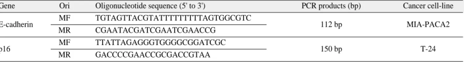
Table 1.Oligonucleotide sequence used in methylation and unmethylation of 5'-UTR Promotor Region CpG island
Gene Ori Oligonucleotide sequence (5' to 3') PCR products (bp) Cancer cell-line
E-cadherin MF TGTAGTTACGTATTTTTTTTTAGTGGCGTC
112 bp MIA-PACA2
MR CGAATACGATCGAATCGAACCG
p16 MF TTATTAGAGGGTGGGGCGGATCGC
150 bp T-24
MR GACCCCGAACCGCGACCGTAA
(UTR: untranslated region, PCR: polymerase chain reaction)
methylation and unmethylation were discribed in Table 1, respectively. The reactions were hotstarted at 95℃ for 15 min- utes, and were carried out for 40 cycels (45 seconds at 95℃, variable annealing time and temperatures according to the primer, and 60 seconds at 72℃) and a final 10 minutes exten- sion at 72℃ in a thermal cycler (MJ Research, Waltham, MA, USA). PCR products were separated by electrophoresis on a 2% agarose gel, then visualized under ultraviolet (UV) illumi- nation using an ethidium bromide stain.
4) Sequencing
The PCR products suggested to harbor mutation were cloned, and recombinant double-stranded plasmid DNA was sequenced by automatic sequencer using an AutoRead 100 sequencing kit (Pharmacia, Stockholm, Sweden).
4. Immunohistochemical stain for p16 and E-cadherin
Formalin-fixation and paraffin-embedding tissues were sec- tioned at 4-6 μm and deparaffinized in xylene and rehydrated in ethanol. The slides were incubated with pretreatment for 30 minutes at 99℃. The sections were incubated with 3% peroxi- dase blocking solution for 10 minutes at room temperature for endogenous peroxidase and incubated with primary antibody at appropriate dilution in primary antibody dilution buffer for 1 hour at room temperature. Commercially available primary antibodies directed against p16 (1:2,000, Abcam, Cambridge, UK) and E-cadherin (1:25, Abcam, Cambridge, UK) were applied. Biotinylated secondary antibody and streptavidin- HRP was carried out using the Envision system (Dako, Glostrup, Denmark). Chromogen was incubated in diaminobenzidine (DAB) (Dako, Glostrup, Denmark) for 10 minutes at room temperature. The slides was developed with Mayer’s hematoxylin for counter staining, dehydrated through alcohol, and cleared in xylene prior to cover slipping for
microscopic examination.
Immunohistochemical stains for p16 and E-cadherin were interpretated as partial loss or total loss of expressions accord- ing to the extent of loss of expressions. If more than half of tumor tissue were negative, it was defined as total loss, and if the less than half of tumor tissue were negative, it was catego- rized as partial loss.
Ⅲ. Results
1. Methylation study for p16 and E-cadherin
Aberrant CpG island methylation of p16 was detected in 4 of 8 cases.(Figs. 1, 2) Four cases showing p16 methylation are benign ameloblastoma, and 3 of them showed no recurrence and 1 case was recurred. None of 2 cases of carcinoma ex- ameloblastoma showed aberrant CpG island methylation of p16.
Aberrant CpG island methylation of E-cadherin was found in 1 of 8 cases.(Figs. 3, 4) One case showing methylation of E- cadherin was benign ameloblastoma with no recurrence.
(Table. 2)
2. Immunohistochemical findings for p16 and E-cadherin
Total or partial loss of p16 expression were noted 4 of 8 cas- es.(Fig. 5) Two cases showing total loss of p16 expression were one benign ameloblastoma and one carcinoma arising in benign ameloblastoma. Two cases exhibiting partial loss of p16 expression were benign ameloblastoma cases, and 1 of them was recurred case.(Fig. 6, Table. 2)
Loss of E-cadherin expression was noted in 5 of 8 cases.
Total loss of E-cadherin expression was noted in 2 cases: one was recurred ameloblastoma and the other carcinoma ex- ameloblastoma. Partial loss of E-cadherin expression was not-
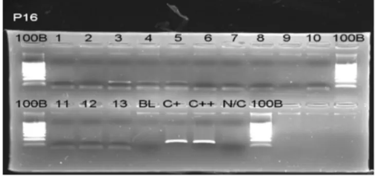
Fig. 2. SSCP analysis of the p16 gene in ameloblastoma.
Lane 1, 3, 4, 5 show corresponding band to C+, C++ line.
(BL: blank, C+, C++: positive cell line, N/C: negative control, SSCP: single strand conformational polymorphism) Fig. 1. Bisulfite genomic sequencing result of 5’CpG island
of the p16 gene.
ed in 2 of 4 benign ameloblastoma cases and 1 of carcinoma ex-ameloblastoma.
3. Comparison of methylation and immunohistochemical studies
Among 4 benign ameloblastoma cases, methylation of p16 gene was noted in 3 cases. One of 3 cases with aberrant p16 gene methylation showed partial loss of p16 expression. One benign ameloblastoma case showing aberrant E-cadherin
methylation revealed partial loss of E-cadherin expression.
In 2 cases of benign ameloblastoma with recurrence, 1 case showed aberrant p16 gene methylation, but loss of p16 expres- sion was not accompanied. Another 1 case of benign ameloblastoma with recurrence showed loss of E-cadherin
Fig. 3.Bisulfite genomic sequencing result of 5’CpG island of the E-cadherin gene.
Fig. 4.SSCP analysis of the E-cadherin gene in ameloblas- toma. Lane 2 shows corresponding band to C+ line.
(BL: blank, C+: positive cell line, N/C: negative control, SSCP: single strand conformational polymorphism)
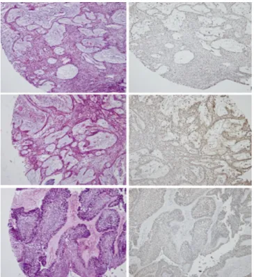
Fig. 5. Loss of p16 expression in benign ameloblastoma without recurrence (upper) and with recurrence (middle), and carcinoma ex-ameloblastoma (lower).
Fig. 6.Loss of E-cadherin expression in benign ameloblas- toma without recurrence (1st lane) and with recurrence (2nd lane), and in carcinoma ex-ameloblastoma (3rd and 4th lane).
expression, even though aberrant E-cadherin gene methylation was not noted.
In 2 cases showing carcinoma arising in ameloblastoma, 1 case revealed loss of p16 expression but aberrant methylation of p16 gene was not associated in this case. Two cases show- ing carcinoma arising in ameloblastoma exhibited loss of E- cadherin expression even though no aberrant methylation of E- cadherin.(Table. 3)
Ⅳ. Discussion
Ameloblastoma is a locally infiltrative, benign, odontogenic neoplasm typically arising in the posterior regions of the jaws.
Approximately 80% of ameloblastomas were reported to occur in the mandible, and 20% in the maxilla12,13. A rare, malignant variant of ameloblastoma was first described in the medical lit- erature in 195014. The World Health Organization separated this cariant into its current subtypes, ie, maligmant ameloblas- toma and ameloblastic carcinoma, in 197215. This classification was further revised by Slootweg and Muller16 in 1984. The World Health Organization described malignant ameloblas- toma as a tumor showing the histopathologic features of classic ameloblastoma with metastatic deposits, and defined
ameloblastic carcinoma as an epithelial proliferation with histopathologic features of malignancy, either associated with an ameloblastoma (carcinoma ex-ameloblastoma) or represent- ing a de novo carcinoma that resembles ameloblastoma histopathologically1.
Epigene alterations such as hypermethylation of CpG islands of tumor suppressor gene may lead to suppression of transcrip- tion, which induces malignant transformation2. Hypermethy- lation of tumor suppressor genes such as p16 and E-cadherin affect DNA repair. DNA methylation could change in response to environmental, physiological and biological signals.
The DNA methylation profile of cancer cells is characterized by global hypomethylation and simultaneous hypermethylation of selected CpG island gene promoters. Recently, the epigenet- ic phenomenon of DNA promoter methylation has gained increasing recognition as an important mechanism for tran- scriptional inactivation of cancer related genes. The identifica- tion of these methylation alterations and elucidation of the mechanistic events are important, as the methylation status of cancer cells can now be manipulated in vivo with demethylat- ing chemotherapeutics5.
The p16 gene encodes a cell cycle protein which inhibits cyclin dependent kinase 4 and 6, and genetic alteration of the Table 3. Comparison of methylation specific PCR and immunohistochemical findings for p16 and E-cadherin in the ameloblastomas
p16 E-cadherin
n MSP IHC
MSP IHC
Partial loss Total loss Partial loss Total loss
Benign A. without recurrence 4 3 1 1 1 2 0
Benign A. with recurrence 2 1 1 0 0 0 1
Carcinoma ex-A. 2 0 0 1 0 1 1
Total 8 4 2 2 1 3 2
(PCR: polymerase chain reaction, MSP: methylation specific PCR, IHC: immunohistochemical staining, A: ameloblastoma)
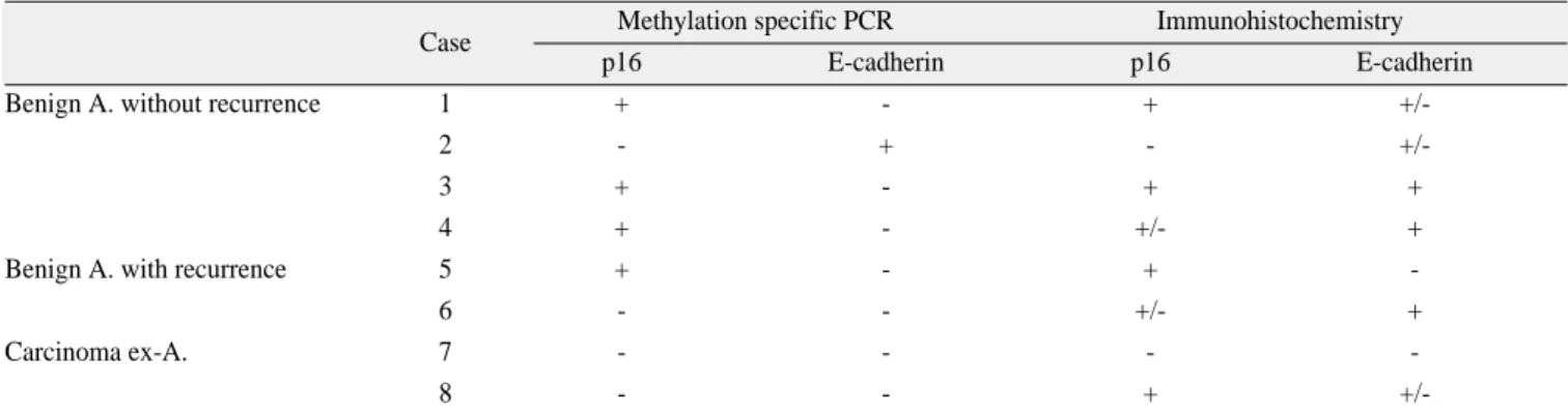
Table 2. Profiles of methylation specific PCR and immunohistochemical findings for p16 and E-cadherin in the ameloblastomas
Case Methylation specific PCR Immunohistochemistry
p16 E-cadherin p16 E-cadherin
Benign A. without recurrence 1 + - + +/-
2 - + - +/-
3 + - + +
4 + - +/- +
Benign A. with recurrence 5 + - + -
6 - - +/- +
Carcinoma ex-A. 7 - - - -
8 - - + +/-
(PCR: polymerase chain reaction, A: ameloblastoma)
p16 genes, leading to its inactivation, may result in degenera- tion of cell proliferation and tumorigenesis. The p16 tumor suppressor gene acts as a negative regulator of cellular prolif- eration, and inactivation of p16, related with homozygous deletions, mutations and hypermethylation. is a frequent event in human oral squamous cell carcinoma. Hypermethylation of tumor suppressor genes p15 and p16 can be detected in several types of malignant tumors including oral squamous cell carci- noma6,7.
According to Zhao et al.17, the p16 gene was altered and inactivated by methylation or mutation in a significant propor- tion of maxillofacial SCC. Twenty two cases (34%) were methylated and 7 (11%) displayed point mutations and the total frequency of alteration of the p16 gene was 43% (28/65).
Loss of p16 protein expression was noted in 17 (67%) of 27 SCC cases, Among 17 cases with loss of p16 protein expres- sion, 10 cases showed methylation and 3 cases revealed muta- tions, which may suggest that methylation and mutation of the p16 gene might account for the loss of p16 expression.
The hypermethylation of p16 may cause deregulation of cel- lular proliferation in ameloblastoma. Abiko et al.8reported that hypermethylation of p16 may have been involved in the malig- nant transformation of the ameloblastoma in the present case.
Hypermethylation of CpG islands of the p16 gene was detected in the malignant parts of the tumor. Nodit et al.18reported alle- ic loss of p16 genes in ameloblastic tumors, but no significant difference between ameloblastic carcinomas and benign ameloblastomas, so other genetic or epigenetic mechanism (not alleic loss) might be responsible for malignant behavior in ameloblastic carcinomas.
In present study, aberrant CpG island methylation of p16 was detected in 4 of 8 cases. Three of 4 cases were benign ameloblastoma without recurrence and 1 case was recurred.
None of carcinoma ex-ameloblastoma revealed aberrant CpG island methylation of p16. These findings suggest that aberrant methylation of p16 gene might be associated with ameloblas- toma but not related to the malignant transformation of ameloblastoma. Loss of p16 expression were noted 4 of 8 cas- es. One of 2 cases showing total loss of p16 protein expression was benign ameloblastoma and the other carcinoma ex ameloblastoma. Partial loss of p16 expression were observed in benign ameloblastoma with or without recurrence. These findings indicated that loss of p16 expression was associated with benign to malignant ameloblastoma, and was not a mark- er of malignant transformation. Only one of 4 cases showing aberrant methylation of p16 gene showed partial loss of p16 expression. There were no overt relationship between aberrant methylation of p16 gene and loss of p16 protein expression.
According to these results, loss of p16 protein expression in the ameloblastomas might be related to the aberrant methyla- tion and another genetic alterations.
In oral squamous cell carcinoma, hypermethylation of the E- cadherin promoter has recently been demonstrated7,9,10. Considerable differences have been observed in the incidence alteration by mutations or aberrant methylation of the CpG islands of different regulator genes. The incidence of lost expression due to methylation in E-cadherin is more occasion- al than for p1611. Methylation is one possible step towards DNA damage, and the same evolutionary pressure will favor all processes of silencing same genes in carcinogenesis3,4. Loss of E-cadherin is related to the increased invasiveness and metastatic potential of neoplastic cells, but several alternative routes to the loss of E-cadherin can be expected in the human tumors. Similarly, there must be a high number of genes with relatively low incidence of aberrant silencing by methylation or mutation, reflecting abundant alternative routes in the multi- step process of carcinogenesis11.
According to Abiko et al8, hypermethylation of E-cadherin was not observed in the benign and malignant ameloblastoma.
In this study, aberrant CpG island methylation of E-cadherin was found in only one case among 8 cases, and this case was benign ameloblastoma with no recurrence, These results sug- gest that aberrant methylation of E-cadherin gene seems to be a very rare event in the development of ameloblastoma, and not a suggesting finding of malignant transformation in the ameloblastoma. In contrast to methylation results, loss of E- cadherin expression was noted in 5 cases, and 3 cases showed partial loss of E-cadherin expression, and 2 of them were benign ameloblastoma and another one was carcinoma ex- ameloblastoma, and complete loss of E-cadherin expression, was noted in 2 cases, 1 recurred ameloblastoma and 1 carcino- ma ex-ameloblastoma. Based on E-cadherin expression find- ings, complete loss of E-cadherin expression might be an adju- vant marker of recurrence or malignant transformation in the ameloblastoma.
Aberrant E-cadherin methylation was noted in only one benign ameloblastoma case, accompanying partial loss of E- cadherin expression. Remaining 4 cases showing loss of E- cadherin expression revealed no evidence of aberrant methyla- tion of E-cadherin gene.
Aberrant methylation of E-cadherin gene may be a rare event in the development of ameloblastoma, in contrast, loss of E- cadherin expression is relatively common. These results suggest that loss of E-cadherin expression related to the other genetic pathway, not to methylation, might be a adjuvant indicator to predict the malignant transformation of ameloblastoma.
This study had some limitations. Because ameloblastic carci- noma is very rare entity of the cancer, too small samples were included. And treatment methods were not defined in all sam- ple, the relation between clinical and pathologic results were not included. Further study need to be continued with proper sample size and clinical follow-up.
Ⅴ. Conclusion
Aberrant methylation of p16 gene or loss of p16 expression might be associated with ameloblastoma but not related to the malignant transformation of ameloblastoma, and loss of p16 expression in the ameloblastomas might be related to the aber- rant methylation and another genetic alterations. Aberrant methylation of E-cadherin gene might be a rare event in the development of ameloblastoma, in contrast, loss of E-cadherin expression is relatively common. These results suggest that loss of E-cadherin expression related to the other genetic path- way, not to methylation, might be a adjuvant indicator to pre- dict the malignant transformation of ameloblastoma.
References
1. Barnes L, Eveson JW, Reichart P, Sidransky D, eds. World Health Organization classification of tumours: pathology and ge- netics of head and neck tumours. Lyon, France: International Agency for Research on Cancer Press; 2005.
2. Baylin SB, Esteller M, Rountree MR, Bachman KE, Schuebel K, Herman JG. Aberrant patterns of DNA methylation, chromatin formation and gene expression in cancer. Hum Mol Genet 2001;
10:687-92.
3. French SW, Dawson DW, Miner MD, Doerr JR, Malone CS, Wall R, et al. DNA methylation profiling: a new tool for evaluat- ing hematologic malignancies. Clin Immunol 2002;103:217-30.
4. Ho A, Dowdy SF. Regulation of G(1) cell-cycle progression by oncogenes and tumor suppressor genes. Curr Opin Genet Dev
2002;12:47-52.
5. Rush LJ, Plass C. Alterations of DNA methylation in hematolog- ic malignancies. Cancer Lett 2002;185:1-12.
6. Kresty LA, Mallery SR, Knobloch TJ, Song H, Lloyd M, Casto BC, et al. Alterations of p16(INK4a) and p14(ARF) in patients with severe oral epithelial dysplasia. Cancer Res 2002;62:5295- 300.
7. Viswanathan M, Tsuchida N, Shanmugam G. Promoter hyperme- thylation profile of tumor-associated genes p16, p15, hMLH1, MGMT and E-cadherin in oral squamous cell carcinoma. Int J Cancer 2003;105:41-6.
8. Abiko Y, Nagayasu H, Takeshima M, Yamazaki M, Nishimura M, Kusano K, et al. Ameloblastic carcinoma ex-ameloblastoma:
report of a case-possible involvement of CpG island hypermethy- lation of the p16 gene in malignant transformation. Oral Surg Oral Med Oral Pathol Oral Radiol Endod 2007;103:72-6.
9. Chen Q, Lipkina G, Song Q, Kramer RH. Promoter methylation regulates cadherin switching in squamous cell carcinoma.
Biochem Biophys Res Commun 2004;315:850-6.
10. Pho SW, Kim YS, Park JY, Kim CH, Lee W, Park MK. The hy- permethylation of E-cadherin gene in oral squamous cell carcino- ma. J Korean Assoc Oral Maxillofac Surg 2008;34:135-40.
11. Auerkari EI. Methylation of tumor suppressor genes p16(INK4a), p7(Kip1) and E-cadherin in carcinogenesis. Oral Oncol 2006;42:5-13.
12. Sastre J, Muñoz M, Naval L, Adrados M. Ameloblastic carcinoma of the maxilla: report of a case. J Oral Maxillofac Surg 2002;60:
102-4.
13. Verneuil A, Sapp P, Huang C, Abemayor E. Malignant amelo- blastoma: classification, diagnostic and therapeutic challenges.
Am J Otolaryngol 2002;23:44-8.
14. Thoma KH. Oral pathology: a histological, roentgenological, and clinical study of the diseases of the teeth, jaws, and mouth. 3rd ed. St. Louis: Mosby; 1950.
15. Pindborg JJ, Kramer I, Torloni H. Histological typing of odonto- genic tumors, jaw cysts, and allied lesions. Geneva, Switzerland:
World Health Organization; 1972.
16. Slootweg PJ, Muller H. Malignant ameloblastoma or ameloblas- tic carcinoma. Oral Surg Oral Med Oral Pathol 1984;57:168-76.
17. Zhao Y, Zhang S, Fu B, Xiao C. Abnormalities of tumor suppres- sor genes P16 and P15 in primary maxillofacial squamous cell carcinomas. Cancer Genet Cytogenet 1999;112:26-33.
18. Nodit L, Barnes L, Childers E, Finkelstein S, Swalsky P, Hunt J.
Allelic loss of tumor suppressor genes in ameloblastic tumors.
Mod Pathol 2004;17:1062-7.