Introduction
Endometrial hyperplasia (EH) was classified by the World Health Organization in 2014 into two categories based on the presence of cytological atypia [1]. Approximately, 200,000 new cases of EH are diagnosed annually in devel- oped countries [2,3]. EH is of significant clinical importance, given that it is the precursor of endometrial carcinoma [4], the most common gynecological cancer in developed coun- tries [5]. The risk of progression to carcinoma is less than 5%
Non-atypical endometrial hyperplasia: risk factors for occult endometrial atypia and malignancy in patients managed with hysterectomy
Lee Shi Hui, MBBS 1 , Selina Hui Men Chin, MBBS 1 , Charissa Goh, MBBS 1 , Lin Xiao Hui, MBBS 2 , Manisha Mathur, MBBS, MRCOG, FRCOG 1 , Timothy Lim Yong Kuei, MBBS, MRCOG, FRCOG 2 , Felicia Chin Hui Xian, MBBS, MRCOG, MMed 2
1
Department of Obstetrics & Gynecology,
2Department of Gynaecological Oncology, KK Women’s and Children’s Hospital, Singapore
Objective
To determine the risk factors for occult endometrial atypia and malignancy in patients diagnosed with non-atypical endometrial hyperplasia (NEH) on endometrial biopsy.
Methods
All new cases of NEH diagnosed between April 2015 and March 2016 at KK Women’s and Children’s Hospital, who underwent hysterectomy as first-line treatment, were included in the study. Patients with a history of endometrial hyperplasia or malignancy were excluded from the study. Patient demographics (e.g., age, parity, body mass index [BMI]), medical history, and clinical presentation were obtained for analysis.
Results
In total, 262 patients were diagnosed with NEH, of which 18.3% (n=48) underwent hysterectomy as first-line management. The average time to surgery was 77.0±35.7 days. All cases were diagnosed by dilation and curettage, and hysteroscopy. The mean age was 51 years, and the mean BMI was 26.9±5.8 kg/m
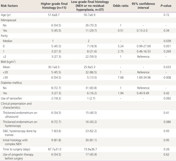
2. Histology from the hysterectomy specimen showed 9 (18.8%) patients with atypical hyperplasia and 2 (4.2%) with grade 1, stage 1A endometrioid adenocarcinoma. Patients with higher grade final pathology had significantly lower median parity (1 vs. 2, P=0.039), higher mean BMI (30.1±6.5 vs. 25.9±5.3 kg/m
2, P=0.033), and BMI ≥30 kg/m
2(54.5% vs. 13.5%, P=0.008, odds ratio 7.68), compared to patients whose final histology showed NEH or no residual hyperplasia.
Conclusion
Occult endometrial atypia and malignancy were found in 18.8% and 4.2% of patients with an initial diagnosis of NEH, respectively. High BMI and low parity were identified as significant risk factors for high-grade endometrial lesions in patients with NEH.
Keywords: Endometrial hyperplasia; Endometrial neoplasms; Dilation and curettage
Received: 2020.09.29. Revised: 2020.12.07. Accepted: 2021.02.21.
Corresponding author: Lee Shi Hui, MBBS
Department of Obstetrics & Gynecology, KK Women’s and Children’s Hospital, 100 Bukit Timah Road, Singapore 229899, Singapore
E-mail: [email protected] https://orcid.org/0000-0001-8369-9336
Articles published in Obstet Gynecol Sci are open-access, distributed under the terms of the Creative Commons Attribution Non-Commercial License (http://creativecommons.
org/licenses/by-nc/3.0/) which permits unrestricted non-commercial use, distribution, and reproduction in any medium, provided the original work is properly cited.
Copyright © 2021 Korean Society of Obstetrics and Gynecology https://doi.org/10.5468/ogs.20294
eISSN 2287-8580
for non-atypical endometrial hyperplasia (NEH) and up to 30% in atypical hyperplasia (AH) [4,6].
A diagnosis of EH requires histological analysis of endo- metrial tissue via endometrial biopsy, which can be obtained from outpatient endometrial sampling or endometrial curet- tage. A comparison of histology from these methods to the corresponding hysterectomy specimen has found a concor- dance rate ranging from 60% to 70% [7-9] for outpatient endometrial sampling and approximately 70% [8,9] for dila- tion and curettage (D&C).
The first-line treatment for NEH is medical therapy with oral or local intrauterine progestins. Successful regression rates with different forms of progestins vary, with reported regression rates ranging from 69% to 92% [10]. Surgery for NEH is generally reserved for patients who have progression or persistence despite medical management or relapse of hy- perplasia on follow-up [11]. Some patients undergo surgery as first-line management upon diagnosis of NEH due to con- traindications to progestins, other gynecological indications for surgery, or patient preferences.
Patients with an initial diagnosis of NEH may be diagnosed with AH or endometrial malignancy from their final hyster- ectomy specimen. The risk of high-grade lesions in patients with NEH has not been well established. The aim of this study was to determine the risk of concurrent atypia and en- dometrial malignancy in patients with NEH and to establish the risk factors for higher grade final pathology.
Materials and methods
This retrospective study was conducted at a single tertiary hospital in Singapore. All new cases of NEH diagnosed by histopathological analysis of endometrial biopsy conducted between April 2015 and March 2016 were considered for inclusion. Patients with a history of EH or malignancy were excluded. Only patients who underwent hysterectomy as a first-line treatment were included in the analysis. All preop- erative and postoperative histological slides were assessed by gynecology pathologists at our tertiary institution.
Patient demographics (e.g., age, ethnicity, parity, body mass index [BMI], and menopausal status), medical history, and use of tamoxifen were obtained. Their clinical presenta- tion, method of diagnosis of NEH (e.g., outpatient endome- trial sampling vs. D&C with hysteroscopy), ultrasound and
hysteroscopic findings, and subtype of NEH (e.g., simple vs.
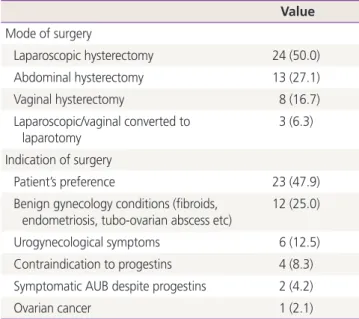
complex) were obtained. We also collected data on the indi- cations and routes of surgery, as well as postoperative com- plications.
Continuous data were presented as mean±standard devia- tion, or median and data range. The independent samples t-test or independent-samples Mann-Whitney U test were used for comparisons of continuous variables between the groups. Categorical data were presented as counts and per- centages, and comparisons were performed using the Pear- son χ
2test or Fisher’s exact test, as appropriate. Simple logis- tic regression analysis was applied to screen several clinical parameters, including age, menopausal status, parity, BMI, diabetes, use of tamoxifen, and ultrasound or hysteroscopic findings of a thickened endometrium. Statistical significance
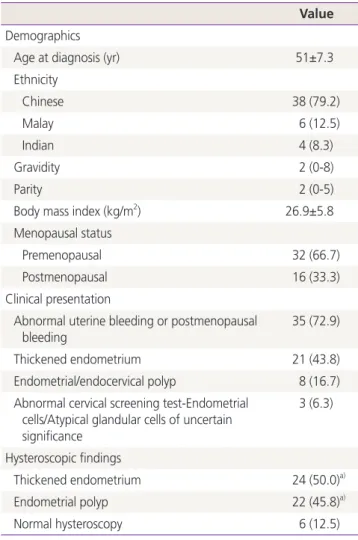
Table 1. Demographics and clinical presentation of study group Value Demographics
Age at diagnosis (yr) 51±7.3
Ethnicity
Chinese 38 (79.2)
Malay 6 (12.5)
Indian 4 (8.3)
Gravidity 2 (0-8)
Parity 2 (0-5)
Body mass index (kg/m
2) 26.9±5.8
Menopausal status
Premenopausal 32 (66.7)
Postmenopausal 16 (33.3)
Clinical presentation
Abnormal uterine bleeding or postmenopausal bleeding
35 (72.9)
Thickened endometrium 21 (43.8)
Endometrial/endocervical polyp 8 (16.7) Abnormal cervical screening test-Endometrial
cells/Atypical glandular cells of uncertain significance
3 (6.3)
Hysteroscopic findings
Thickened endometrium 24 (50.0)
a)Endometrial polyp 22 (45.8)
a)Normal hysteroscopy 6 (12.5)
Values are presented as mean±standard deviation or number (%).
a)