IS THE EXPRESSION OF P16 INK4A AND GALECTIN‐3 CORRELATED WITH DISEASE PROGRESSION OF CERVICAL NEOPLASIA?
Sun‐suk Kim, MD, Hye‐yon Cho, MD, Sung‐won Kang, MD, Hong‐bae Kim, MD, Sung‐ho Park, MD
Department of Obstetrics and Gynecology, Hallym University College of Medicine, Seoul, Korea
Objective
The aim of this study is to investigate whether the expressions of p16INK4A and galectin-3 are associated with the progression of cervical neoplasia and to evaluate its usefulness as a diagnostic tool.
Methods
Eighty-seven formalin-fixed paraffin-embedded cervical specimens (20 normal, 17 LSILs, 26 HSILs, 24 invasive cervical cancers) collected between 2005 and 2009 were selected. We examined the expression of p16INK4A and galectin-3 using immunohistochemical stains with the scoring system.
Results
The mean proportion of p16INK4A and galectin-3 was 0.1 and 1.70 in normal lesions, 1.35 and 2.17 in LSILs, 3.42 and 3.11 in HSILs, 3.79 and 3.08 in invasive cancers. The mean proportion of p16INK4A and galectin-3 was correlated signifi cantly with the degree of neoplasia (P<0.05).
Conclusion
The increased immunohistochemical staining expressions of the p16INK4A and galectin-3 are associated with the progression of cervical neoplasia. Therefore immunohistochemical staining of p16INK4A and galectin-3 can be a useful biomarker for the diagnosis of a progression of cervical neoplasia.
Keywords: p16INK4A, Galectin-3, Uterine Cervical neoplasms, Immunohistochemistry
Received: 2010.12. 8. Revised: 2011. 3. 9. Accepted: 2011. 3.15.
Corresponding author: Sung-ho Park, MD
Department of Obstetrics & Gynecology, Kangnam Sacred Heart Hospital, 949‐1 Daerim‐1 dong, Yeongdeungpo‐gu,
Seoul 150‐950, Korea
Tel: +82-2-829-5151 Fax: +82-2-833-5323 E-mail: [email protected]
Th is is an Open Access article distributed under the terms of the Creative Commons Attribution Non-Commercial License (http://creativecommons.org/licenses/
by-nc/3.0/) which permits unrestricted non-commercial use, distribution, and reproduction in any medium, provided the original work is properly cited.
Uterine cervical cancer is the second most common malignant tumor, accounting for about 15% of all female cancer worldwide.
Annually 400,000 patients are newly diagnosed, and 250,000 die from this disease globally. Although the overall incidence of the disease is decreasing, 3,616 new cases were diagnosed in 2007 in Korea, and the incidence of cervical cancer is still higher in Korea than in other developed countries according to the research data of the National Cancer Information Center from May 2010 [1].
Although Papanicolaou (Pap) smear has signifi cantly contributed to the decreasing incidence and mortality of cervical cancer [2], a large number of women still suffer from this disease in both developing and developed countries. Furthermore, uterine cervical cancer is also found in women who have received regular periodic testing [3]. It is thought that the low sensitivity of the PAP smear
atypical cells [4].Therefore, it is clear that development of a more accurate biologic marker is badly needed to improve the sensitivity doi: 10.5468/KJOG.2011.54.4.192
pISSN 2233-5188 · eISSN 2233-5196
and specifi city of the screening test process in diagnosing uterine cervical cancer.
Cervical cancer develops by multilevel cancer steps. It is well known that the high risk human papilloma virus (HPV) is closely related to the onset of cervical cancer. Persistent HPV infection can cause cervical dysplasia and contribute to cancer development by affecting the restoration & apoptosis of damaged cells. That is, HPV E6 destroys p53 [5] and HPV E7 inactivates products of renti- noblastoma (pRB), and this can increase the expression of p16INK4A [6].p16INK4A is involved in the G1 cell cycle which is an important stage for the repair and extinction of damaged cells. Accordingly, it is suggested that expression of p16INK4A can help in screening tumor cells.
Galectin-3 is one of the genetic families widely distributed in car- bohydrate binding hormones, expressed variably in tumor cells and involved in cellular growth, differentiation, inflammation, apop- tosis and metastasis [7-12].It has not yet been shown defi nitively how galectin-3 affects tumor progression but it is clear that ex- pression of galectin-3 differs according to the type of tissue from which a tumor arises [13,14]. The author compared the expression of p16INK4A and galectin-3 in normal uterine cervix, precancerous lesions including low grade squamous intraepithelial lesions (LSILs), high grade squamous intraepithelial lesions (HSILs) and invasive cancer to determine if there is some difference between these le- sions.
Materials and Methods
1. Tissue samples
We obtained tissue samples from patients who underwent uterine cervical biopsy or hysterectomy in Kangnam Sacred Heart Hospital between 2005 and 2009. The tissue was prepared with formalin and embedded in paraffi n, and then an H&E stain was done. All tissue was thoroughly examined by pathologists. We selected 20 cases of normal tissue, 17 cases of LSIL, 26 cases of HSIL, and 24 cases of invasive cancer at random.
We selected squamous cell lesions rather than other cell lesions because squamous cell lesions are the most common pathologic type in the cervix. Normal cervical tissue was obtained from 20 patients who had undergone hysterectomy due to conditions other than cervical lesions.
2. Immunohistochemical staining of p16INK4A & galectin-3 We obtained sections 4 μm thick from paraffi n-embedded tissue
samples. These sections were attached to slides and deparaffi nat- ed with xylen, and then hydrated by degrees with 100%, 90%, and 75% alcohol.
The slides were inserted in 0.1 M citric acid solution (pH 6.0) and microwaved 4 times for 20 minutes. All slides were then treated with 3% hydrogen peroxide for 15 minutes, to inactivate the hy- droperoxide enzyme. Then, galectin-3 and p16INK4A were treated with a protein blocking solution for 10 minutes and 20 minutes each and washed with a pH 7.6 Tris buffer solution.
A primary antibody was applied to p16INK4A (monoclonal, Neo- marker, Fremont, CA, USA; 1:100) and galectin-3 (Abcam, Cam- brige, UK; 1:50) for 1 hour then washed 3 times with a Tris buffer solution.
HRP EMV kit (Dako Co., Glostrup, Denmark) was applied for 30 minutes at normal temperature, washed 3 times with Tris buffer solution. Then, ager was done by applying 3, 3-diaminobenzene for 5 minutes. The stained slides were cleaned with distilled wa- ter and the contrast stain was done by hematoxylin, before the mounting was done.
3. Interpretation of staining results
We examined all the slides with an optical microscope at ×100 fi eld and also examined the checked number of cells, which pre- sented positive at ×400 fi eld. Two independent observers evaluated the staining of the slides and we analyzed the proportion and in- tensity of the staining responses in the cell and nucleus. In p16INK4A, a proportion of stained cells < 1% means 0, 1-4% means 1 +, 5-25%
means 2 +, 25-75% means 3 +, and > 75% means 4 +. In galec- tin-3, proportion of stained cells < 5% means 0, 5-25% means 1 +, 25-50% means 2 +, 51-75% means 3 +, and > 75% means 4.
4. Statistical analysis
A one-way analysis of variance (ANOVA) was used to evaluate the differences in the expression of p16INK4A and galectin-3 between normal and abnormal tissue, including LSIL, HSIL, and invasive cer- vical cancer. Analysis was done by SPSS 15.0. P-value < 0.05 was statistically signifi cant.
Results
1. Expression of p16INK4A protein
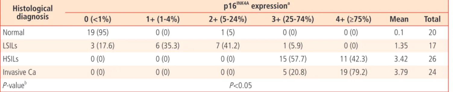
The proportion of p16INK4A expression was negative in 95% cases of normal epithelium, and the mean value was 0.1. In LSILs, the proportion was 1 + or 2 + in 76.5% and the mean value was 1.35.
There was no negative response, 1+ or 2+ in HSILs. 3 + comprised 57.7% and 4 + was 42.3% of cases of HSILs, and the average val- ue was 3.42. In invasive cancer, = 3 + was 100%, 3 + was 20.8%,
4 + was 79.2%, and the average value was 3.79 (Fig. 1). There was a statistical signifi cance between the proportion of p16INK4A protein expression and the grade of uterine cervical lesion (P < 0.05) (Table 1).
Table 1. Proportion of p16INK4A expression versus original hstological diagnosis Histological
diagnosis
p16INK4A expressiona
0 (<1%) 1+ (1-4%) 2+ (5-24%) 3+ (25-74%) 4+ (≥75%) Mean Total
Normal 19 (95) 0 (0) 1 (5) 0 (0) 0 (0) 0.1 20
LSILs 3 (17.6) 6 (35.3) 7 (41.2) 1 (5.9) 0 (0) 1.35 17
HSILs 0 (0) 0 (0) 0 (0) 15 (57.7) 11 (42.3) 3.42 26
Invasive Ca 0 (0) 0 (0) 0 (0) 5 (20.8) 19 (79.2) 3.79 24
P-valueb P<0.05
Values are presented as number (%).
LSILs, low grade squamous intraepithelial lesions; HSILs, high grade squamous intraepithelial lesions; Ca, cancer.
aTheproportion of p16INK4A expression based on <1%, 1-4%, 5-24%, 25-74%, and ≥75% of cells immunohistochemical stained in a lesion.
bThe expression of p16INK4A was positively associated with the grade of cervical neoplasia.
(A) Normal cervix (B) LSILs
(C) HSILs (D) Invasive cancer
Fig. 1. Immunohistochemical staining of cervical neoplasia for p16INK4A (×100).
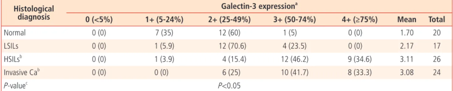
Table 2. Proportion of galectin-3 expression versus original histological diagnosis Histological
diagnosis
Galectin-3 expressiona
0 (<5%) 1+ (5-24%) 2+ (25-49%) 3+ (50-74%) 4+ (≥75%) Mean Total
Normal 0 (0) 7 (35) 12 (60) 1 (5) 0 (0) 1.70 20
LSILs 0 (0) 1 (5.9) 12 (70.6) 4 (23.5) 0 (0) 2.17 17
HSILsb 0 (0) 1 (3.9) 4 (15.4) 12 (46.2) 9 (34.6) 3.11 26
Invasive Cab 0 (0) 0 (0) 6 (25) 10 (41.7) 8 (33.3) 3.08 24
P-valuec P<0.05
Values are presented as number (%).
LSILs, low grade squamous intraepithelial lesions; HSILs, high grade squamous intraepithelial lesions; Ca, cancer.
aProportion of galectin-3 expression based on < 5%, 5-24%, 25-49%, 50-74%, and ≥75% of cells Immunohistochemical stained in a lesion.
bNo major difference between HSILs and invasive cancers exist at Scheffer test (P=1.00).
cThe expression was positively associated with the grade of cervical neoplaisa (ANOVA test).
(A) Normal cervix (B) LSILs
(C) HSILs (D) Invasive cancer
Fig. 2. Immunohistochemical staining of cervical neoplasia for galectin-3 (×100).
2. Expression of galectin‐3 protein
The proportion of galectin‐3 expression was negative, 1 +, or 2 + in 95% cases of normal epithelium, and the average value was 1.70.
In LSILs, it was 2 + or 3 + in 94.1% of cases, and the average value was 2.17. It was ≥ 3 + in 80.8% of HSILs, and the average value was 3.11. Although the proportion of galectin‐3 was ≥ 3 + in 75%
of invasive cancers, the average value was 3.08 (Fig. 2). Therefore no major difference between HSILs and invasive cancers existed in regard to the proportion of galectin‐3 expression (P = 1.00) (Table 2).
However there was a statistical signifi cance between the propor- tion of galectin‐3 expression and the grade of cervical lesions like p16INK4A (P < 0.05) (Table 2, Fig. 3).
Discussion
Cervical cancer arises from carcinoma in situ, which originates from precancerous lesions in normal cervical epithelium, or cervi- cal dysplasia.
HPV infection is recognized as the most prevalent cause of cervical cancer. HPV E6 destroys p53 [5] and HPV E7 inactivates pRB, po- tentially increasing the expression of p16INK4A with persistent HPV infection [6].
p16INK4A gene, a cyclin‐dependent kinase (cdk) suppressor factor, regulates the activity of cdk4 and cdk6, and is inactivated by ge- netic defect or hypermethylation in many types of cancers [15,16].
The role of p16INK4A as a tumor suppressor gene has been reported in many types of malignant tumors such as malignant melanoma, gastrointestinal cancers, and lung cancers. Expression of p16INK4A
related to HPV has a different mechanism of expression from other types of malignant tumors. The E7 tumor protein in high‐risk types of HPV combines with and inactivates the pRb, tumor sup- pressor protein, and releases a transcription factor resembling E2F.
This transcription factor activates the cell cycle by advancing G1/
S phase [17]. p16INK4A expression which is controlled by a negative feedback mechanism, is relatively increased by the inactivation of pRb [18]. There are many reports that p16INK4A is over‐expressed in cervical cancer and precancerous lesions infected with high risk types of HPV [18-20]. Since the relationship between HPV infection and p16INK4A expression in immunohistochemical results was fi rst reported by Sano et al. [15] the role of p16INK4A has been widely studied [21,22].Klases et al. [19] noted the expression of p16INK4A in cervical tissue samples using immunohistochemical staining. They also found that all cervical intraepithelial neoplasia (CIN) lesions other than low risk types of HPV infected CIN, and almost all invasive cervical cancers had a strong expression of p16INK4A. However there was no expression of p16INK4A in normal cervical epithelial cells, inflammatory lesions and low risk types of HPV infected CIN. Furthermore, they also found that there was same expression of p16INK4A in PAP smear of cervical dysplasia.
Therefore p16INK4A expression has been considered as a definite biomarker, which can confi rm tumor cells in cervical tissue samples and PAP smears. Similarly, in our study, p16INK4A protein was not expressed in 95% of normal epithelium, LSILs had < 25% ex- pression in 94.1%, and HSILs and invasive cancers had ≥ 25%
expression in 100%. More than 75% of expression was of higher frequency in invasive cancer than HSIL (78.2% vs. 42.3%). In our results, p16INK4A protein was mainly expressed in HSILs and inva- sive cancers, therefore immunohistochemial staining of p16INK4A would be helpful in diagnosing cervical lesions.
However Wong et al. [23] have insisted that the p16INK4A expres- sion rate is the result of p16INK4A over‐expression by HPV infection.
Therefore, further analysis is needed to make clear the relevance of HPV infection to p16INK4A expression.
Galectin‐3, 31‐kDa, a kind of carbohydrate binding protein, has a chemical affi nity to β‐galactosides, and has the homology of ge- netic arrangement in carbohydrate binding lesion [7].
Galectin‐3 had been known as Carbohydrate binding protein-35 (CBP‐35), Mac‐2, L‐29, L‐34, or L‐31. It consists of aminoterminal including repetitive genetic arrangement, which is abundant in leucine, tyrosine & proline, and globular structure including carbo- hydrate binding lesion [7‐14]. Galectin‐3 responds to special kind of ligands, and is present in a number of biological steps such as cellular growth, differentiation, inflammation, modification, me- Fig. 3. The correlation between histologic diagnosis and immunohisto-
chemical staining proportion. LSILs, low grade squamous intraepithelial lesions; HSILs, high grade squamous intraepithelial lesions; Ca, cancer.
p16INK4A Galectin-3
Normal LSILs HSILs Invasive Ca.
Histologic diagnosis
Proportion
4 3.5 3 2.5 2 1.5 1 0.5 0
tastasis and adhesion. Galectin‐3 is secreted by tumor cells and macrophages, induces chemotaxis by affecting vascular intraepi- thelium, and accelerates motility in early stage of tumor formation [11].
The importance of galectin-3 expression in many types of human tumors has been studied. Galectin-3 expression has been found to be higher in pancreatic, gastric, thyroid, head & neck, renal, colon, rectal, liver cancer and leiomyosarcoma than in normal cells. On the other hand, there are conflicting results in regard to other types of tumors. Galectin-3 expression, for example, is lower in breast, ovarian and endometrial cancer than normal cells. In meta- static cancer, the frequency of galectin-3 expression is higher than in primary cancer [24].
The only report regarding the relationship between grades of cer- vical lesions and galectin‐3 expression is by Lee et al. [25] which concluded that galectin‐3 expression decreases as the cervical le- sion progresses [25].
But our conclusion is contrary to Lee’s fi ndings. In our study, more than 50% expression was only found at 5% in normal epithelium, 23.5% in LSILs, and 70‐80% in HSILs & invasive cancers.
Additionally, the intensity of galectin‐3 expression increased as the cervical lesion progressed to invasive cancer. Thus, we could use galectin‐3 immunohistochemical staining as a tool in diagnosing and supposing uterine cervical lesions. But it is thought that larger scale research is needed, in order to consider the rise and fall in galectin‐3 expression as cervical lesions progress.
The author used immunohistochemical stains of p16INK4A and galectin‐3. p16INK4A is a gene associated with controlling the cell cycle in canceration. Galectin‐3 is a kind of carbohydrate binding protein, that is diversely expressed in tumors, and affects cellular growth, differentiation, infl ammation, apoptosis, and metastasis.
Expression of p16INK4A & galectin‐3 increases as the cervical lesion progresses to invasive cancers. This suggests that the expression of p16INK4A and galectin‐3 using immunohistochemical stains is as- sociated with the progression of cervical lesions. Therefore, immu- nohistochemical staining of p16INK4A & galectin‐3 would be valuable when used with PAP smear in diagnosing cervical lesions and pre- dicting disease progression. But further research including research focusing on the association between these biomarkers and biopsy results (mild, moderate, severe dysplasia) is needed in the future.
References
1. National Cancer Information Center [Internet]. Goyang (KR):
National Cancer Information Center; c2008 [cited 2010 May 31]. Available from: http://www.cancer.go.kr/cms/index.html.
2. Anderson GH, Boyes DA, Benedet JL, Le Riche JC, Matisic JP, Suen KC, et al. Organisation and results of the cervical cytol- ogy screening programme in British Columbia, 1955-85. Br Med J (Clin Res Ed) 1988;296:975-8.
3. Sasieni PD, Cuzick J, Lynch-Farmery E. Estimating the ef- ficacy of screening by auditing smear histories of women with and without cervical cancer. The National Co-ordinating Network for Cervical Screening Working Group. Br J Cancer 1996;73:1001-5.
4. Fahey MT, Irwig L, Macaskill P. Meta-analysis of Pap test ac- curacy. Am J Epidemiol 1995;141:680-9.
5. Scheffner M, Huibregtse JM, Vierstra RD, Howley PM. The HPV- 16 E6 and E6-AP complex functions as a ubiquitin-protein ligase in the ubiquitination of p53. Cell 1993;75:495-505.
6. Khleif SN, DeGregori J, Yee CL, Otterson GA, Kaye FJ, Nevins JR, et al. Inhibition of cyclin D-CDK4/CDK6 activity is associ- ated with an E2F-mediated induction of cyclin kinase inhibitor activity. Proc Natl Acad Sci U S A 1996;93:4350-4.
7. Barondes SH, Cooper DN, Gitt MA, Leffl er H. Galectins. Struc- ture and function of a large family of animal lectins. J Biol Chem 1994;269:20807-10.
8. Konstantinov KN, Robbins BA, Liu FT. Galectin-3, a beta-galac- toside-binding animal lectin, is a marker of anaplastic large- cell lymphoma. Am J Pathol 1996;148:25-30.
9. Moutsatsos IK, Wade M, Schindler M, Wang JL. Endogenous lectins from cultured cells: nuclear localization of carbohy- drate-binding protein 35 in proliferating 3T3 fi broblasts. Proc Natl Acad Sci U S A 1987;84:6452-6.
10. Woo HJ, Shaw LM, Messier JM, Mercurio AM. The major non- integrin laminin binding protein of macrophages is identical to carbohydrate binding protein 35 (Mac-2). J Biol Chem 1990;265:7097-9.
11. Inohara H, Akahani S, Koths K, Raz A. Interactions between galectin-3 and Mac-2-binding protein mediate cell-cell adhe- sion. Cancer Res 1996;56:4530-4.
12. Frigeri LG, Liu FT. Surface expression of functional IgE bind- ing protein, an endogenous lectin, on mast cells and macro- phages. J Immunol 1992;148:861-7.
13. Liu FT. S-type mammalian lectins in allergic infl ammation. Im- munol Today 1993;14:486-90.
14. Inohara H, Raz A. Functional evidence that cell surface galectin-3 mediates homotypic cell adhesion. Cancer Res 1995;55:3267-71.
P16
INK4A와 Galectin-3의 면역 염색발현과 자궁경부 신생물의 진행과의 관련성 연구
한림대학교 의과대학 산부인과학교실 김선숙, 조혜연, 강성원, 김홍배, 박성호
목적
이 연구의 목적은 자궁경부의 신생물의 진행에 따른 p16INK4A과 galectin-3의 면역 염색 발현 정도를 비교하여 자궁경부 병변의 진단상의 도움이 될 수 있는지를 확인하고자 하였다.
연구방법
2005년부터 2009년 사이의 총 87개의 포르말린에 포매된 자궁경부조직(정상, 20; low grade intraepithelial lesions [LSILs], 17; high grade intraepithelial lesions [HSILs], 26; 침윤암, 24)을 이용하여 p16INK4A와 galectin-3 면역화학염색을 시행하여 발현정도를 비교하였으며
p16INK4A와 galecen-3염색정도에 따라 점수를 준 후에 병변의 진행정도와 점수를 비교 평가하였다.
결과
p16INK4A와 galectin-3의 평균발현 정도는 정상에서 0.1과 1.70, LSILs에서는 1.35와 2.17, HSILs에서는 3.42와 3.11, 침윤암에서는 3.79와
3.08이었다. p16INK4A와 galectin-3의 발현빈도는 자궁경부신생물의 진행 정도에 따라 증가되는 소견을 보였다(P<0.05).
결론
p16INK4A와 galectin-3의 발현정도는 면역화학염색 시행 시 자궁경부 신생물의 진행에 따라 증가되었다. 따라서 p16INK4A와 galectin-3의 면
역화학염색은 자궁경부신생물의 진행을 진단하는데 도움이 될 수도 있으리라 생각된다.
중심단어: p16INK4A, Galectin-3, 자궁경부 신생물, 면역화학염색 15. Sano T, Oyama T, Kashiwabara K, Fukuda T, Nakajima T. Ex-
pression status of p16 protein is associated with human pap- illomavirus oncogenic potential in cervical and genital lesions.
Am J Pathol 1998;153:1741-8.
16. Nakao Y, Yang X, Yokoyama M, Ferenczy A, Tang SC, Pater MM, et al. Induction of p16 during immortalization by HPV 16 and 18 and not during malignant transformation. Br J Cancer 1997;75:1410-6.
17. Nevins JR. E2F: a link between the Rb tumor suppressor pro- tein and viral oncoproteins. Science 1992;258:424-9.
18. Brown DC, Gatter KC. Monoclonal antibody Ki-67: its use in histopathology. Histopathology 1990;17:489-503.
19. Klaes R, Friedrich T, Spitkovsky D, Ridder R, Rudy W, Petry U, et al. Overexpression of p16(INK4A) as a specifi c marker for dysplastic and neoplastic epithelial cells of the cervix uteri. Int J Cancer 2001;92:276-84.
20. Keating JT, Cviko A, Riethdorf S, Riethdorf L, Quade BJ, Sun D, et al. Ki-67, cyclin E, and p16INK4 are complimentary sur- rogate biomarkers for human papilloma virus-related cervical
neoplasia. Am J Surg Pathol 2001;25:884-91.
21. Negri G, Egarter-Vigl E, Kasal A, Romano F, Haitel A, Mian C.
p16INK4a is a useful marker for the diagnosis of adenocarci- noma of the cervix uteri and its precursors: an immunohisto- chemical study with immunocytochemical correlations. Am J Surg Pathol 2003;27:187-93.
22. Kim JR, Kim SY, Kim MJ, Kim JH. Alterations of CDKN2 (MTS1/
p16INK4A) gene in paraffi n-embedded tumor tissues of hu- man stomach, lung, cervix and liver cancers. Exp Mol Med 1998;30:109-14.
23. Wong YF, Wang W, Wong FWS, Chanc AMZ. Genetic studies in gynecologic malignancies. J Pract Gynecol Oncol 1993;9:101-2.
24. Schoeppner HL, Raz A, Ho SB, Bresalier RS. Expression of an endogenous galactose-binding lectin correlates with neoplas- tic progression in the colon. Cancer 1995;75:2818-26.
25. Lee JW, Song SY, Choi JJ, Choi CH, Kim TJ, Kim J, et al. De- creased galectin-3 expression during the progression of cervi- cal neoplasia. J Cancer Res Clin Oncol 2006;132:241-7.