Received: 2012.12.16. Revised: 2013.2.8. Accepted: 2013.2.18.
Corresponding author: Sung Shin Shim
Department of Obstetrics and Gynecology, CHA Gangnam Medical Center, CHA University, 566 Nonhyeon-ro, Gangnam-gu, Seoul 135-913, Korea
Tel: +82-2-3468-3000 Fax: +82-2-558-1119 E-mail: [email protected]
Articles published in Obstet Gynecol Sci are open-access, distributed under the terms of the Creative Commons Attribution Non-Commercial License (http://creativecommons.
org/licenses/by-nc/3.0/) which permits unrestricted non-commercial use, distribution, and reproduction in any medium, provided the original work is properly cited.
Copyright © 2013 Korean Society of Obstetrics and Gynecology
Introduction
Pregnancy in advanced age is defined as childbirth at the age of 35 years or order regardless of whether the childbirth is the first or not. Recently, the percentages of pregnancies in advanced age have shown a gradual increase as women have entered the workforce in increasing numbers. In South Korea, the percentages of pregnancies in women of advanced age have more than doubled, increasing from 6.2% in 1999 to 15.4% in 2009 [1].
Fetal chromosomal abnormalities may be caused by a non-
Maternal age-specific rates of fetal chromosomal
abnormalities in Korean pregnant women of advanced maternal age
Young Joo Kim, Jee Eun Lee, Soo Hyun Kim, Sung Shin Shim, Dong Hyun Cha
Department of Obstetrics and Gynecology, CHA Gangnam Medical Center, CHA University, Seoul, Korea
Objective
To evaluate the association of maternal age with occurrence of fetal chromosomal abnormalities in Korean pregnant women of advanced maternal age (AMA).
Methods
A retrospective review of the amniocentesis or chorionic villous sampling (CVS) database at Gangnam and Bundang CHA Medical Centers, between January 2001 and February 2012, was conducted. This study analyzed the incidence of fetal chromosomal abnormalities according to maternal age and the correlation between maternal age and fetal chromosomal abnormalities in Korean pregnant women ≥35 years of age. In addition, we compared the prevalence of fetal chromosomal abnormalities between women of AMA only and the others as the indication for amniocentesis or CVS.
Results
A total of 15,381 pregnant women were selected for this study. The incidence of aneuploidies increased exponentially with maternal age (P<0.0001). In particular, the risk of trisomy 21 (standard error [SE], 0.0378; odds ratio, 1.177;
P<0.001) and trisomy 18 (SE, 0.0583; odds ratio, 1.182; P=0.0040) showed significant correlation with maternal age. Comparison between women of AMA only and the others as the indication for amniocentesis or CVS showed a significantly lower rate of fetal chromosomal abnormalities only in the AMA group, compared with the others ( P<0.0001).
Conclusion
This study demonstrates that AMA is no longer used as a threshold for determination of who is offered prenatal diagnosis, but is a common risk factor for fetal chromosomal abnormalities.
Keywords: Fetal chromosome aberrations; Maternal age; Prenatal diagnosis http://dx.doi.org/10.5468/ogs.2013.56.3.160
pISSN 2287-8572 · eISSN 2287-8580
disjunction phenomenon that occurs in the period of meiosis during maternal oogenesis, which has been reported to have a direct association with maternal age. Therefore, pregnancy in advanced age is a critical risk factor for fetal chromosomal abnormalities [2-4].
Currently, fetal chromosomal abnormalities due to maternal age have been reported to include trisomy 21, trisomy 18, trisomy 13, triple X syndrome, and Klinefelter’s syndrome [5].
As the percentages of pregnancies in women of advanced age have increased, there has been increasing demand for prenatal genetic counseling that helps to predict the risk of fetal chromosomal abnormalities depending on age. Most of the data were obtained from studies of women in North America or Europe. However, rare data from study of fetal chromosomal abnormalities depending on age targeting Ko- rean women have been reported [6].
Against this background, the intention of this study was to examine the incidence rate of fetal chromosomal abnormali- ties depending on maternal age and to investigate correlation between fetal chromosomal abnormalities and maternal age, which targeted Korean pregnant women who underwent am- niocentesis or chorionic villous sampling (CVS). In addition, this study examined if there was any meaningful difference in the incidence rate of fetal chromosomal abnormalities between cases where only advanced age of pregnant women was con- sidered as an indication for amniocentesis or CVS and cases where other indications were considered for such examinations.
Materials and methods
This study targeted 15,454 pregnant women who underwent amniocentesis or CVS in Gangnam and Bundang CHA Medi- cal Centers during the period from January 2001 to February 2012 and was approved by Institutional Review Board in CHA Medical Center. This study was conducted as a retrospective study based on genetic information and inpatient and out- patient charts. However, 71 persons whose age could not be determined and 2 persons whose indication for amniocentesis or CVS could not be confirmed were excluded from this study.
As a result, the cases of 15,381 pregnant women were in- cluded. Age of the cases was the age at the time of delivery.
Fetal chromosomal abnormalities were classified according to numeric and structural chromosomal abnormalities. The numeric chromosomal abnormalities were divided again ac-
cording to autosomal and sex chromosomal abnormalities. In regard to structural chromosomal abnormalities, normal varia- tions such as heterochromatin, satellite chromosomes and pericentric inversion of chromosome 9 were excluded from the abnormal category.
For analysis of maternal age-specific rates of fetal chro- mosomal abnormalities in pregnant women of advanced maternal age (AMA), maternal age beyond 35 years was segmented with a one-year interval. In addition to, among women ≥35 years of age, when only AMA was considered as indication of amniocentesis or CVS was performed, analysis was also performed to determine whether there was any dif- ference in incidence rate of fetal chromosomal abnormalities compared to cases where other indications were considered.
Logistic regression analysis was performed in order to inves- tigate correlation between increase in maternal age and fetal chromosomal abnormalities. In addition, SPSS ver. 16.0 (SPSS Inc., Chicago, IL, USA) was used for statistical analysis of the study results. Significance probability of less than 0.05 was considered to show statistical significance.
Results
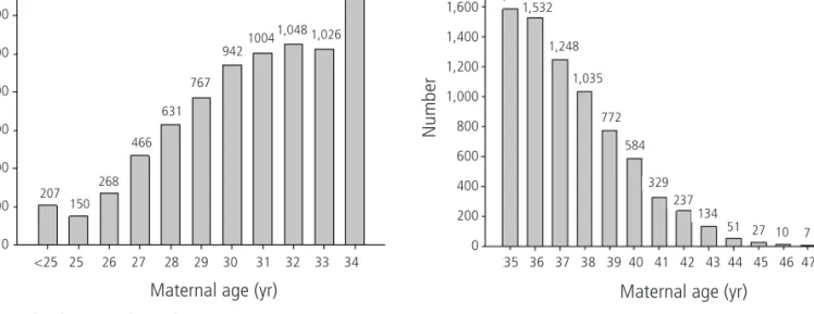
Among pregnant women who underwent amniocentesis or CVS during the study period 15,381 persons were selected for inclusion in this study. Age of the cases ranged from 19 years to 52 years. Average age was 34.0 years (±4.3); age distribu- tion of the entire group of study subjects is shown in Fig. 1.
According to the results of fetal chromosomal examination, 14,818 cases (96.34%) were found to have normal fetus chromosomes while 563 cases (3.66%) were found to have abnormal fetus chromosomes. Among the abnormal cases (3.66%), 357 cases (2.32%) had numeric chromosomal ab- normalities, while 206 cases (1.34%) had structural chromo- somal abnormalities. With respect to numeric chromosomal abnormalities, trisomy 21 was found in 181 cases (1.18%), which was the highest frequency. Then, trisomy 18 was found in 72 cases (0.47%), Turner’s syndrome in 41 cases (0.27%), Klinefelter’s syndrome in 28 cases (0.18%), trisomy 13 and triple X syndrome in 14 cases for each (0.09%), and XYY syn- drome in 7 cases (0.05%), in this order.
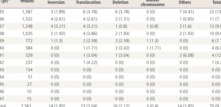
Tables 1, 2 show the analyses of the incidence rate of nu-
meric and structural chromosomal abnormalities when age
was increased by a one-year interval among women older
than 35 years at the time of delivery. Trisomy 21 showed an incidence rate of 11.34 out of 1,000 cases at the age of 35 years, 15.41 cases at the age of 40, and 37.04 cases at the age of 45. Trisomy 18 showed an incidence rate of 1.89 out of 1,000 cases at the age of 35 years, 5.14 cases at the age of 40, and 37.04 cases at the age of 45, which is a sharp in- crease.
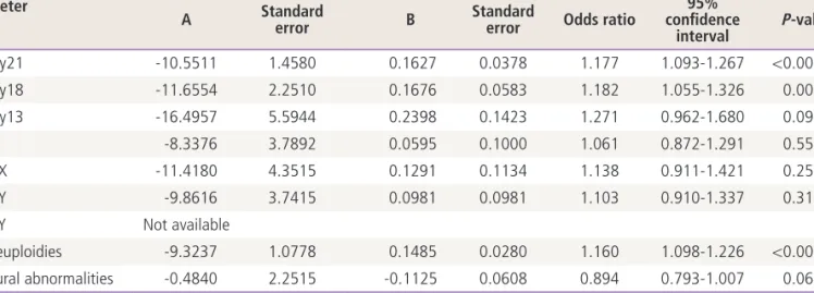
According to the results of logistic regression analysis, tri-
somy 21 and trisomy 18 showed close correlation with age.
In other words, as age increased by one year, odds ratio of trisomy 21 tended to increase by 1.177 times (Table 3, Fig. 2).
According to increase in age by one year, odds ratio of trisomy 18 also tended to increase by 1.182 times (Table 3, Fig. 3). In addition, in regard to all of the fetal aneuploidies, odds ratio also tended to increase by 1.160 times as age increased by one year (Table 3, Fig. 4).
Table 1. Incidences of numeric chromosomal abnormalities found in amniocentesis or chorionic villus sampling Maternal
age (yr)
a)No. of fetuses
Numeric chromosomal abnormalities
All 47,+21 47,+18 47,+13 45,X 47,XXX 47,XXY 47,XYY
35 1,587 31 (19.53) 18 (11.34) 3 (1.89) 0 (0) 2 (1.26) 3 (1.89) 3 (1.89) 2 (1.26) 36 1,532 21 (13.71) 9 (5.87) 6 (3.92) 1 (0.65) 2 (1.31) 1 (0.65) 2 (1.31) 0 (0) 37 1,248 24 (19.23) 8 (6.41) 10 (8.01) 0 (0) 3 (2.4) 0 (0) 3 (2.4) 0 (0) 38 1,035 31 (29.95) 14 (13.53) 5 (4.83) 1 (0.97) 6 (5.8) 3 (2.9) 2 (1.93) 0 (0) 39 772 21 (27.2) 14 (18.13) 1 (1.3) 1 (1.3) 1 (1.3) 2 (2.59) 2 (2.59) 0 (0) 40 584 13 (22.26) 9 (15.41) 3 (5.14) 0 (0) 0 (0) 0 (0) 1 (1.71) 0 (0) 41 329 15 (45.59) 7 (21.28) 2 (6.08) 1 (3.04) 3 (9.12) 1 (3.04) 1 (3.04) 0 (0) 42 237 12 (50.63) 6 (25.32) 4 (16.88) 1 (4.22) 0 (0) 0 (0) 1 (4.22) 0 (0) 43 134 7 (52.24) 5 (37.31) 2 (14.93) 0 (0) 0 (0) 0 (0) 0 (0) 0 (0) 44 51 5 (98.04) 3 (58.82) 1 (19.61) 0 (0) 0 (0) 0 (0) 1 (19.61) 0 (0) 45 27 2 (74.07) 1 (37.04) 1 (37.04) 0 (0) 0 (0) 0 (0) 0 (0) 0 (0)
46 10 0 (0) 0 (0) 0 (0) 0 (0) 0 (0) 0 (0) 0 (0) 0 (0)
47 15 1 (66.67) 0 (0) 0 (0) 0 (0) 0 (0) 1 (66.67) 0 (0) 0 (0)
Total 7,561 183 (24.2) 94 (12.43) 38 (5.03) 5 (0.66) 17 (2.25) 11 (1.45) 16 (2.12) 2 (0.26) Values are presented as number (incidences per 1,000).
a)
Maternal age at the time of delivery.
Fig. 1. Age distribution in the study group.
Number
Maternal age (yr)
<25 25 26 27 28 29 30 31 32 33 34 207 150
268 466
631 767
942 1004 1,048 1,026 1,311 1,400
1,200
1,000
800
600
400
200
0
Number
Maternal age (yr)
35 36 37 38 39 40 41 42 43 44 45 46 47 48 1,587
1,532
1,248 1,035
772 584
329 237 134
51 27 10 7 8 1,800
1,600
1,400
1,200
1,000
800
600
400
200
0
According to the results of this study, trisomy 13, Turner’s syndrome, triple X syndrome, Klinefelter’s syndrome, and structural chromosomal abnormalities did not show any significant correlation with maternal age (Table 3). XYY syn- drome was found in two of the study subjects. Therefore, due to the very small number, analysis of the correlation between fetal chromosomal abnormalities and age was not possible.
When only advanced age of women aged 35 years or older
was considered as indication of amniocentesis or CVS were performed, the incidence rate of fetal chromosomal abnor- malities was 1.80%. When other indications, including AMA were considered, the incidence rate of fetal chromosomal ab- normalities was 6.45%. As a result, incidence of fetal chromo- somal abnormalities was found to be significantly low when only advanced age was considered as an indication. However, both the AMA only group and other indication groups showed Table 2. Incidences of structural chromosomal abnormalities found in amniocentesis or chorionic villus sampling
Maternal
age (yr)
a)No. of fetuses
Structural chromosomal abnormalities Inversion Translocation Deletion Marker
chromosome Others Total
35 1,587 3 (1.89) 6 (3.78) 6 (3.78) 0 (0) 7 (4.41) 22 (13.86)
36 1,532 4 (2.61) 4 (2.61) 2 (1.31) 0 (0) 1 (0.65) 11 (7.18)
37 1,248 4 (3.21) 4 (3.21) 1 (0.8) 1 (0.8) 2 (1.6) 12 (9.62)
38 1,035 2 (1.93) 4 (3.86) 2 (1.93) 0 (0) 2 (1.93) 10 (9.66)
39 772 1 (1.3) 2 (2.59) 2 (2.59) 1 (1.3) 0 (0) 6 (7.77)
40 584 0 (0) 1 (1.71) 2 (3.42) 1 (1.71) 0 (0) 4 (6.85)
41 329 0 (0) 1 (3.04) 1 (3.04) 0 (0) 2 (6.08) 4 (12.16)
42 237 0 (0) 1 (4.22) 0 (0) 0 (0) 0 (0) 1 (4.22)
43 134 0 (0) 0 (0) 0 (0) 0 (0) 0 (0) 0 (0)
44 51 0 (0) 0 (0) 0 (0) 0 (0) 0 (0) 0 (0)
45 27 0 (0) 0 (0) 0 (0) 0 (0) 0 (0) 0 (0)
46 10 0 (0) 0 (0) 0 (0) 0 (0) 0 (0) 0 (0)
47 15 0 (0) 0 (0) 0 (0) 0 (0) 0 (0) 0 (0)
Total 7,561 14 (1.85) 23 (3.04) 16 (2.12) 3 (0.4) 14 (1.85) 70 (9.26)
Values are presented as number (incidences per 1,000).
a)
Maternal age at the time of delivery.
Trisomy 21
Maternal age at the time of delivery (years)
35 36 37 38 39 40 41 42 43 44 45 46 47R ate p er 1,0 00
1 10 100
Fig. 2. Observed rates per 1,000 of trisomy 21 based on maternal age.
Maternal age at the time of delivery (yr) Trisomy 21
Rate per 1,000
35 36 37 38 39 40 41 42 43 44 45 46 47 100
10
1
Fig. 3. Observed rates per 1,000 of trisomy 18 based on maternal age.
Maternal age at the time of delivery (yr) Trisomy 18
35 36 37 38 39 40 41 42 43 44 45 46 47 100
10
1
Rate per 1,000
an increasing tendency in the expected rate of fetal chromo- somal abnormalities with the increase in age (Table 4).
Discussion
This study targeted pregnant women who underwent amnio- centesis or CVS in South Korea in order to analyze correlation between fetal chromosomal abnormalities and maternal age.
As the percentages of pregnancies in advanced age have increased in Korea, demand for prediction of risk of fetal chromosomal abnormalities depending on age in prenatal
genetic counseling has also increased. However, data from studies targeting Korean women have rarely been reported.
Consequently, this study has significant meaning in that it tar- geted Korean women and it utilized a large quantity of data that had accumulated over the past 10 years. Because fetal chromosomal abnormalities were examined among women in high-risk groups who underwent amniocentesis or CVS, inci- dence rate of fetal chromosomal abnormalities was relatively higher than incidence rate in the normal group.
A number of studies have reported that fetal chromosomal abnormalities showing a close association with maternal age included trisomy 21, trisomy 18, trisomy 13, triple X syn- drome, and XYY syndrome. In 2010, Park et al. [6] published a research paper on a study of fetal chromosomal abnormali- ties depending on maternal age among Korean women. In a study targeting 2,032 women who underwent amniocentesis due to the sole indication of AMA, they analyzed correlation between fetal chromosomal abnormalities and maternal age.
They reported that risk of trisomy 21, trisomy 18, triple X syndrome, and all aneuploidies showed a significant increase according to increase in maternal age. In this study, risk of trisomy 21, trisomy 18, and all of the fetal aneuploidies was found to show a significant increase according to increase in maternal age. In other words, as maternal age increased by one year, odds ratio of trisomy 21 tended to increase by 1.177 times, odds ratio of trisomy 18 tended to increase by 1.182 times, and odds ratio of all of the fetal aneuploidies tended to increase by 1.160 times. In addition, a sharp increase Table 3. Parameters and standard errors of logistic regression equations for fetal chromosomal abnormalities diagnosed at amniocentesis or chorionic villus sampling
Parameter
Constant term Maternal term
A Standard
error B Standard
error Odds ratio 95%
confidence
interval P-value
Trisomy21 -10.5511 1.4580 0.1627 0.0378 1.177 1.093-1.267 <0.0001
Trisomy18 -11.6554 2.2510 0.1676 0.0583 1.182 1.055-1.326 0.0040
Trisomy13 -16.4957 5.5944 0.2398 0.1423 1.271 0.962-1.680 0.0918
45,X -8.3376 3.7892 0.0595 0.1000 1.061 0.872-1.291 0.5514
47,XXX -11.4180 4.3515 0.1291 0.1134 1.138 0.911-1.421 0.2552
47,XXY -9.8616 3.7415 0.0981 0.0981 1.103 0.910-1.337 0.3172
47,XYY Not available
All aneuploidies -9.3237 1.0778 0.1485 0.0280 1.160 1.098-1.226 <0.0001
Structural abnormalities -0.4840 2.2515 -0.1125 0.0608 0.894 0.793-1.007 0.0641
Fig. 4. Observed rates per 1,000 of all aneuploidies based on ma- ternal age.
Maternal age at the time of delivery (yr) All aneuploidies
35 36 37 38 39 40 41 42 43 44 45 46 47 1,000
100
10
Rate per 1,000
was found in incidence rate per 1,000 cases of trisomy 21 and trisomy 18 at the age of 45. However, because the number of cases at the age of 46 or older was very small, analysis of cor- relation between fetal chromosomal abnormalities and extreme maternal age was not possible. No significant correlation with maternal age was found in trisomy 13, Turner’s syndrome, triple X syndrome, XYY syndrome, and structural chromosomal ab- normalities. In particular, according to the statistics, trisomy 13, Turner’s syndrome, and triple X syndrome were not found to show any significant correlation with maternal age. However, there was limitation in that the number of study subjects with each type of abnormality was too small to determine whether the resulting value was significant.
Amniocentesis is usually performed after 16 weeks, while CVS is generally performed within 10 to 12 weeks. If sponta- neous abortion occurs before 12 weeks, more than 50% of cases are likely to be attributable to fetal chromosomal ab- normalities. According to the results of studies on analysis of abnormal karyotype frequency depending on gestational age,
the frequency was higher as the gestational age was lower [7].
In this study, a separate adjustment was not made for cases where fetal chromosomal abnormalities caused spontaneous abortion before amniocentesis or CVS was performed. There- fore, it is considered that there may be a slight difference from the actual incidence rate of fetal chromosomal abnormalities.
In addition, this study compared incidence rate of fetal chro- mosomal abnormalities between when only pregnancy in ad- vanced age was considered as an indication for performance of amniocentesis or CVS and when other indications as well as AMA were considered among women older than 35 years.
The results of comparison demonstrated that the incidence rate of fetal chromosomal abnormalities was significantly high when other indications, including pregnancy in advanced age were considered for performance of genetic testing. Recently, remarkable development has been achieved not only in ultra- sonography but also various prenatal screening tests. Accord- ing to Malone et al. [8] in 2005, when prenatal screening test was performed using the combined method (i.e., sequential Table 4. Comparison with incidences of fetal chromosomal abnormalities between the advanced maternal age (AMA) only group and the others groups as an indication for amniocentesis or chorionic villus sampling among women older than 35 years
Maternal age (yr)
a)Indication
Chi-square test P-value
AMA Others
Abnormal All Abnormal All
35 6 (153.7) 922 47 (14.1) 665 111.2386 <0.0001
36 8 (122.5) 980 24 (23.0) 552
37 16 (53.1) 850 20 (19.9) 398
38 18 (40.4) 728 23 (13.3) 307
39 17 (31.9) 542 10 (23.0) 230
40 4 (106.5) 426 13 (12.2) 158
41 7 (35.4) 248 12 (6.8) 81
42 6 (29.0) 174 7 (9.0) 63
43 5 (21.0) 105 2 (14.5) 29
44 3 (11.7) 35 2 (8.0) 16
45 1 (21.0) 21 1 (6.0) 6
46 - 8 - 2
47 - 13 1 (2.0) 2
48 - 2 - -
49 - 1 - -
50 - - - 1
Total 91 (1.80%) 5,055 162 (6.45%) 2,510
Values are presented as number (incidences per 1,000).
a)