https://doi.org/10.20307/nps.2018.24.4.259
259
Isolation of Flavonoid Glycosides with Cholinesterase Inhibition Activity and Quantification from Stachys japonica
Agung Nugroho
1, Jae Sue Choi
2, Su Hui Seong
2, Byong-Min Song
3, Kyoung-Sik Park
4, and Hee-Juhn Park
5,*
1
Department of Agro-industrial Technology, Lambung Mangkurat University, Banjarbaru 70714, Indonesia.
2
Department of Food Nutrition, Pukyong National University, Busan 48513, Korea
3
Deparment of Forest Science, Sangji University, Wonju 26339, Korea
4
Deparment of Oriental Medicine, Sangji University, Wonju 26339, Korea
5
Department of Pharmaceutical Engineering, Sangji University, Wonju 26339, Korea.
Abstract − The three flavone glycosides, 4'-O-methylisoscutellarein 7-O-(6'''-O-acetyl)-β-D-allopyranosyl(1 → 2)- β-D-glucopyranoside (1), isoscutellarein 7-O-(6'''-O-acetyl)-β-D-allopyranosyl(1 → 2)-β-D-glucopyranoside (3), and isoscutellarein 7-O- β-D-allopyranosyl(1 → 2)-β-D-glucopyranoside (4) in addition to a flavonol glycoside, kaempferol 3-O-β-D-glucopyranoside (astragalin, 2), were isolated from Stachys japonica (Lamiaceae). In cholinesterase inhibition assay, compound 1 significantly inhibited aceylcholinesterase (AChE) and butyrylcholinesterase (BChE) activities (IC
50s, 39.94 μg/ml for AChE and 86.98 μg/ml for BChE). The content of isolated compounds were evaluated in this plant extract by HPLC analysis. Our experimental results suggest that the flavonoid glycosides of S. japonica could prevent the memory impairment of Alzheimer’s disease.
Keywords − Stachys japonica, Lamiaceae, isoscutellarein glycoside, cholinesterase, Alzheimer’s disease
Introduction
Alzheimer’s disease can be caused by the loss of acetylcholine which is a neurotransmitter responsible for memory or cognition (Balkis et al., 2015). Many researchers attempt to search for therapeutic agents capable of inhibiting acetylcholinesterase (AChE) or butyrylcholine- sterase (BChE) from natural sources to develop anti- Alzheimer’s drugs.
1In Korea, Stachys japonica (Lamiaceae) is used to treat the Alzheimer’s disease,
2though S. sieboldii is done for the same purposes.
3It is also said that S. japonica is effective against mainly CNS disease like insomnia, anxiety, neurosis, and hypertension.
3Phytochemical and pharmacological studies of S. sieboldiihave demonstrated the presence of phenylethanoid glycosides
4and its anti- Alzheimer’s activities.
5Flavonoid glycosides or other phenolic substances were known from some Stachys species.
6,7However, the components of S. japonica have not been elucidated. In the present phytochemical research, the four
flavonoid glycosides including 4'-O-methylisoscutellarein glycoside (1), isoscutellarein glycoside (3 and 4) and a astragalin glycoside (2) were isolated. In the present study, the NMR data of compound 1 was assigned for the first time, though it has been identified by LC-MS method.
8,9See comment in PubMed Commons below
We attempted to find natural glycosides with the anti- cholinesterase activity, since a lot of substances exist in the form of glycosides rather than of their aglycones. The aglycones and their glycosides were also tested for the cholinesterase inhibition activity, because glycosides are often hydrolyzed in the gastrointestinal tract.
10Furthermore, the four compounds were quantitatively analyzed using an HPLC method.
Experimental
Instruments and reagents – The melting point (mp) was measured using an Electrothermal digital melting point apparatus (Bibby Scientific Limited, Staffordshire, UK).UV spectra were measured on a UV-160A UV- visible recording spectrophotometer. IR spectra were recorded with a KBr disk method on a JASCO 4200 FT- IR spectrometer.
1H- and
13C-nuclear magnetic resonance (NMR) spectra were taken on a Bruker AM-600 spectro-
*Author for correspondence
Hee-Juhn Park, Ph.D, Department of Pharmaceutical Engineering, Sangji University, Wonju 26339, Republic of Korea.
Tel: +82-33-730-0564; E-mail: [email protected]
meter using an internal standard tetramethylsilane (TMS).
The ion exchange resin used for column chromatography was Diaion HP-20 (Mitsubishi Chemical Co.).
Plant material – The herbs of S. japonica (Lamiaceae) was collected in the field of Hoengsung-gun, Gangwon- do, Korea. Thisplant was dried cut before extraction. The plant was identified by Prof. Byong-Min Song (Depart- ment of Forest Science, Sangji University, Korea). The voucher specimen (natchem #54) was deposited in the laboratory of Natural Product Chemistry, Department of Pharmaceutical Engineering, Sangji University, Korea.
Extraction and fractionation – The plant material (1.95 kg) was extracted with aqueous MeOH (80%
MeOH) three times under reflux. The extracted solution was filtered and evaporated under reduced pressure on a rotatory evaporator to give 203 g of an aq. MeOH extract.
For fractionation, this extract (193 g) was suspended in H
2O, and partitioned with CHCl
3three times in a separating funnel. Concentration of the CHCl
3-soluble portion yielded a CHCl
3fraction (9.8 g). The residual H
2O-soluble portion was further fractionated with BuOH three times. The BuOH-soluble portion was driedin vacuo on a rotatory evaporator to give aBuOH fraction (23 g).
Further fractionation of the BuOH fraction was performed overdiaion HP-20 column to remove unnecessary sugars or inorganic substances. The BuOH fraction (23 g) was passed into diaion HP-20 column with H
2O (2 L), 20%
MeOH, and then 80% MeOH, respectively. Since we observed high content of the glycosides in the 80%
MeOH fraction (11.0 g), this fraction was chosen for successive isolation.
Isolation – For isolation of flavonoid glycosides, the 80% MeOH fraction (8.0 g) was subjected to silica gel column chromatography (200 g SiO
2, Ø 4.5 × 33 cm) using the eluent of CHCl
3-MeOH-H
2O (7:3:1, lower phase) and collected by each 60 ml. The aliquots were grouped into four sub-fractions (SJ-#1, SJ-#2, SJ-#3, and SJ-#4) after checking TLC. For purification, in the separate columns, these four sub-fractions were further subjected to silica gel column (80 g SiO
2, Ø3.0 × 33 cm) chromatography using the solvent CHCl
3-MeOH-H
2O (7:2:1, lower phase). From the fractions of SJ-#1, SJ-#2, SJ-#3, and SJ-#4, the isolated compounds, 1 (32 mg), 2 (28 mg), 3 (85 mg), and 4 (39 mg), were obtained. Physical and spectroscopic data of the isolated compounds were described as below.
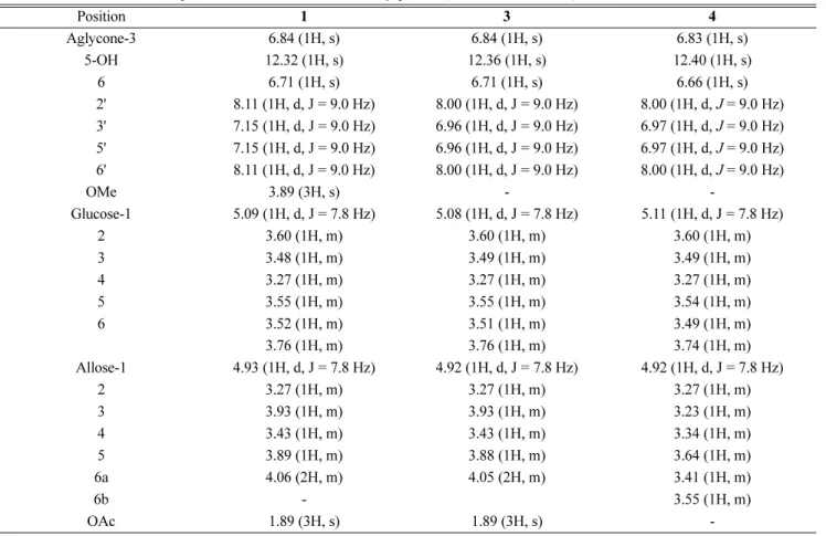
Table 1.
1H-NMR data of compounds 1, 3, and 4 isolated from S. japonica (600 MHz, DMSO-d
6)
Position 1 3 4
Aglycone-3 6.84 (1H, s) 6.84 (1H, s) 6.83 (1H, s)
5-OH 12.32 (1H, s) 12.36 (1H, s) 12.40 (1H, s)
6 6.71 (1H, s) 6.71 (1H, s) 6.66 (1H, s)
2' 8.11 (1H, d, J = 9.0 Hz) 8.00 (1H, d, J = 9.0 Hz) 8.00 (1H, d, J = 9.0 Hz) 3' 7.15 (1H, d, J = 9.0 Hz) 6.96 (1H, d, J = 9.0 Hz) 6.97 (1H, d, J = 9.0 Hz) 5' 7.15 (1H, d, J = 9.0 Hz) 6.96 (1H, d, J = 9.0 Hz) 6.97 (1H, d, J = 9.0 Hz) 6' 8.11 (1H, d, J = 9.0 Hz) 8.00 (1H, d, J = 9.0 Hz) 8.00 (1H, d, J = 9.0 Hz)
OMe 3.89 (3H, s) - -
Glucose-1 5.09 (1H, d, J = 7.8 Hz) 5.08 (1H, d, J = 7.8 Hz) 5.11 (1H, d, J = 7.8 Hz)
2 3.60 (1H, m) 3.60 (1H, m) 3.60 (1H, m)
3 3.48 (1H, m) 3.49 (1H, m) 3.49 (1H, m)
4 3.27 (1H, m) 3.27 (1H, m) 3.27 (1H, m)
5 3.55 (1H, m) 3.55 (1H, m) 3.54 (1H, m)
6 3.52 (1H, m) 3.51 (1H, m) 3.49 (1H, m)
3.76 (1H, m) 3.76 (1H, m) 3.74 (1H, m)
Allose-1 4.93 (1H, d, J = 7.8 Hz) 4.92 (1H, d, J = 7.8 Hz) 4.92 (1H, d, J = 7.8 Hz)
2 3.27 (1H, m) 3.27 (1H, m) 3.27 (1H, m)
3 3.93 (1H, m) 3.93 (1H, m) 3.23 (1H, m)
4 3.43 (1H, m) 3.43 (1H, m) 3.34 (1H, m)
5 3.89 (1H, m) 3.88 (1H, m) 3.64 (1H, m)
6a 4.06 (2H, m) 4.05 (2H, m) 3.41 (1H, m)
6b - 3.55 (1H, m)
OAc 1.89 (3H, s) 1.89 (3H, s) -
4'-O-Methylisoscutellarein 7-O-(6'''-O-acetyl)-β-D- allopyranosyl(1 →2)-β-D-glucopyranoside] (1) – Mp 247 ºC, yellowish powder; UV λ
maxnm (log ε): (MeOH) 280 (4.29), 307 (4.37), 326 (4.28); (MeOH + NaOH) 319 (4.16), 373 (4.00); (MeOH + AlCl
3) 284 (4.23), 323 (4.41), 346 (4.34); (MeOH + AlCl
3+ HCl) 283 (4.24), 322 (4.36), 347 (4.25); (MeOH + NaOAc) 280 (4.33), 307 (4.40), 326 (4.33); (MeOH + NaOAc + H
3BO
3) 280 (4.30), 307 (4.37), 326 (4.28); IR ν
max(KBr) cm
−1: 3400 (O-H, broad), 2937 (C-H), 1734 (C=O), 1658 ( α,β- unsaturated ketone), 1606, 1505, 1440 (aromatic C=C), 1221 (C-O), 1087, 1031 (glycosidic C-O); HR-FAB-MS (m/z): 665.1685 [M-H]
−(calculated,m/z665.1718);
1H- NMR (600 MHz, DMSO-d
6) and
13C-NMR (150 MHz, DMSO-d
6): Table 1 and Table 2.
Kaempferol 3- O-β-D-glucopyranoside (astragalin, 2) – Mp 230 - 233 ºC, UV λ
max(MeOH) nm: 267, 300 (sh), 352; IR ν
max(KBr) cm
−1: 3620 – 3000 (broad, OH), 1655 ( α,β-unsaturated ketone), 1606, 1562, 1506 (aromatic C=C), 1360, 1291 (aromatic C-O), 1179, 1056, 1011 (glycosidic C-O);
1H-NMR (600 MHz, DMSO-d
6) and
13
C-NMR (150 MHz, DMSO-d
6): Literature.
11Isoscutellarein 7-O-(6'''-O-acetyl)-β-D-allopyranosyl- (1 →2)-β-D-glucopyranoside (3) – Mp 235 ºC, orange yellow powder; UV λ
max(MeOH) nm (log ε): (MeOH) 278 (4.29), 307 (4.15), 328 (sh, 4.28); (MeOH + NaOH) 278 (sh, 4.29), 378 (4.41); (MeOH + AlCl
3) 282 (4.15), 323 (4.28), 350 (4.28); (MeOH + AlCl
3+ HCl) 282 (4.13), 323 (4.26), 350 (4.25); (MeOH + NaOAc) 277 (4.26), 307 (4.29), 328 (sh, 4.25); (MeOH + NaOAc + H
3BO
3) 278 (4.28), 307 (4.32), 328 (4.27); IR ν
max(KBr) cm
−1: 3374 (O-H, broad), 2937 (C-H), 1723 (C=O), 1659 ( α,β- unsaturated ketone), 1608, 1582 m 1449 (aromatic C=C), 1220 (C-O), 1087, 1032 (glycosidic C-O); HR-FAB-MS (m/z): 651.1538 [M-H]
−(calculated m/z651.1561);
1H- NMR (600 MHz, DMSO-d
6) and
13C-NMR (150 MHz, DMSO-d
6): Table 1 and Table 2.
Isoscutellarein 7-O- β-D-allopyranosyl-(1→2)-β-D- glucopyranoside (4) – Mp 239 ºC, orange yellow powder, UV λ
maxnm (log ε): (MeOH) 279 (4.06), 305 (4.09), 328 (4.07); (MeOH + NaOH) 274 (4.06), 377 (4.10); (MeOH + AlCl
3) 274 (4.07), 324 (4.07), 349 (4.08); (MeOH + AlCl
3+ HCl) 274 (4.07), 324 (4.07), 349 (4.08); (MeOH + NaOAc) 276 (4.09), 307 (4.08), 326 (4.05); (MeOH + NaOAc + H
3BO
3) 276 (4.10), 307 (4.09), 326 (4.07); IR ν
max(KBr) cm
−1: 3425 (O-H, broad), 2931 (C-H), 1660 ( α,β-unsaturated ketone), 1607, 1508, 1454 (aromatic C=C), 1223 (C-O), 1084, 1030 (glycosidic C-O);
1H- NMR (600 MHz, DMSO-d
6) and
13C-NMR (150 MHz, DMSO-d
6): Table 1 and Table 2.
Hydrolysis of compounds 2 and 3 – Compound 3 (60 mg) dissolved in 5% H
2SO
4in MeOH-H
2O (60:40) was heated under reflux for 5 h. The cooled reaction mixture was partitioned with EtOAc. The aglycone (3a) was obtained from the concentration of the EtOAc-soluble portion to be identified as isoscutellarein by the interpretation of spectroscopic data (Teles et al., 2015).
Acid hydrolysis of compound 2 was performed in the same way with compound 3. The aglycone (2a) of 2 was produced by the concentration of the EtOAc-soluble portion and identified as kaempferol by direct comparison with authentic specimen.
Kaempferol (2a) – Mp 277 - 279 ºC, UV λ
max(MeOH) nm: 267, 364; IR ν
max(KBr) cm
−1: 3350 (broad, OH), 1667 ( α,β-unsaturated ketone), 1620, 1575, 1510, Table 2.
13C-NMR data of compounds 1, 3, and 4 isolated from S.
japonica (150 MHz, DMSO-d
6)
Position 1 3 4
Isoscutellarein 2 164.1 164.1 164.6
3 103.0 103.1 103.1
4 182.9 182.8 182.8
5 152.7 152.7 152.9
6 100.0 100.0 99.3
7 151.1 151.0 151.7
8 128.0 128.0 127.7
9 144.3 144.2 144.8
10 106.1 106.1 105.7
1' 123.4 121.7 121.7
2' 129.0 129.1 129.1
3' 115.1 116.4 116.5
4' 163.0 161.9 161.9
5' 115.1 116.4 116.5
6' 129.0 129.1 129.1
OMe 56.1 -
Glc 1 100.6 100.6 100.2
2 83.0 83.1 81.8
3 77.7 77.7 77.6
4 69.8 69.8 69.8
5 76.1 76.1 76.2
6 61.1 61.1 61.1
All 1 103.9 103.2 102.2
2 72.0 72.0 71.5
3 71.3 71.3 72.0
4 67.4 67.3 67.7
5 72.0 72.0 75.0
6 64.0 64.0 61.5
COCH
320.9 20.9 -
COCH
3170.8 170.8 -
(Aromatic C=C), 1375, 1245, 1175 (C-O);
1H-NMR (600 MHz, DMSO-d
6) and
13C-NMR (150 MHz, DMSO-d
6):
Literature.
12Isoscutellarein (3a) – Mp 257 - 259 ºC, yellow powder, IR ν
max(KBr) cm
−1: 3367 (O-H, broad), 3028, 2954 (C- H), 1661 ( α,β-unsaturated ketone), 1610, 1582, 1509, 1445 (aromatic C=C), 1243, 1178 (C-O), 831; UV λ
maxnm (log ε): (MeOH) 292 (4.26), 329 (4.21); (MeOH + NaOH) 295 (4.11), 377 (4.33); (MeOH + AlCl
3) 308 (4.26), 363 (4.14); (MeOH + AlCl
3+ HCl) 305 (4.20), 358 (4.17); (MeOH + NaOAc) 293 (4.28), 346 (sh, 4.05);
(MeOH + NaOAc + AlCl
3) 295 (4.30), 342 (sh, 4.11);
1H- NMR (600 MHz, DMSO-d
6) and
13C-NMR (150 MHz, DMSO-d
6): Literature.
13Cholinesterase inhibition assay – Cholinesterase activity was assayed modifying the method described by Ellman et al..
14The inhibitory activities of AChE and BChE serving for ACh and BCh as the substrate, respectively, were measured. In brief, the solution added with 100 mM sodium phosphate buffer (pH 8.0) 140 μl, each sample 20 μl, and AChE (0.36 U) or BChE (0.36 U) 20 μl, respectively, in 96 well plates, was incubated for 15 min at room temperature, and then the reaction mixture was adjusted to 200 μl adding 10 μl DTNB [5,5'-dithiobis(2- nitrobenzoic acid)] and the substrate, 10 μl of ACh or BCh, in 96 well plate. After 15 min, the yellow 5-thio-2- nitrobenzoate anion formed by the reaction between DTNB and thiocholine resulted from the hydrolysis of ACh or BCh was measured at 412 nm by a microplate reader VERSA max (Molecular Devices, CA, USA). The cholinesterase inhibition activity was expressed as the IC
50value ( μg/ml) that is the concentration inhibiting the hydrolysis of ACh and BCh by 50%. The cholinesterase inhibition rate (%) was calculated by the following equation: Inhibition (%) = [1 − (A
samp/ A
con) / A
std] × 100, where A
samp, A
con, and A
stdare the absorbance when added with the sample and enzyme, with the sample but without the enzyme, and without the sample, respectively.
HPLC analysis – As the two mobile solvents, 0.05%
acetic acid in MeOH (solvent A) and H
2O (solvent B) were used. ACapcell Pak C18 column (5 μm, 4.6 mm × 250 mm, Shiseido, Japan) was used as a HPLC column. The programmed elution was performed in the following gradient elution: 0 – 20 min (20 → 65% B), 20 – 21 min (65 → 100% B), 21 – 25 min (100% B), 25 – 27 min (100 → 20% B), and 27 – 30 min (20% B), at the flow rate of 1.0 ml/min at the column temperature of 40 ºC. The detection wavelength was fixed at 254 nm. For the sensitivity for detection and quantification, the LOD (limit-of-detection) and LOQ (limit-of-quantification) were
determined by anS/N (signal-to-noise) method, respectively.
Result and Discussion
One flavonol glycoside and three flavone glycosides were isolated from the BuOH fraction obtained from 80%
MeOH extract of S. japonica. By the interpretation of
1H- and
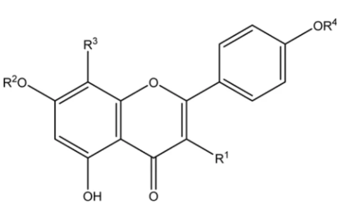
13C-NMR spectral data, compounds 1, 3, and 4 were identified as 4'-O-methylisoscutellarein 7-(6'''-O-acetyl)- O-β-D-allopyranosyl(1 →2)-β-D-glucopyranoside] (1), isoscutellarein 7-O-(6'''-O-acetyl)-β-D-allopyranosyl(1 → 2)- β-D-glucopyranoside] (3), and isoscutellarein 7-O-β-D- allopyranosyl-(1 →2)-β-D-glucopyranoside] (4). Meanwhile, the flavonol glycoside (2) was astragalin (kaempferol3-O- β-D-glucopyranoside) (Fig. 1).
The UV spectroscopic data of compounds 1, 3 and 4 measured by the addition of shift reagent were noted in the experimental section. No significant shifts of absorption bands were observed by the addition of NaOAc and H
3BO
3, though considerable shifts due to 5-hydroxy-4- keto system were shown by the addition of AlCl
3. These results suggest that the three compounds are the flavone 7-O-glycosides with no o-dihydroxy group in the B-ring.
IR spectra of the three compounds demonstrated the presence of hydroxy group, aliphatic C-H, α, β-unsaturated ketone, aromatic C=C, aromatic C-O, aliphatic C-O, and glycosidic C-O in the functional group region.
As shown in Table 1, the singlet peaks of C-3 of 1, 3, 1 : R
1= H, R
2= (6-O-acetyl)-β-D-allopyranosyl(1 → 2)-β-D- glucopyranosyl, R
3= OH, R
4=Me
2 : R
1= O-β-D-glucopyranosyl, R
2= R
3= R
4= H 2a : R
1= OH, R
2= R
3= R
4= H
3 : R
1= R
4= H, R
2= (6- O-acetyl)-β-D-allopyranosyl(1 →2)-β-D- glucopyranosyl, R
3= OH
3a : R
1= R
2= R
4= H, R
3= OH
4 : R
1= R
4= H, R
2= β-D-allopyranosyl(1→2)-β-D-glucopyranosyl, R
3= OH
Fig. 1. Structure of flavonoid glycosides (1 – 4) isolated from S.
japonica and their aglycones (2a and 3a).
and 4 shown at δ 6.84, 6.84 and 6.83, respectively, suggest that the aglyconesare the flavone type. In particular, chemical shifts of C-5 and C-7 are shown in δ
C149.5 and 150.8 ppm in the literature data (
13C-NMR) of isoscutellarein 5-O-glucoside. As shown in Table 2, the peaks of C-5 in 1, 3, and 4 were observed at δ
C152.7, 152.7, and 152.9, and those of C-7 were shown in 151.1, 151.0, and 151.7. These results indicate that the sugar moiety is linked not to the C-5 position of the aglycone but to the C-7. The identification of 4'-O-methylisos- cutellarein, the aglycone of compound 1, and isoscutellarein, that of compounds 3 and 4, was supported by literature data.
13,15The presence of the two sugars, D-glucopyranosyl and D-allopyranosyl, in the glycosides was identifiable in the course of comparison with the literature NMR data of the flavonoid glycosides 1, 3, 4, isolated from other Stachys species.
6,7,16In the
1H-NMR spectrum of compound 1, β- configuration of D-glucose can be identified from the coupling constant (d, J = 7.8 Hz) of the anomeric proton shown at δ 5.09. The β-configuration of D-allose was also observed from the coupling constant (d, J = 7.8 Hz) at δ 4.92. The anomeric proton of D-allose shown at δ 4.93 is long-range coupled to δ
C83.1 in the HMBC spectrum, indicating that the second sugar is attached to the C-2 position in the first sugar. The presence of acetyl group at the C-6''' of D-allose was determined by the HMBC spectral interpretation.
The NMR data of compounds 1, 3, and 4 were successfully assigned, as shown in Table 1 and Table 2.
Therefore, the three compounds were identified to be 4'- O-methylisoscutellarein 7-(6'''-O-acetyl)-O-β-D-allopyranosyl (1 →2)-β-D-glucopyranoside] (1), isoscutellarein 7-O-(6'''- O-acetyl)-β-D-allopyranosyl(1→2)-β-D-glucopyranoside]
(3), and isoscutellarein 7-O-β-D-allopyranosyl-(1 →2)-β- D-glucopyranoside] (4) by comparing with literature data.
6,7,16The four compounds were isolated for the first time from S. japonica.
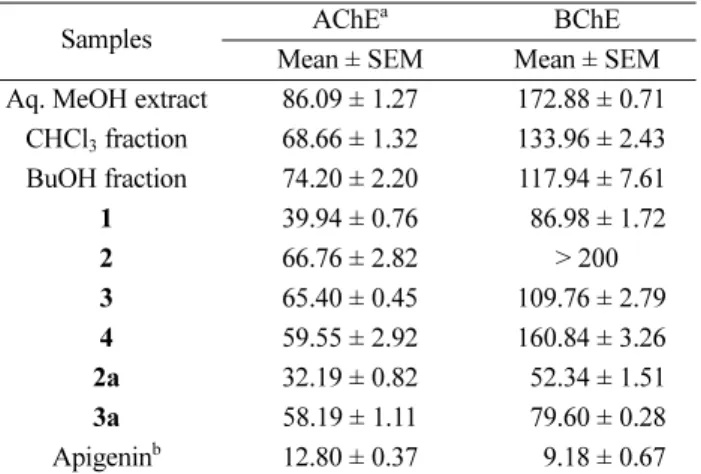
Cholinesterases divided into AChE and BChE are key enzymes that play significant roles in cholinergic trans - mission by hydrolyzing ACh.
17Cholinesterase inhibitors can treat Alzheimer’s disease, since the inhibition of cholinesterase increases this neurotransmitter responsible for brain’s memory. In our tests, the samples of the aq.
MeOH extract, its two fractions (CHCl
3- and BuOH fractions), the four flavonoid glycosides isolated from the BuOH fraction, and the aglycones (2a and 3a) obtained by hydrolysis of glycosides were assayed for cholinesterase inhibition activity. The aq. MeOH extract displayed the data of 86.09 ± 1.27, 172.88 ± 0.71 μg/ml as the IC
50value. The activities of CHCl
3- and BuOH fractions were similar each other.
As shown in Table 3, the IC
50s of AChE inhibition were observed in the range of 39.94 – 66.76 μg/ml whereas those of BChE were in the range of 52.34 – 160.84 μg/ml. Compound 1 possessing the aglycone of 4'-O-methylisoscutellarein was more active (IC
50, 39.94 μg/ml for AChE and 86.98 μg/ml for BChE) than other compounds with isoscutellarein. Astragalin (3) was less active than its aglycone (kaempferol). In this assay, apigenin which is known to have potent anti-cholinesterase activity
1,18Table 3. Cholinesterase inhibitory activities of the S. japonica extract, its fractions and isolated compounds
Samples AChE
aBChE
Mean ± SEM Mean ± SEM
Aq. MeOH extract 86.09 ± 1.27 172.88 ± 0.71 CHCl
3fraction 68.66 ± 1.32 133.96 ± 2.43 BuOH fraction 74.20 ± 2.20 117.94 ± 7.61
1 39.94 ± 0.76 86.98 ± 1.72
2 66.76 ± 2.82 > 200
3 65.40 ± 0.45 109.76 ± 2.79
4 59.55 ± 2.92 160.84 ± 3.26
2a 32.19 ± 0.82 52.34 ± 1.51
3a 58.19 ± 1.11 79.60 ± 0.28
Apigenin
b12.80 ± 0.37 9.18 ± 0.67
a
The values (μg/ml) indicate 50% cholinesteraseinhibitory effects.
These data represent the average values of three repeated experi- ments.
bPositive control.
Fig. 2. HPLC chromatogram of standard compounds and 80%
MeOH fraction of S. japonica.
and anxiolytic activity
19was used as the positive control.
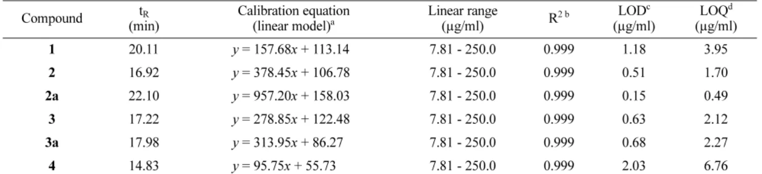
Six compounds including the four isolated ones and the two aglycones were able to be identified on the HPLC chromatogram by direct comparison with standard ones (Fig. 2). As shown in Table 4, the regression equations which the linearity was verified by the R
2value more than 0.999 were established as the HPLC analytical method.
Limits of detection and quantification were also sufficiently low. In the HPLC chromatogram of the 80% MeOH fraction, compound 3 exhibited the highest peak of the tested compounds. The content of compound 3 was 154.54 mg/g 80% MeOH fraction (Table 5). However, the concentration of the two aglycones, 2a and 3a, were low.
In consequence, the four flavonoids present in the hydrophilic fraction (BuOH fraction) of the S. japonica extract may contribute to the prevention of memory impairment of Alzheimer’s disease. Furthermore, com- pound 1 with apotent cholinesterase inhibition activity were identified as 4'-O-isoscutellarein 7-O-(6'''-O-acetyl)- β-D-allopyranosyl(1→2)-β-D-glucopyranoside and quantified in S. japonica.
Acknowledgements
This research was supported by the Sangji University Research Fund, 2016.
References
(1) Balkis, A.; Tran, K.; Lee, Y. Z.; Ng, K. See comment in PubMed Commons below J. Agric. Sci. 2015, 7, 1916-9760.
(2) Luo, W.; Chen, Y.; Wang, T.; Hong, C.; Chang, L. P.; Chang, C. C.;
Yang, Y. C.; Xie, S. Q.; Wang, C. J. Bioorg. Med. Chem. 2016, 24, 672- 680.
(3) Kim, T. J. Korean Plant Resources; Publishing Center of Seoul National University: Korea, 1996, pp 54-55.
(4) Nishimura, H.; Sasaki, H.; Inagaki, N.; Chin, M.; Mitsuhashi, H.
Phytochemistry 1991, 30, 965-969.
(5) Harada, S.; Tsujita, T.; Ono, A.; Miyagi, K.; Mori, T.; Tokuyama, S.
J. Nutr. Sci. Vitaminol. 2015, 61, 167-174.
(6) Rahimi Khoigani, S.; Rajaei, A.; Goli, S. A. Nat. Prod. Res. 2017, 31, 355-358.
(7) Venditti, A.; Serrilli, A. M.; Di Cecco, M.; Ciaschetti, G.; Andrisano, T.; Bianco, A. Nat Prod Res. 2013, 27, 190-193.
(8) Petreska, J.; Stefova, M.; Ferreres, F.; Moreno, D. A.; Tomás- Barberán, F. A.; Stefkov, G.; Kulevanova, S.; Gil-Izquierdo, A. Food Chem. 2011, 125, 13-20.
(9) Pereira, O. R.; Domingues, M. R. M.; Silva, A. M. S.; Cardoso, S.
M. Food Res. Intern. 2012, 48, 330-335.
(10) Cui, Q.; Pan, Y.; Xu, X.; Zhang, W.; Wu, X.; Qu, S.; Liu, X.
Fitoterapia 2016, 109, 67-74.
(11) Park, H. J.; Nugroho, A.; Jung, B. R.; Won, Y. H.; Jung, Y. J.; Kim, W. B.; Choi, J. S. Kor. J. Plant Res. 2010, 23, 393-398.
(12) Park, H. J.; Young, H. S.; Park, K. Y.; Rhee, S. H.; Chung, H. Y.;
Choi, J. S. Arch. Pharm. Res. 1991, 14, 167-171.
(13) Teles, Y. C. F.; Horta, C. C. R.; de Fátima Agra, M.; Siheri, W.;
Boyd, M.; Igoli, J. O.; Gray, A. I.; de Fátima Vanderlei de Souza, M.
Molecules 2015, 20, 20161-20172.
(14) Ellman, G. L.; Courtney, K. D.; Andres, V. Jr.; Feather-stone, R. M.
Biochem. Pharmacol. 1961, 7, 88-95.
(15) Demirtas, I.; Gecibesler, I. H.; Yaglioglu, A. S. Phytochemistry Table 4. Linearity of standard curves and limits of detection and quantification for the standard compounds
Compound t
R(min)
Calibration equation (linear model)
aLinear range
(µg/ml) R
2 bLOD
c(µg/ml)
LOQ
d(µg/ml)
1 20.11 y = 157.68x + 113.14 7.81 - 250.0 0.999 1.18 3.95
2 16.92 y = 378.45x + 106.78 7.81 - 250.0 0.999 0.51 1.70
2a 22.10 y = 957.20x + 158.03 7.81 - 250.0 0.999 0.15 0.49
3 17.22 y = 278.85x + 122.48 7.81 - 250.0 0.999 0.63 2.12
3a 17.98 y = 313.95x + 86.27 7.81 - 250.0 0.999 0.68 2.27
4 14.83 y = 95.75x + 55.73 7.81 - 250.0 0.999 2.03 6.76
a