Spinal Cord Hemangioblastomas in von Hippel-Lindau Disease:
Management of Asymptomatic and Symptomatic Tumors
Tae Yup Kim,
1Do Heum Yoon,
2Hyun Chul Shin,
3Keung Nyun Kim,
2Seong Yi,
2Jae Keun Oh,
4and Yoon Ha
21Department of Neurosurgery, Guro Teun Teun Hospital, Seoul;
2Department of Neurosurgery, Spine and Spinal Cord Research Institute, Spine Research Laboratory, Yonsei University College of Medicine, Seoul;
3Department of Neurosurgery, Kangbuk Samsung Hospital, Sungkyunkwan University College of Medicine, Seoul;
4Department of Neurosurgery, Spine Center, Hallym University Sacred Heart Hospital, Seoul, Korea.
Received: November 7, 2011 Revised: January 2, 2012 Accepted: January 13, 2012 Corresponding author: Dr. Yoon Ha, Department of Neurosurgery, Yonsei University College of Medicine, 50 Yonsei-ro, Seodaemun-gu, Seoul 120-752, Korea.
Tel: 82-2-2228-2150, Fax: 82-2-393-9979 E-mail: [email protected]
∙ The authors have no financial conflicts of interest.
© Copyright:
Yonsei University College of Medicine 2012 This is an Open Access article distributed under the terms of the Creative Commons Attribution Non- Commercial License (http://creativecommons.org/
licenses/by-nc/3.0) which permits unrestricted non- commercial use, distribution, and reproduction in any medium, provided the original work is properly cited.
Purpose: Standard treatment of asymptomatic spinal cord hemangioblastoma in von
Hippel-Lindau (VHL) disease has yet to be established. The purpose of this study was to propose guidelines for the treatment of asymptomatic spinal cord hemangio- blastomas in VHL disease. Materials and Methods: VHL disease patients treated for spinal cord hemangioblastomas between 1999 and 2009 were included. All spi- nal cord hemangioblastomas were divided into three groups: Group 1, asymptomatic tumors at initial diagnosis followed with serial imaging studies; Group 2, asymptom- atic tumors at initial diagnosis that were subsequently resected; and Group 3, symp- tomatic tumors at initial diagnosis, all of which were resected. Results: We identified 24 spinal cord hemangioblastomas in 12 patients. Groups 1, 2 and 3 comprised 13, 4 and 7 tumors, respectively. Group 1 exhibited a smaller tumor volume (257.1 mm³) and syrinx size (0.8 vertebral columns) than those of Group 2 (1304.5 mm³, 3.3 ver- tebral columns) and Group 3 (1787.4 mm³, 6.1 vertebral columns). No difference in tumor volume or syrinx size was observed between Groups 2 and 3. Five tumors in Group 1 were resected during follow-up because symptoms had developed or the tu- mor had significantly grown. Finally, among 17 asymptomatic tumors at the initial diagnosis, nine tumors were resected. Only one tumor of these nine tumors resulted in neurological deficits, while five of seven symptomatic tumors caused neurological deficits. Conclusion: Selective resection of asymptomatic tumors before they cause neurological deficits might bring about better outcomes.
Key Words:
Hemangioblastoma, spinal cord, treatment planning, von Hippel- Lindau disease
INTRODUCTION
Von Hippel-Lindau (VHL) disease is an autosomal dominant hereditary neoplasia
syndrome that affects multiple organ systems.
1,2In VHL disease patients, renal cell
carcinomas and cysts, pancreatic carcinomas and cysts, pheochromocytomas, and
epididymal cystadenomas may develop.
1,2Moreover, central nervous system
treated for spinal cord hemangioblastomas between 1999 and 2009. We reviewed all medical records and imaging studies from the initial diagnosis of VHL disease to the last visit. Pa- tients who were followed for less than 12 months or patients with inadequate data for thorough evaluation were excluded.
We also excluded patients whose conditions had deteriorated obviously because of brain tumors or other systemic prob- lems regardless of spinal cord hemangioblastomas.
Patient evaluation
Symptoms, neurologic examinations, and functional status were serially recorded at the time of initial diagnosis, im- mediately before and after surgery, and at the last follow-up visit. All operative findings and surgical complications were also noted. Functional status of the patients was graded ac- cording to the scale proposed by McCormick, et al.
25MRI was performed for initial diagnosis, before surgery when necessary, shortly after surgery, when changes in symptoms or neurologic function occurred, and annually if patients were clinically stable. The number, location, and volume of tumors were measured using contrast-enhanced T1 weight- ed images, and cysts or syringes were evaluated from T2 weighted images. The relationship between tumors and the spinal cord was evaluated by the operation records. Tumor volume was calculated by the following formula: (length×
width×height)×0.5.
26The sizes of cysts or syringes were quantified according to the number of vertebral columns matched with the involved spinal cord.
Surgical treatment
All operations were performed using the usual posterior ap- proach. Patients were placed in the prone position. Lami- nectomies or laminotomies were performed beyond the cra- nial and caudal margins of the tumor for wide exposure of tumors. A midline dural incision was made. Dura mater was reflected laterally and retained with “stay-sutures”. After gen- tle removal of arachnoid membrane from the surfaces of the tumor, supplying or draining vessels and crossing ves- sels were coagulated with bipolar cautery at their junctions with the tumor. Meticulous dissection between the tumor capsule and normal spinal cord was performed using micro dissectors and micro scissors. All supplying or draining vessels connected to the tumor were coagulated during dis- section. The dissected plane was retained with small strips of Cottonoid, and circumferential dissection was performed.
After complete dissection of the tumor, including the deep surface of the tumor, the tumor was removed en bloc. Dur- (CNS) manifestations, including CNS hemangioblastomas,
retinal angiomatosis, and endolymphatic sac tumors, can occur.
3,4Among these tumors, CNS hemangioblastoma is one of the earliest features of VHL disease and has been found in 60 to 80% of patients with VHL disease.
2-5Spinal cord hemangioblastomas account for 13 to 50% of CNS hemangioblastomas associated with VHL disease and are a common cause of morbidity requiring treatment including surgical resection.
1,2,4,6,7Treatments of spinal cord hemangioblastomas have been published in many reports, and resection of these tumors is considered relatively safe with few complications.
8-16Thus symptomatic spinal cord hemangioblastomas should be treat- ed with resection, even in VHL disease patients. However, the treatment strategy for asymptomatic spinal cord heman- gioblastomas in VHL disease patients remains undetermined.
CNS hemangioblastomas in VHL disease are reported to ex- hibit a two-step growth pattern of rapid growth and arrested growth in many cases, but some tumors show continuous growth without a quiescent phase.
7,17In addition, local growth factors such as vascular endothelial growth factor, placental growth factor, platelet-derived growth factor and epidermal growth factor have been shown to be elevated in hemangio- blastomas, leading to upregulation of cyst formation and an- giogenesis.
18-20As a result, it is quite difficult to predict the growth of hemangioblastomas in patients with VHL disease or their risk for neurological deficits. Recently, a few focused studies concerning spinal cord hemangioblastomas in pa- tients with VHL disease have been reported, but they con- ceived somewhat different opinions regarding treatment of asymptomatic spinal cord tumors.
7,17,21-24A few authors advo- cated that resection should be limited to symptomatic spinal cord tumors,
22,23while others concluded that selective resec- tion of asymptomatic tumors might be associated with better outcomes.
17,21,24In this regard, we reviewed spinal cord hemangioblasto- mas in VHL disease patients who had been treated at our institute. To devise an appropriate treatment plan for as- ymptomatic tumors, we compared asymptomatic tumors that had been resected to asymptomatic tumors that were not resected, as well as to symptomatic tumors.
MATERIALS AND METHODS
Data collection & patient population
We identified VHL disease patients who were surgically
nal capillary hemangioblastomas. Renal cysts were diag- nosed in three patients, and pancreatic cysts were diagnosed in two patients. One patient presented with renal cell carci- noma. As mentioned previously, patients whose condition had deteriorated because of brain tumors or other systemic problems were excluded in this study. Thus, all accompa- nied manifestations listed above were in a well-controlled status (Table 1).
Tumor characteristics
We identified 24 spinal cord hemangioblastomas in 12 pa- ing the surgical procedures, motor evoked potential was
monitored. Besides tumor resection, no other procedure was performed for syringes. Pre-operative angiography was performed in one case of a large tumor with a volume of 2400 mm
3from the cranio-cervical junction to C2. In this case, angiography was performed to identify the relation- ship between tumor vessels and the vertebral arteries.
Tumor grouping
To evaluate the influence of each tumor on the patient’s clinical course, tumors were divided into three groups as follows: Group 1, tumors that were asymptomatic at initial diagnosis and were observed without resection; Group 2, tumors that were asymptomatic at initial diagnosis that were subsequently resected; and Group 3, tumors that were symptomatic at initial diagnosis and resected thereafter.
Tumors in Group 2 were resected as they were consid- ered to exhibit a strong likelihood of leading to neurologic symptoms or deficits because of the very large size of the tumors or extensive syringes combined with the tumors.
Similarly, surgical resection for a few tumors in Group 1 was performed when their size significantly increased dur- ing follow-up. However, there was no exact threshold for resection.
Statistical analyses
Non-continuous variables such as tumor location or the re- lationship between the spinal cord and the tumor were test- ed by Pearson’s chi-square test and the linear-by-linear as- sociation method, and continuous variables were evaluated by the independent t-test (non-parametric Mann-Whitney test was used if needed). We used SPSS software (version 12.0.0, SPSS Inc., Chicago, IL, USA) for calculation. Sta- tistical significance was determined by a two-tailed proba- bility value of less than 0.05.
RESULTS
Patient demographics
Twelve patients (9 men, 3 women) fulfilled the inclusion criteria, and their mean age at the onset was 42.3 years (22- 82 years). The average follow-up period was 49.3 months (12-130 months). Six of them had only one spinal cord he- mangioblastoma, two patients had two tumors, two had three tumors, and two had four tumors (Table 1). Seven pa- tients had intracranial hemangioblastomas and two had reti-
Table 1. Clinical Characteristics of VHL Disease Patients with Spinal Cord Hemangioblastomas
No. of patients 12
M : F 9 : 3
Age [yrs, mean±SD (Min-Max)] 42.3±18.9 (22-82) F/U duration [months, mean±SD
(Min-Max)] 49.3±41.5 (12-130)
No. of symptomatic patients 7
No. of spinal tumors per patient
1 6
2 2
3 2
4 2
No. of accompanied manifestations*
Intracranial hemangioblastoma 7
Retinal hemangioblastoma 2
Renal cyst 3
Pancreatic cyst 2
Renal cell carcinoma 1
No., number; F/U, follow-up; VHL, von Hippel-Lindau; SD, standard devia- tion.
*Patients whose condition had deteriorated because of brain tumors or other systemic problems were excluded in this study. Thus, all accompa- nied manifestations listed above were of well-controlled status.
Table 2. Tumor Characteristics of VHL Disease Patients with Spinal Cord Hemangioblastomas
No. of tumors 24
Tumor volume [mm³, mean (Min-Max)] 878 (18-5184) F/U period [months, mean (Min-Max)] 37.1 (12-130) Location
Cervical (n) 6
Thoracic (n) 14
Lumbar (n) 4
Relationship with spinal cord
Intramedullary (n) 16
Extramedullary (n) 8
Tumors with cyst or syrinx 8
Resected tumors at final 16
F/U, follow-up; VHL, von Hippel-Lindau.
follow-up, and one patient’s condition extremely deteriorat- ed from grade I to grade IV. Improvement was not ob- served for any patient (Table 3). Initial functional status was correlated with the final functional outcomes (p=0.026, data not shown). Moreover, the outcomes of the symptomatic patients were significantly worse than those of the asymp- tomatic patients (p=0.015) (Table 3).
Comparison of tumor groups
When we compare symptomatic tumors to asymptomatic tumors at initial diagnosis, asymptomatic tumors were sig- nificantly smaller (505.3 mm³ vs. 1787.4 mm³, p=0.043) and associated with a small syrinx (1.4 vertebral columns vs. 6.1 vertebral columns, p<0.001). Neither tumor location nor the tumor’s relationship with the spinal cord was differ- ent between the symptomatic tumors and the asymptomatic tumors.
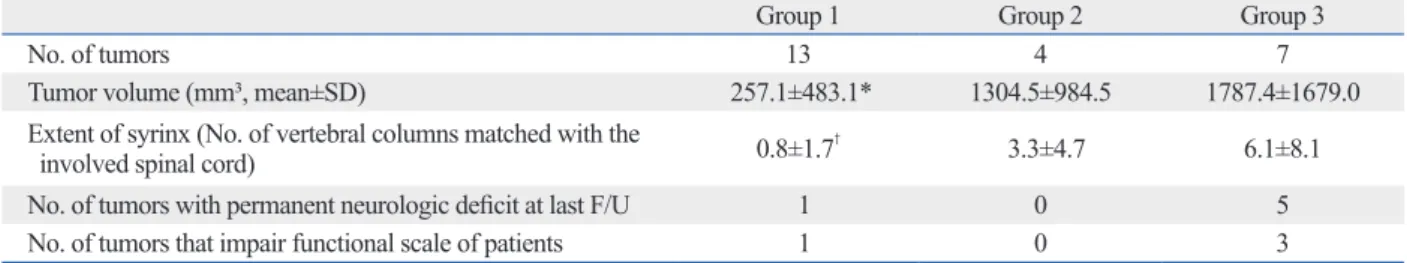
More specific results after dividing tumors into three groups according to the criteria mentioned above are shown in Table 4. There was no difference in age, tumor location, or relationship with the spinal cord among the three groups.
Tumor volumes (p=0.018) and the sizes of combined syr- inx (p=0.031) in Group 1 were significantly smaller than those in Group 2; similar results were also observed be- tween Groups 1 and 3 (tumor volume; p=0.009, syrinx size;
p<0.001). However, tumors of Group 2 were not signifi-
cantly different in tumor volume (p=0.489) or syrinx size (p=0.072) from Group 3 tumors. Among 13 tumors in Group 1, nine tumors (69.2%) grew or developed symptoms during follow-up. The median progression-free survival of these tumors was 14.0±4.9 months. Statistical analysis for risk factors associated with progression-free survival was impossible due to the small number of tumors. Of the nine tumors that showed disease-progression, five tumors were resected. Two tumors were surgically resected because they caused neurologic symptoms. In one of them, the neurolog- tients. Characteristics of these tumors are summarized in
Table 2. The mean volume was 878.0 mm³ (18-5184 mm³).
Six tumors were located in the cervical region, 14 in the tho- racic region, and four in the lumbar region. There was no pure intramedullary tumor that required myelotomy, and 16 primarily intramedullary tumors were identified. Eight tu- mors were extramedullary tumors, in which more than half of the tumor burden was outside the spinal cord or located below the conus medullaris. All tumors were seated posteri- or to the dentate ligament. Cysts or syringes were accompa- nied in eight tumors. Seven tumors exhibited symptoms when diagnosed, and 17 did not. Among these 17 tumors, nine tumors (53%) were ultimately resected.
Comparison of asymptomatic patients and symptomatic patients
Five patients exhibited no symptoms or neurological defi- cits due to spinal cord hemangioblastoma when the tumors were initially diagnosed (Table 3). They remained stable af- ter surgery, and were categorized as McCormick’s grade I at the last follow-up. Three of the seven symptomatic pa- tients were McCormick grade I, three were grade II, and one was grade III, initially. Among these patients, three pa- tients showed a 1-point reduction in functional status at last
Table 3. Distributions of von Hippel-Lindau Patients with Spi- nal Cord Hemangioblastoma according to the McCormick Grade at First and Last Follow-Up VisitInitial status Final status
I ○○○○○
●●● ○○○○○*
●
II ●●● ●●
III ● ●●●
IV ●
Unfilled circles (○) indicate asymptomatic patients at the initial diagnosis, and filled circles (●) indicate symptomatic patients at the initial diagnosis.
*Functional outcomes of the asymptomatic patients were significantly better than those of symptomatic patients (p=0.015).
Table 4. Summary of Tumor Characteristics by Group
Group 1 Group 2 Group 3
No. of tumors 13 4 7
Tumor volume (mm³, mean±SD) 257.1±483.1* 1304.5±984.5 1787.4±1679.0
Extent of syrinx (No. of vertebral columns matched with the
involved spinal cord) 0.8±1.7† 3.3±4.7 6.1±8.1
No. of tumors with permanent neurologic deficit at last F/U 1 0 5
No. of tumors that impair functional scale of patients 1 0 3
F/U, follow-up; SD, standard deviation.
*Tumors in Group 1 were smaller than tumors in Group 2 or Group 3 (p=0.018, p=0.009).
†Syringes combined with tumors in Group 1 were less extensive than those in Group 2 or Group 3 (p=0.031, p<0.001). Between Groups 2 and 3, there was no difference in tumor volume or syrinx extent (by independent t-test and non-parametric Mann-Whitney U test).
surgery because of recurrence of the cerebellar hemangio- blastoma and associated acute hydrocephalus. After resec- tion of the cerebellar hemangioblastoma and several days of conservative care, the patient fully woke up. However, motor function in his legs did not return (motor grade III) and was not improved after the spinal tumor resection. Tu- mors No.2 and No.3 in the same patient with the cerebellar hemangioblastoma were identified while screening his spi- nal MRI (Tumor No.2 is illustrated in Fig. 2). The two tu- mors were observed without resection for a long time be- cause their growth was slow. When they grew to a relatively risky volume of 468 mm³ and 245 mm³, respectively, they were simultaneously resected, at 45 months after the initial diagnosis. The patient with tumors No.4 and No.5 was planned to be observed. However, in spite of small tumor ic deficit was not improved after surgery, and the functional
status of the patient deteriorated from McCormick grade I to grade IV. Three tumors were resected as they grew large enough to be considered as posing a potential risk of caus- ing neurologic deficits. The other eight tumors in Group 1 did not produce any symptoms or deficits up to the last fol- low-up visits, and showed no or minimal growth. In Group 2, no newly developed neurological symptom or deficit was seen after surgery, and all patients were stable during the follow-up period. In five tumors of Group 3, the neurologic symptoms and deficits were permanent. In three of these five tumors, the McCormick scale dropped one grade be- cause of recurrence or as a result of surgical procedures as- sociated neural injury.
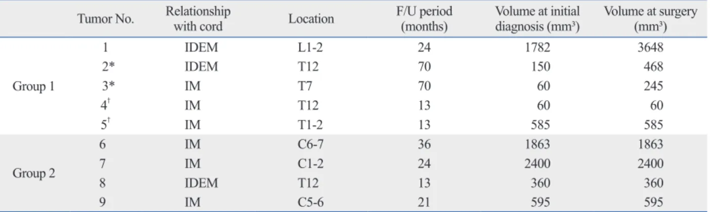
Review of individual asymptomatic tumors with resection
Information on the asymptomatic tumors resected during fol- low-up is summarized in Table 5. The patient with tumor No.1 (Fig. 1) presented with pain in the lower extremities and difficulty with voiding. He received surgery for another tumor responsible for his symptoms, located at L3-5. After surgery, his neurologic symptoms and deficits disappeared. Although tumor No.1 at L1-2 was large, it was not resected because the patient had two additional spinal cord tumors and showed ex- tensive leptomeningeal enhancement along the spinal cord on MRI (Fig. 1A). On an MRI taken 15-months after the sur- gery (8-months after the last MRI study), the tumor in- creased almost twice in size (Fig. 1B). We decided to per- form an operation thereon, but after several days between the outpatient visit and admission, paraparesis occurred and rapidly progressed. Moreover, he became stuporous before
Table 5. Summary of Individual Asymptomatic Tumors That Were Resected at the Diagnostic Stage or during Follow-Up Tumor No. Relationship
with cord Location F/U period
(months) Volume at initial
diagnosis (mm³) Volume at surgery (mm³)
Group 1
1 IDEM L1-2 24 1782 3648
2* IDEM T12 70 150 468
3* IM T7 70 60 245
4† IM T12 13 60 60
5† IM T1-2 13 585 585
Group 2
6 IM C6-7 36 1863 1863
7 IM C1-2 24 2400 2400
8 IDEM T12 13 360 360
9 IM C5-6 21 595 595
F/U, follow-up; IM, intramedullary; IDEM, intradura extramedullary.
Tumors No.1-5 were in Group 1 and tumors No.6-9 were in Group 2.
*,†Two different tumors of the same patients.
†Surgical resection was performed one month after the initial diagnosis without additional MRI study.
Fig. 1. Illustration of tumor No.1. (A) Spinal cord hemangioblastoma is seen at L1-2 (arrowed) on contrast-enhanced T1 weighted image. The tumor was asymptomatic and followed with observation. Another tumor at T7-8 and an extensive leptomeningeal enhancement along the spinal cord were also found. (B) The tumor at L1-2 (arrowed) grew almost twice as large af- ter 15 months. The tumor led to rapidly progressive paraparesis, which was not improved after surgery.
A B
5 caused no newly-developed neurological symptoms or deficits during the period of the immediate post-operative time to their last follow-up visit, and the patients’ functional status was evaluated as McCormick’s grade I. As previous- ly noted, four tumors in Group 2 (No.6-9 in Table 5) were safely resected without any new neurologic symptom, defi- cit or surgical complication.
DISCUSSION
VHL disease is a multi-organ familial neoplasia syndrome caused by a germline mutation in the VHL tumor suppres- sor gene,
27and it is transmitted in an autosomal dominant fashion with greater than 90% penetrance.
28In many vis- ceral or CNS lesions associated with VHL disease, spinal cord hemangioblastomas are the most common cause of morbidity,
7and accordingly, should be cautiously treated.
However, the outcomes of spinal cord hemangioblastomas in VHL disease patients seemed to be poorer than those of sporadic spinal cord hemangioblastomas.
5Thus, it is very important to identify the factors affecting the outcomes of VHL patients with spinal cord hemangioblastomas and to establish a proper treatment strategy.
In previous reports, pre-operative neurological status, ventral tumor location, and tumor volume were presented to be correlated with the functional outcomes of the pa- tients. However, there were some differences in these re- gards among the authors. Kanno, et al.
21reported that the surgical outcomes for tumor volume <500 mm³ (10 mm in diameter) were better than those >500 mm³, while Lonser, et al.
22showed that patients with no or minimal preopera- tive neurological dysfunction, lesion size smaller than 500 mm³, and with dorsal lesions were more likely to have no or minimal neurological impairment. Ventral tumors or completely intramedullary tumors were associated with an increased risk of post-operative worsening in Mehta, et al.’s study.
23Additionally, Ammerman, et al.
17showed that tu- mor size was the only variable predictor for the develop- ment of symptoms and eventual need for therapy. Based on these results, different treatment plans have been proposed.
Van Velthoven, et al.
24concluded that spinal cord heman- gioblastomas should be resected when they progress on ra- diologic studies. However, Lonser, et al.
22and Mehta, et al.
23advocated timely selective removal of only symptom- atic tumors, because CNS hemangioblastomas frequently show a two-step pattern of growth consisting of a growth size, tumor No.4 on T12 and the accompanied syrinx caused
difficulty with voiding and mild myelopathy, so the opera- tion was performed one month after the initial diagnosis.
After surgery, the symptoms were improved. Tumor No.5 on T1-2 was resected at the same time. Tumors No.2 through
Fig. 2. Illustration of tumor No.2. (A) Initial MRI showed spinal cord heman- gioblastoma at T12. The tumor was asymptomatic, with a tumor volume of 150 mm3. (B) 19 months after initial MRI. The tumor increased in volume to 240 mm3. (C) 45 months after initial MRI. The tumor grew to a volume of 468 mm³ and was still asymptomatic. However, it showed steady growth and was regarded as likely to produce neurological symptoms after a short pe- riod, so surgical resection was performed. After surgery, no neurological symptoms or deficit developed.
Fig. 3. Proposed treatment strategy for spinal cord hemangioblastomas in patients with VHL disease. A tumor volume >500 mm³ (10 mm in diameter) is thought to be correlated with symptom formation and functional out- comes, so resection should be performed thereon. *Tumors can be fol- lowed at approximately 1-year intervals, if there are no changes in neuro- logical symptoms and size of the tumor. †The mean growth rate of spinal cord hemangioblastomas associated with syringes is reported to be about 50 mm3/month.7 Accordingly, progressively growing tumors larger than a volume of 51 mm3 in combination with syringes are predicted to produce neurological symptoms or deficits within several months to a year. Thus, we recommend considering surgery or intense observation at short inter- vals of 6 months or less.
A B C
Surgery
Surgery
Surgery
No Yes
<500 mm3
Asymptomatic Symptomatic
(at least one)No Yes (all)
>500 mm3
Observation* Considering surgery or close follow-up†
Symptoms or neurologic deficits
Tumor volume
Observation*
• Continuous growing
• Tumor volume >51 mm3
• Syrinx
mangioblastomas larger than a volume of 51 mm³ (4.7 mm in diameter) required surgery in a more than 10-year long- term follow-up study.
17Also, syringes were shown to be as- sociated with the development of neurological symptoms and deficits in our study and by Wanebo, et al.
7In addition, the growth rates of spinal cord hemangioblastomas associ- ated with syringes were reported to be much higher than those of spinal cord hemangioblastomas not associated with syringes, and the mean growth rate of spinal cord heman- gioblastomas associated with syringes was about 50 mm
3/ month.
7Therefore, progressively growing tumors larger than a volume of 51 mm³ in combination with syringes are predicted to produce neurological symptoms or deficits within several months to a year, and resection of these tu- mors might result in better outcomes when surgical treat- ment is performed before they cause neurological symp- toms or deficits. Therefore, we recommend considering surgery for these tumors or intense observation at short in- tervals of 6 months or less (Fig. 3). In conclusion, the clini- cal features of VHL disease are very complicated, and VHL disease patients should be thoroughly evaluated. Moreover, spinal cord hemangioblastomas without symptoms must be closely observed, and, in particular, if they are significantly large or combined with an extensive syrinx, surgical resec- tion should be performed before neurological deficits occur.
ACKNOWLEDGEMENTS
This study was suported by National Research Foundation of Korea Grant funded by the Korean Government (2012- 0001560) and 2009 National Agenda Project (NAP), fund- ed by the Korea Research Council of Fundamental Science
& Technology (P-09-JC-LU63-C01).
REFERENCES
1. Maher ER, Yates JR, Harries R, Benjamin C, Harris R, Moore AT, et al. Clinical features and natural history of von Hippel-Lindau disease. Q J Med 1990;77:1151-63.
2. Lonser RR, Glenn GM, Walther M, Chew EY, Libutti SK, Line- han WM, et al. von Hippel-Lindau disease. Lancet 2003;361:
2059-67.
3. Filling-Katz MR, Choyke PL, Oldfield E, Charnas L, Patronas NJ, Glenn GM, et al. Central nervous system involvement in Von Hippel-Lindau disease. Neurology 1991;41:41-6.
4. Neumann HP, Eggert HR, Scheremet R, Schumacher M, Mohad- jer M, Wakhloo AK, et al. Central nervous system lesions in von Hippel-Lindau syndrome. J Neurol Neurosurg Psychiatry 1992;
phase and a quiescent phase. In addition, Kanno, et al.
21suggested that surgical treatment should be considered be- fore the tumor volume exceeds 500 mm³ on MRI during fol- low-up. Ammerman, et al.
17reported that surgical treatment of asymptomatic tumors might be associated with less risk and improved neurological outcomes when the tumors are smaller and when surgical treatment is performed before tu- mors cause neurologic deficits, if they are predicted to pro- duce symptoms and eventually require treatment within the next few years. They also recommended the combined vol- ume of the tumor and cyst to be used as a predictive marker for the development of symptoms and eventual need for therapy.
In this study, five asymptomatic patients and seven symp- tomatic patients were markedly different in their final func- tional outcomes, and the outcomes were firmly correlated with initial neurological status. Also, neurological symp- toms and deficits were shown to be affected by tumor vol- ume and the extent of the combined syrinx. In other words, VHL patients with a large tumor and an extensive syrinx were at a greater risk of neurological deficits, some of which may be irreversible. Accordingly, asymptomatic spinal cord hemangioblastomas that posed a greater risk of causing neurological deficits were resected at the time of initial diag- nosis at our institute, and they were evaluated in this study as Group 2 tumors. These tumors were as large as symptom- atic tumors (Group 3), whereas the corresponding patients (Group 2) showed better outcomes. Therefore, it was thought reasonable to conclude that the functional outcomes of pa- tients with a large tumor are affected more by the presence of neurological symptoms and deficits than by the tumor volume itself, and thus resection of significantly large as- ymptomatic tumors might bring about better outcomes.
Based on the results of this study and previous reports, we recommend selective surgery for asymptomatic spinal cord hemangioblastomas in VHL disease before the tumors cause neurologic symptoms. Our proposal for the treatment of spinal cord hemangioblastomas in VHL disease is illus- trated in Fig. 3. Large tumors are strongly associated with the generation of symptoms, and a tumor volume of >500 mm³ is regarded to be correlated with poor functional out- comes.
17,21,22Thus, tumors >500 mm³ should be resected re- gardless of whether they are asymptomatic. Tumors smaller than a volume of 500 mm³ can be followed at approximate- ly 1-year intervals. However, if symptoms or neurological deficits occur during follow-up, resection must be performed.
It was previously shown that 98% of the spinal cord he-
with von Hippel-Lindau disease: implications for treatment. J Neurosurg 2006;105:248-55.
18. Böhling T, Hatva E, Kujala M, Claesson-Welsh L, Alitalo K, Hal- tia M. Expression of growth factors and growth factor receptors in capillary hemangioblastoma. J Neuropathol Exp Neurol 1996;
55:522-7.
19. Stratmann R, Krieg M, Haas R, Plate KH. Putative control of an- giogenesis in hemangioblastomas by the von Hippel-Lindau tumor suppressor gene. J Neuropathol Exp Neurol 1997;56:1242-52.
20. Wizigmann-Voos S, Breier G, Risau W, Plate KH. Up-regulation of vascular endothelial growth factor and its receptors in von Hip- pel-Lindau disease-associated and sporadic hemangioblastomas.
Cancer Res 1995;55:1358-64.
21. Kanno H, Yamamoto I, Nishikawa R, Matsutani M, Wakabayashi T, Yoshida J, et al. Spinal cord hemangioblastomas in von Hippel- Lindau disease. Spinal Cord 2009;47:447-52.
22. Lonser RR, Weil RJ, Wanebo JE, DeVroom HL, Oldfield EH. Sur- gical management of spinal cord hemangioblastomas in patients with von Hippel-Lindau disease. J Neurosurg 2003;98:106-16.
23. Mehta GU, Asthagiri AR, Bakhtian KD, Auh S, Oldfield EH, Lonser RR. Functional outcome after resection of spinal cord he- mangioblastomas associated with von Hippel-Lindau disease. J Neurosurg Spine 2010;12:233-42.
24. Van Velthoven V, Reinacher PC, Klisch J, Neumann HP, Gläsker S. Treatment of intramedullary hemangioblastomas, with special attention to von Hippel-Lindau disease. Neurosurgery 2003;53:
1306-13.
25. McCormick PC, Torres R, Post KD, Stein BM. Intramedullary ep- endymoma of the spinal cord. J Neurosurg 1990;72:523-32.
26. Lundin P, Pedersen F. Volume of pituitary macroadenomas: as- sessment by MRI. J Comput Assist Tomogr 1992;16:519-28.
27. Latif F, Tory K, Gnarra J, Yao M, Duh FM, Orcutt ML, et al. Iden- tification of the von Hippel-Lindau disease tumor suppressor gene.
Science 1993;260:1317-20.
28. Maher ER, Iselius L, Yates JR, Littler M, Benjamin C, Harris R, et al. Von Hippel-Lindau disease: a genetic study. J Med Genet 1991;28:443-7.
55:898-901.
5. Conway JE, Chou D, Clatterbuck RE, Brem H, Long DM, Rigamonti D. Hemangioblastomas of the central nervous system in von Hippel-Lindau syndrome and sporadic disease. Neurosur- gery 2001;48:55-62.
6. Lamiell JM, Salazar FG, Hsia YE. von Hippel-Lindau disease af- fecting 43 members of a single kindred. Medicine (Baltimore) 1989;68:1-29.
7. Wanebo JE, Lonser RR, Glenn GM, Oldfield EH. The natural his- tory of hemangioblastomas of the central nervous system in pa- tients with von Hippel-Lindau disease. J Neurosurg 2003;98:82-94.
8. Boström A, Hans FJ, Reinacher PC, Krings T, Bürgel U, Gilsbach JM, et al. Intramedullary hemangioblastomas: timing of surgery, microsurgical technique and follow-up in 23 patients. Eur Spine J 2008;17:882-6.
9. Dwarakanath S, Sharma BS, Mahapatra AK. Intraspinal heman- gioblastoma: analysis of 22 cases. J Clin Neurosci 2008;15:1366-9.
10. Huang JS, Chang CJ, Jeng CM. Surgical management of heman- gioblastomas of the spinal cord. J Formos Med Assoc 2003;102:
868-75.
11. Lee DK, Choe WJ, Chung CK, Kim HJ. Spinal cord hemangio- blastoma: surgical strategy and clinical outcome. J Neurooncol 2003;61:27-34.
12. Lonser RR, Oldfield EH. Microsurgical resection of spinal cord hemangioblastomas. Neurosurgery 2005;57(4 Suppl):372-6.
13. Mandigo CE, Ogden AT, Angevine PD, McCormick PC. Opera- tive management of spinal hemangioblastoma. Neurosurgery 2009;65:1166-77.
14. Pietilä TA, Stendel R, Schilling A, Krznaric I, Brock M. Surgical treatment of spinal hemangioblastomas. Acta Neurochir (Wien) 2000;142:879-86.
15. Shin DA, Kim SH, Kim KN, Shin HC, Yoon DH. Surgical man- agement of spinal cord haemangioblastoma. Acta Neurochir (Wien) 2008;150:215-20.
16. Wang C. Spinal hemangioblastoma: report on 68 cases. Neurol Res 2008;30:603-9.
17. Ammerman JM, Lonser RR, Dambrosia J, Butman JA, Oldfield EH. Long-term natural history of hemangioblastomas in patients