INTRODUCTION
Atherosclerosis, a chronic multi-factorial vascular disease char-
acterized by chronic inflammation of the arterial wall, is a lead- ing health issue worldwide among older adults.1 Endothelial dysregulation and apoptosis have been considered as the ini- tiating factors in the development and pathogenesis of athero- sclerosis.2 Epidemiological studies have suggested that oxidized low-density lipoprotein (ox-LDL), a common factor used to establish experimental models of atherosclerosis, is able to in- duce apoptosis of endothelial cells (ECs) and has been consid- ered as a critical risk factor for the development of atheroscle- rosis.3 Therefore, inhibition of ox-LDL-induced EC apoptosis may be utilized as a promising novel therapeutic approach in the treatment of atherosclerosis.4 Although various cardiovas- cular risk factors have been shown to contribute to endothelial
MiR-590 Inhibits Endothelial Cell Apoptosis by Inactivating the TLR4/NF- κB Pathway in Atherosclerosis
Lei Yang
1and Chuanyu Gao
21Department of Emergency, Zhengzhou University People’s Hospital, Zhengzhou;
2Department of Coronary Heart Disease, Zhengzhou University People’s Hospital, Fuwai Central China Cardiovascular Hospital, Zhengzhou, China.
Purpose: Previous study has well documented the anti-apoptotic effects of miR-590 on oxidized low-density lipoprotein (ox-LDL)- treated endothelial cells (ECs). However, the mechanism underlying the anti-apoptotic effects of miR-590 in ox-LDL-treated ECs remains to be further addressed.
Materials and Methods: ApoE-/- mice fed with a high-fat diet (HFD) and human aortic endothelial cells (HAECs) treated with ox- LDL were used as in vivo and in vitro models of atherosclerosis. The expressions of miR-590 and toll-like receptor 4 (TLR4) were detected by quantitative real-time PCR and Western blot, respectively. Atherosclerotic lesion analysis was performed using Evans blue and hematoxylin-eosin staining. Cell proliferation was assessed by MTT assay. Apoptosis was examined using flow cytome- try analysis and Western blot analysis of Cleaved poly (ADP-ribose) polymerase (PARP) and Cleaved Caspase-3 levels. The effect of miR-590 on TLR4/nuclear factor kappa B (NF-κB) pathway was evaluated by Western blot. Binding between miR-590 and TLR4 was confirmed by luciferase reporter assay and Western blot.
Results: miR-590 was downregulated in the aorta tissues from HFD-fed apoE-/- mice and ox-LDL-treated HAECs. miR-590 over- expression inhibited atherosclerotic lesion in HFD-induced apoE-/- mice and promoted proliferation and inhibited apoptosis of ox- LDL-treated HAECs. Additionally, TLR4 was identified as a direct target of miR-590 in ox-LDL-treated HAECs. Moreover, anti- miR-590 reversed TLR4 knockdown-mediated promotion of cell proliferation and suppression of apoptosis in ox-LDL-treated HAECs. miR-590 overexpression suppressed the TLR4/NF-κB pathway, and inhibition of the TLR4/NF-κB pathway promoted cell proliferation and impeded apoptosis in ox-LDL-treated HAECs.
Conclusion: miR-590 promoted proliferation and blocked ox-LDL-induced apoptosis in HAECs through inhibition of the TLR4/
NF-κB pathway.
Key Words: MiR-590, ApoE-/- mice, ox-LDL, TLR4/NF-κB pathway, atherosclerosis
pISSN: 0513-5796 · eISSN: 1976-2437
Received: August 30, 2018 Revised: October 30, 2018 Accepted: November 28, 2018
Corresponding author: Chuanyu Gao, MD, Department of Coronary Heart Dis- ease, Zhengzhou University People’s Hospital, Fuwai Central China Cardiovascular Hospital, No.1, Fuwai Avenue, Zhengzhou 450003, China.
Tel: 86-371-58681130, Fax: 86-371-58681130, E-mail: [email protected]
•The authors have no potential conflicts of interest to disclose.
© Copyright: Yonsei University College of Medicine 2019
This is an Open Access article distributed under the terms of the Creative Com- mons Attribution Non-Commercial License (https://creativecommons.org/licenses/
by-nc/4.0) which permits unrestricted non-commercial use, distribution, and repro- duction in any medium, provided the original work is properly cited.
Yonsei Med J 2019 Mar;60(3):298-307 https://doi.org/10.3349/ymj.2019.60.3.298
dysregulation and apoptosis, the molecular mechanisms un- derlying EC apoptosis still remain largely elusive.
In recent years, increasing studies have focused on the roles of microRNAs (miRNAs) in vascular diseases. miRNAs are a group of evolutionarily conserved, small, endogenous, non- coding RNAs of 18–22 nucleotides in length. They negatively regulate gene expression by pairing with the 3’untranslated re- gion (3’UTR) of target mRNA at the post-transcriptional level, resulting in a decrease of protein expression via either mRNA degradation or translation inhibition.5 miRNAs are implicated in diverse pathophysiological processes, including differentia- tion, growth, proliferation, and apoptosis.6 Recent studies have shown that miRNAs are abnormally expressed at different stages of atherosclerosis.7 Moreover, aberrantly expressed miRNAs have been found to contribute to the development and pro- gression of atherosclerosis via multiple target factors or key pathways.8 miR-590, a miRNA located on human genome chro- mosome 7q11.23, is downregulated and plays critical roles in cardiovascular diseases, including atherosclerosis.9,10 Impor- tantly, a previous study has well documented the anti-apop- totic effects of miR-590 on ox-LDL-treated ECs.10 However, the mechanism underlying the anti-apoptotic effects of miR-590 in ox-LDL-treated ECs remains to be further addressed.
In the present study, we revealed that miR-590 is downregu- lated in apolipoprotein E-deficient (apoE-/-) mice fed a high-fat diet (HFD) and ox-LDL-treated ECs. Moreover, miR-590 over- expression inhibited atherosclerotic lesions in HFD-induced apoE-/- mice and promoted the proliferation and inhibited apop- tosis in ox-LDL-treated ECs via inactivation of the toll-like re- ceptor 4 (TLR4)/nuclear factor kappa B (NF-κB) pathway.
Therefore, miR-590 may serve as a potential therapeutic strate- gy for the treatment of atherosclerosis.
MATERIALS AND METHODS
Animal models and quantification of atherosclerotic lesions
The animal experiments were approved by the People’s Hospi- tal of Zhengzhou University and performed in accordance with the National Institutes of Health Guidelines on the Use of Labo- ratory Animals. Male apoE-/- mice of C57BL/6 J background and wild-type C57BL/6 J controls (6-week old; weight, 22±5 g) were obtained from the People’s Hospital of Zhengzhou Uni- versity and housed under sterile animal room conditions under a 12 h light and dark cycle at a controlled temperature of 25°C with free access to food and water. All apoE-/- mice were ran- domly allocated to three groups (n=10/group): the control group, miR-control group, and miR-590 group. The apoE-/- mice were fed HFD (10% lard, 10% cholesterol, 2% cholate, and 78%
basal feed) for 12 weeks to induce atherosclerosis. The experi- mental mice were injected with miR-control or miR-590 agomir (miR-590) at a dose of 40 mg/kg once every 4 weeks via the tail
vein after starting HFD. After 12 weeks, the mice were sacri- ficed, and the whole length of each aorta and aortic sinuses were resected and stored at -80°C for further use.
Histological evaluation of atherosclerotic lesions
Isolated aortic sinuses were immediately fixed with 4% para- formaldehyde, dehydrated, embedded in paraffin, and sec- tioned into 4-µm serial sections. For atherosclerotic lesion ex- amination in aortic sinuses, serial paraffin sections were stained with hematoxylin and eosin (H&E) (Sigma, St. Louis, MO, USA) and observed using an Olympus fluorescent microscope (Olympus Corp., Tokyo, Japan). To assess atherosclerotic plaque formation in atherosclerotic lesions, aortic sinus sec- tions were immersed in 100% isopropanol and examined us- ing Evans blue (Abcam, Cambridge, MA, USA). Atherosclerotic lesion area was quantified using Image Pro Plus 6.0 (Media Cybernetics, Inc., Rockville, MD, USA).Cell culture and transfection
Primary human aortic endothelial cells (HAECs) were obtained from the American Type Culture Collection (Manassas, VA, USA) and cultured in RPMI-1640 medium (Gibco, Carlsbad, CA, USA) supplemented with 10% fetal bovine serum (FBS; GE Healthcare Life Sciences, Logan, UT, USA), 100 U/mL penicil- lin G, and 100 µg/mL streptomycin at 37°C in a 5% CO2 atmo- sphere. miR-590 mimics (miR-590), miRNA negative control (miR-con), miR-590 inhibitors (anti-miR-590), inhibitor nega- tive control (anti-miR-con), siRNA specially against TLR4 (si- TLR4), siRNA negative control (si-con), pcDNA-TLR4 (TLR4), and pcDNA empty vector (pcDNA) were obtained from Ribo- Bio Co., Ltd. (Guangdong, China). When grown to 70–80% con- fluence, HAECs were transiently transfected with miRNAs (100 nM), siRNAs (100 nM), and pcDNA plasmids (1 μg/mL) using Lipofectamine 2000 reagent (Invitrogen, Carlsbad, CA, USA).
One day after transfection, HAECs were exposed to 100 μg/mL of ox-LDL (UnionBiol, Beijing, China) for a further 24 h.
Cell viability assay
The 3-(4, 5-dimethylthiazol-2-yl)-2, 5-diphenyltetrazolium bromide (MTT) assay was conducted to assess cell viability of HAECs. In brief, HAECs were seeded into 96-well plates with 200 μL of culture medium at a density of 2×10⁴ cells per well and transfected with miR-590, miR-con, anti-miR-590, anti-miR- con, si-TLR4, si-con, anti-miR-590+si-TLR4, or anti-miR-con+si- TLR4, or exposed to 50 μM PDTC (Sigma) for 24 h, followed by treatment with 100 μg/mL of ox-LDL for 24 h. Then, 20 µL of MTT solution (5 mg/mL; Sigma) was added to each well and incubated for another 4 h at 37°C. Subsequently, the superna- tants were discarded and 150 µL of dimethyl sulfoxide was added to dissolve the formazan crystals. The optical density at 490 nm was measured by a microplate reader (Molecular De- vices, Sunnyvale, CA, USA).
Flow cytometry analysis
The apoptosis of treated HAECs was assessed using Annexin V/fluorescein isothiocyanate (FITC) and propidium iodide (PI) apoptosis detection kits (BD Biosciences, San Jose, CA, USA).
The HAECs treated as stated above in 1 mL of culture medium were collected, washed with PBS twice, digested, and resus- pended in 200 µL of binding buffer at a density of 2×10⁵ cells/
mL. Then, the cells were double stained with 5 µL of FITC-An- nexin V and 5 µL of PI for 15 min in the dark. Finally, the apop- totic percentage of cells was detected using a FACScan flow cytometer (BD Biosciences). TLR4 protein levels on the cell surface were evaluated by flow cytometry. Briefly, cells after different treatments were collected by incubation with trypsin under cell culture condition for 2 minutes. Excess trypsin was deactivated using cell culture media, and the cells were labeled with FITC-conjugated antibody against TLR4 (Abcam). The cells were then subject to flow cytometry to analyze TLR4 pro- tein levels on the cell surface.
RNA extraction and quantitative real-time PCR
Total RNA was extracted from the aorta or treated HAECs us- ing TRIzol reagent (Invitrogen). For the determination of miR- 590, total RNA was reversely transcribed into cDNA using the TaqMan MicroRNA Reverse Transcription kit (Applied Biosys- tems, Foster City, CA, USA). A TaqMan miRNA assay was per- formed to quantify the expression of miR-590. Quantitative real- time PCR (qRT-PCR) was performed on the CFX96TM Real-TimePCR Detection System (Bio-Rad Laboratories, Hercules, CA, USA). GAPDH were used as the internal controls for miR-590 expression.
Western blot
Whole cell extracts were extracted from treated HAECs or the isolated aorta using RIPA lysis buffer (Invitrogen) containing protease inhibitors (Sigma). Equal amounts of proteins were subjected to 12% sodium dodecyl sulfate polyacrylamide gel electrophoresis and transferred onto polyvinylidene fluoride membranes (Millipore, Bedford, MA, USA). Subsequent to blocking with 5% nonfat milk in TBS containing 0.05% Tween-20 for 2 h, the membranes were incubated at 4°C overnight with the primary antibodies, followed by incubation with horse- radish peroxidase-conjugated secondary antibody (Santa Cruz Biotechnology, Santa Cruz, CA, USA) for 1 h at room tem- perature. The immunoblotting signals were visualized using an Enhanced Chemiluminescence Western Blotting Analysis kit (GE Healthcare, Solingen, Germany). Antibodies against Cleaved poly (ADP-ribose) polymerase (PARP) Cleaved Cas- pase-3, α-smooth muscle actin (α-SMA), and Vimentin were purchased from Cell Signaling Technology (Danvers, MA, USA), and antibodies against TLR4, phosphorylated-p65 (p- p65), p65, p-IκBα, IκBα, and GAPDH were all obtained from Santa Cruz Biotechnology. Pecam-1 antibody was obtained from BD Biosciences.
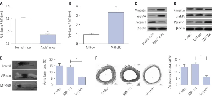
Fig. 1. The effects of miR-590 on atherosclerotic lesion in HFD-induced apoE-/- mice. ApoE-/- mice were fed on a HFD for 12 weeks and injected with miR- 590 or miR-control via tail vein once every 4 weeks after starting HFD. (A) qRT-PCR analysis of miR-590 expression in the aorta derived from apoE-/- mice fed on a HFD or wild-type C57BL/6 J controls fed on a normal diet. U6 was used as the normalization. (B) qRT-PCR analysis of miR-590 expression in HFD- fed apoE-/- mice injected with miR-590 or miR-control. U6 was used as the normalization. (C) Western blot analysis of the protein levels of Pecam-1, α-SMA and Vimentin in apoE-/- mice fed on a HFD or wild-type C57BL/6 J controls fed on a normal diet. (D) Western blot analysis of the protein levels of Pecam-1, α-SMA and Vimentin HFD-fed apoE-/- mice injected with miR-590 or miR-control. (E) Atherosclerotic plaque formation in the resected aortic si- nuses was assessed by Evans blue staining. (F) The atherosclerotic lesion in aortic sinuses was examined by hematoxylin and eosin staining (×40).
*p<0.05. HFD, high-fat diet; α-SMA, α-smooth muscle actin.
1.5
1.0
0.5
0.0
20 15 10 5 0
20 15 10 5 0 4
3 2 1 0
Control
Control Control Control
Normal mice MiR-con
MiR-con MiR-con MiR-con
ApoE
-/- mice
MiR-590
MiR-590 MiR-590 MiR-590
Normal mice ApoE-/- mice MiR-con MiR-590
*
* *
* Vimentin
α-SMA Pecam-1 β-actin Vimentin
α-SMA Pecam-1 β-actin
Control
MiR-con
MiR-590
Relative miR-590 level Aortic lesion area (%) Aortic sinus lesion area (%)
Relative miR-590 level
A
E
B
F
C D
Luciferase reporter assay
The 3’UTR sequences of TLR4 containing the putative binding sites of miR-590 were synthesized and inserted into pMIR-RE- PORT luciferase reporter plasmids (Promega, Madison, WI, USA) to produce TLR4-WT. A pMIR-REPORT plasmid con- taining TLR4 mRNA 3’UTR with a mutant sequence in the miR- 590 binding sites were synthesized by Suzhou GenePharma Co., Ltd. (Jiangsu, China) and named as TLR4-MUT. HAECs cells were seeded into 96-well plates with 200 μL of culture medium and cotransfected with 50 nM miR-590, anti-miR-590, or matched controls and 100 ng of the luciferase vectors using Lipofectamine 2000 reagent (Invitrogen) according to the man- ufacturer’s protocol. The cells were harvested at a point 48-h posttransfection, and relative luciferase activity was detected using a Dual-Luciferase Reporter Assay System (Promega).
Statistical analysis
In vitro experiments were performed in triplicates. All results are shown as the mean±standard deviation from three inde- pendent experiments. All statistical analyses were performed using SPSS version 11.0 software (SPSS Inc., Chicago, IL, USA) with one-way analysis of variance analysis or Student’s t test.
p<0.05 was considered statistically significant.
RESULTS
MiR-590 overexpression inhibits atherosclerotic lesions in HFD-induced apoE
-/-mice
qRT-PCR analysis initially demonstrated that miR-590 expres- sion was abnormally downregulated in the aorta tissue derived from apoE-/- mice, compared with that in normal mice (Fig. 1A).
Additionally, miR-590 expression was upregulated after miR- 590 injection (Fig. 1B). We found that Pecam-1 levels were re- duced and that α-SMA and Vimentin levels were increased in aortic arches from apoE-/- mice in comparison with the normal mice (Fig. 1C), while these changes were significantly reversed following transfection with miR-590 (Fig. 1D). To assess the effects of miR-590 on the development of atherosclerosis in apoE-/- mice, we evaluated atherosclerotic lesions in aortic si- nuses by Evans blue and H&E staining. As shown in Fig. 1E and F, miR-590 overexpression significantly suppressed atheroscle- rotic plaque formation and reduced atherosclerotic lesion area in HFD-induced apoE-/- mice in comparison with the miR-con- trol group, suggesting that forced expression of miR-590 at- tenuated atherosclerotic lesion in HFD-induced apoE-/- mice.
1.5
1.0
0.5
0.0
3
2
1
0
Control MiR-con MiR-590
Anti-miR-conAnti-miR-590 ox-LDL
ox-LDL ox-LDL ox-LDL
0 h 24 h 48 h 72 h 0 h 24 h 48 h 72 h
Cleaved Caspase-3
MiR-con Anti-miR-con MiR-590 Anti-miR-590
Cleaved PARP
*
*
* *
*
*
*
Relative miR-590 expression Relative miR-590 expression
A B C
2.0 1.5 1.0 0.5 0.0
1.5
1.0
0.5
0.0
MTT (OD 490 nm) MTT (OD 490 nm)
MiR-con MiR-590
Anti-miR-con Anti-miR-590
PI
Annexin V-FITC MiR-con MiR-590
Anti-miR-con Anti-miR-590
40 30 20 10 0 MiR-conMiR-590
Anti-miR-conAnti-miR-590 ox-LDL
ox-LDL ox-LDL
* *
*
*
*
*
Apoptosis rate (%)
D
MiR-con MiR-590
Anti-miR-conAnti-miR-590 ox-LDL Cleaved PARP
Cleaved Caspase-3
GAPDH E
1.5
1.0
0.5
0.0
Relative protein expression
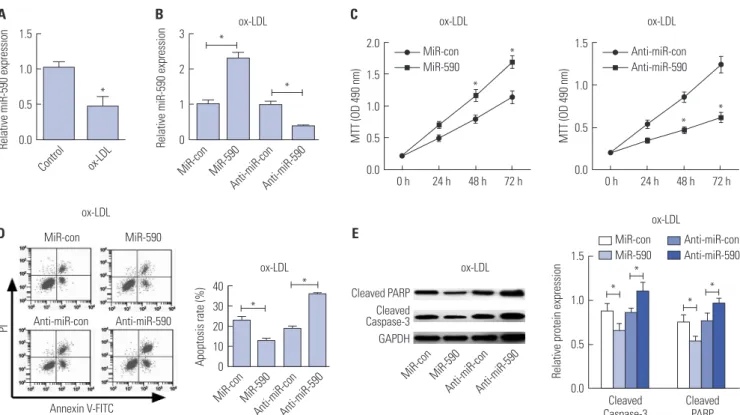
Fig. 2. The effects of miR-590 on the proliferation and apoptosis in ox-LDL-treated HAECs. (A) qRT-PCR analysis of miR-590 expression in HAECs following ox-LDL challenge. U6 was used as the normalization. (B) qRT-PCR analysis of miR-590 expression in HAECs transfected with miR-590, anti-miR-590, or matched controls, followed by ox-LDL treatment. U6 was used as the normalization. (C) MTT assay was performed to evaluate cell proliferation at 24, 48, and 72 h in HAECs transfected with miR-590, anti-miR-590, or matched controls, followed by ox-LDL stimulation. (D) Flow cytometry analysis was con- ducted to determine the percentage of HAEC apoptosis after transfection with miR-590, anti-miR-590, or matched controls, followed by ox-LDL adminis- tration. (E) Western blot was employed to detect the expression levels of Cleaved PARP and Cleaved-Caspase-3 in HAECs transfected with miR-590, anti- miR-590, or matched controls, followed by ox-LDL stimulation. *p<0.05. ox-LDL, oxidized low-density lipoprotein; HAECs, human aortic endothelial cells.
MiR-590 overexpression promotes proliferation and inhibits apoptosis in ox-LDL-treated HAECs
We initially analyzed the expression of miR-590 in HAECs un- der the condition of ox-LDL administration. qRT-PCR analysis demonstrated that miR-590 expression was significantly de- creased in ox-LDL-treated HAECs, compared with control cells (Fig. 2A). To further evaluate the effects of miR-590 on the de- velopment of atherosclerosis in vitro, HAECs were transfected with miR-590, anti-miR-590, or matched controls following ox-LDL stimulation for 24 h. As a result, miR-590 transfection significantly increased the expression of miR-590, while anti- miR-590 transfection significantly decreased the expression of miR-590 in ox-LDL-treated HAECs (Fig. 2B). Subsequently, MTT assay revealed that cell proliferation was significantly fa- cilitated by regained expression of miR-590 in HAECs follow- ing ox-LDL challenge, compared with the miR-con group (Fig.
2C). On the contrary, inhibition of miR-590 by anti-miR-590 led to a significant inhibition of cell proliferation in ox-LDL- treated HAECs (Fig. 2C). Meanwhile, flow cytometry analysis proved that increasing miR-590 expression significantly hin- dered apoptosis of ox-LDL-treated HAECs, while anti-miR-590 triggered the opposite effects (Fig. 2D). Consistent with the flow cytometry analysis, Western blot analysis demonstrated that cleavage of PARP and Caspase-3 was significantly attenuat- ed by miR-590 overexpression, but was enhanced following the transfection of anti-miR-590 in ox-LDL-treated HAECs (Fig. 2E).
These above results indicated that miR-590 overexpression promotes proliferation and inhibits apoptosis in ox-LDL-treated HAECs.
TLR4 is a direct target of miR-590 in ox-LDL-treated HAECs
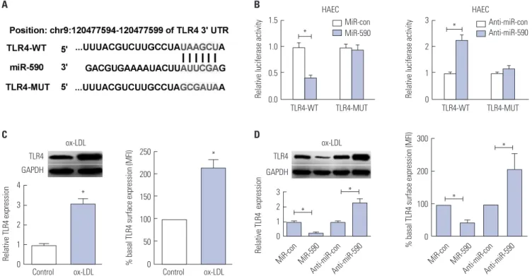
To explore the molecular mechanism by which miR-590 regu- lates the proliferation and apoptosis of ox-LDL-treated HAECs, bioinformatics analyses were employed to predict the poten- tial targets of miR-590. TLR4, which was reported to play a cru- cial role in the development of atherosclerosis,11 was identified as a potential target of miR-590. To verify this prediction, the WT or MUT 3’UTR sequences of TLR4 containing the putative binding sites of miR-590 were inserted into pMIR-REPORT plasmids, as shown in Fig. 3A. The results from luciferase re- porter assay demonstrated that miR-590 overexpression re- sulted in a marked decrease of luciferase activity of the 3’UTR of TLR4, but not that of the TLR4-MUT in HAECs cells (Fig. 3B).
In contrast, anti-miR-590 led to an obvious elevation of lucif- erase activity of TLR4-WT, but had no influence on the lucif- erase activity of TLR4-MUT (Fig. 3B). Additionally, we found that both cellular and cell surface levels of TLR4 protein were significantly increased in ox-LDL-treated HAECs versus their respective controls (Fig. 3C). Moreover, re-expression of miR- 590 notably decreased TLR4 protein levels, and miR-590 inhi- bition distinctly increased TLR4 protein level in ox-LDL-treated
MiR-con MiR-590 MiR-con MiR-590
Anti-miR-conAnti-miR-590 Anti-miR-conAnti-miR-590
TLR4-WT
Control Control
TLR4 GAPDH
TLR4 GAPDH
TLR4-WT MiR-con
MiR-590
Anti-miR-con Anti-miR-590
TLR4-MUT
ox-LDL
ox-LDL ox-LDL
ox-LDL
TLR4-MUT
HAEC HAEC
A B
D C
*
*
* *
*
*
*
1.5 * 1.0
0.5
0.0
4 3 2 1 0
3 2 1 0 250
200 150 100 50 0
300
200
100
0 3
2
1
Relative luciferase activity 0
Relative TLR4 expression Relative TLR4 expression
% basal TLR4 surface expression (MFI) % basal TLR4 surface expression (MFI)Relative luciferase activity
Fig. 3. MiR-590 directly targets TLR4 in ox-LDL-treated HAECs. (A) Bioinformatics analysis of the predicted interaction of miR-590 in the 3’UTR of TLR4. (B) Luciferase activity was determined by luciferase reporter assay in HAECs cells co-transfected with TLR4-WT or TLR4-MUT and miR-590, anti-miR-590, or respective controls. (C) Western blot (left) was performed to detect the cellular protein level of TLR4 in HAECs with or without ox-LDL treatment, while TLR4 protein level on cell surface was evaluated by flow cytometry (right). (D) The cellular protein level of TLR4 in ox-LDL-treated HAECs transfected with miR-590, anti-miR-590, or matched controls was detected by Western blot (left), while TLR4 protein level on cell surface was evaluated by flow cytometry (right). *p<0.05. TLR4, toll-like receptor 4; ox-LDL, oxidized low-density lipoprotein; HAECs, human aortic endothelial cells.
HAECs (Fig. 3D). These results confirmed the authentic bind- ing between miR-590 and TLR4.
MiR-590 overexpression antagonizes the apoptosis- promoting role of TLR4 in ox-LDL-treated HAECs
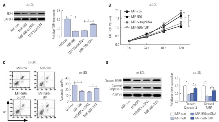
To further address whether miR-590s could attenuate the apop- tosis of ox-LDL induced HAECs by targeting TLR4, HAECs were transfected with TLR4 overexpressing plasmid to restore TLR4 protein levels in miR-590 overexpressing HAECs under ox- LDL treatment. Western blot results indicated that TLR4 pro- tein levels in HAEC under ox-LDL treatment could be signifi- cantly reduced by miR-590 overexpressing, which was largely restored by TLR4 overexpressing plasmid transfection (Fig. 4A).MTT assay results demonstrated that cell proliferation of ox- LDL-treated HAECs was significantly increased by miR-590 overexpression, which was largely abolished by TLR4 restora- tion (Fig. 4B). Furthermore, flow cytometry analysis results showed that miR-590 overexpression significantly attenuated ox-LDL induced apoptosis in HAECs, which was weakened by TLR4 restoration (Fig. 4C). The influence of miR-590 overex- pression and TLR4 restoration on ox-LDL induced HAECs apoptosis was also verified by Western blot analysis of PARP protein cleavage and caspase-3 activation (Fig. 4D). These re- sults indicated that miR-590 could attenuate ox-LDL induced
cell apoptosis in HAECs by targeting TLR4.
MiR-590 overexpression suppresses the TLR4/NF-κB pathway in ox-LDL-treated HAECs
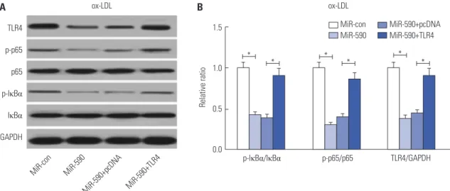
Since the TLR4/NF-κB pathway has attracted great attention due to its crucial role in atherosclerosis,12 we further analyzed the effects of miR-590 on the TLR4/NF-κB pathway. Western blot analyses revealed that miR-590 overexpression specifically suppressed the protein levels of TLR4, p-IκBα/IκBα ratio, and p-p65/p65 ratio, which was partially recuperated by forced expression of TLR4 (Fig. 5A and B) in ox-LDL-treated HAECs, suggesting that ectopic expression of miR-590 inhibited the activation of the TLR4/NF-κB pathway in ox-LDL-treated HAECs.
Inhibition of the TLR4/NF-κB pathway promotes cell proliferation and impedes apoptosis
in ox-LDL-treated HAECs
PDTC, a NF-κB inhibitor, was used to suppress the TLR4/NF- κB pathway. As shown in Fig. 6A and B, PDTC treatment trig- gered a substantial decline of the ratio of p-IκBα/IκBα and p-p65/p65 in ox-LDL-treated HAECs, suggesting the suppres- sion of the TLR4/NF-κB pathway by PDTC. Additionally, PDTC exposure dramatically promoted cell proliferation in ox-LDL- treated HAECs, compared with the control group (Fig. 6C).
TLR4 GAPDH
MiR-con
MiR-con MiR-590
MiR-590 MiR-590+pcDNA
MiR-590+pcDNA MiR-590+TLR4
MiR-590+TLR4
ox-LDL ox-LDL
1.5 1.0 0.5 0.0
2.0 1.5 1.0 0.5 0.0
*
* * *
Relative TLR4 expression MTT (OD 490 nm)
A B
0 h 24 h 48 h 72 h MiR-con
MiR-590 MiR-590+pcDNA MiR-590+TLR4
Cleaved Caspase-3
MiR-con MiR-590+pcDNA MiR-590 MiR-590+TLR4
Cleaved PARP
PI
V-FITC
MiR-con MiR-590
MiR-590+
pcDNA MiR-590+
TLR4
40 30 20 10 0
MiR-con MiR-con
MiR-590 MiR-590
MiR-590+pcDNAMiR-590+TLR4 MiR-590+pcDNAMiR-590+TLR4
ox-LDL ox-LDL
ox-LDL
* * * *
*
*
Apoptosis rate (%)
C
ox-LDL Cleaved PARP
Cleaved Caspase-3
GAPDH D
1.5 1.0 0.5 Relative protein expression 0.0
Fig. 4. Anti-miR-590 reverses TLR4 knockdown-mediated promotion of cell proliferation and suppression of apoptosis in ox-LDL-treated HAECs. HAECs were transfected with si-TLR4, si-con, or cotransfected with si-TLR4 and anti-miR-590 or anti-miR-con, following ox-LDL stimulation. (A) Western blot analysis of TLR4 protein level in the treated HAECs. (B) Cell proliferation at 24, 48, and 72 h in the treated HAECs was evaluated by MTT assay. (C) Flow cy- tometry analysis was performed to detect the apoptotic rates in the treated HAECs. (D) Western blot was applied to analyze the protein levels of Cleaved PARP and Cleaved Caspase-3 in the treated HAECs. *p<0.05. ox-LDL, oxidized low-density lipoprotein; TLR4, toll-like receptor 4; HAECs, human aortic en- dothelial cells.
Moreover, PDTC treatment exerted a significant inhibitory role in apoptosis in ox-LDL-treated HAECs relative to the con- trol group (Fig. 6D), as demonstrated by flow cytometry anal- ysis. Furthermore, PDTC also greatly diminished the protein levels of Cleaved PARP and Cleaved Caspase-3 in ox-LDL-treat- ed HAECs with respect to the control group (Fig. 6E). Together, these results demonstrated that inhibition of the TLR4/NF-κB pathway facilitated cell proliferation and restrained apoptosis in ox-LDL-treated HAECs.
DISCUSSION
Currently, substantive studies have revealed that the progres- sion of atherosclerosis is a complex, multiple-step process.13 EC dysfunction, which may be affected by EC proliferation and apoptosis, is regarded as a major contributor to the pathogen- esis of atherosclerosis.14 Up to now, the precise mechanism underlying the pathogenesis of atherosclerosis still remains largely unknown. In the present study, we demonstrated the anti-apoptotic effects of miR-590 in ox-LDL-treated HAECs.
Fig. 5. The effects of miR-590 on the TLR4/NF-κB pathway in ox-LDL-treated HAECs. (A) Western blot analysis of TLR4, p-IκBα, IκBα, p-p65, and p65 in HAECs after transfection with miR-590, miR-con, miR-590+TLR4, or miR-590+pcDNA, following ox-LDL challenge. (B) Quantification analysis of the protein level of TLR4, p-IκBα/IκBα ratio and p-p65/p65 ratio in the treated HAECs. *p<0.05. ox-LDL, oxidized low-density lipoprotein; TLR4, toll-like receptor 4; NF- κB, nuclear factor kappa B; HAECs, human aortic endothelial cells.
A B
MiR-con MiR-590
MiR-590+pcDNA MiR-590+TLR4
ox-LDL ox-LDL
TLR4 p-p65 p65 p-IκBα IκBα GAPDH
p-IκBα/IκBα p-p65/p65 TLR4/GAPDH
* * * * *
* 1.5
1.0
0.5
0.0
Relative ratio
MiR-con MiR-590+pcDNA MiR-590 MiR-590+TLR4
Fig. 6. TInhibition of the TLR4/NF-κB pathway facilitates cell proliferation and restrained apoptosis in ox-LDL-treated HAECs. HAECs were exposed to 50 μM PDTC for 24 h, followed by treatment with 100 μg/mL ox-LDL for 24 h. (A) The protein levels of p-p65, p65, p-IκBα and IκBα in the treated HAECs were detected by Western blot. (B) Quantification analysis of the ratio of p-IκBα/IκBα and p-p65/p65 in the treated HAECs. (C) MTT assay was performed to as- sess cell proliferation at 24, 48, and 72 h in the treated HAECs. (D) Flow cytometry analysis was conducted to examine the apoptosis of treated HAECs. (E) Western blot was employed to determine the protein levels of Cleaved PARP and Cleaved Caspase-3 in the treated HAECs. *p<0.05. ox-LDL, oxidized low- density lipoprotein; TLR4, toll-like receptor 4; NF-κB, nuclear factor kappa B; HAECs, human aortic endothelial cells.
A B C
Control
Control Control
p-IκBα/IκBα Control PDTC
Control PDTC p-p65/p65
PDTC
PDTC PDTC
ox-LDL ox-LDL ox-LDL
p-p65 p65 p-IκBα IκBα GAPDH
1.5
1.0
0.5
0.0
2.0 1.5 1.0 0.5 0.0
* *
Relative ratio MTT (OD 490 nm)
0 h 24 h 48 h 72 h
*
* Control
PDTC
PI
Control PDTC 30
20
10
0
1.5
1.0
0.5
0.0
ox-LDL ox-LDL
ox-LDL
*
* *
Apoptosis rate (%) Relative protein expression
D ox-LDL
Cleaved PARP Cleaved Caspase-3
GAPDH E
V-FITC Cleaved Caspase-3 Cleaved PARP
Mechanistically, the anti-atherosclerotic effect of miR-590 was mediated through inhibition of the TLR4/NF-κB pathway.
Therefore, miR-590 may be a novel target for the prevention and treatment of atherosclerosis.
Recently, miRNAs are attracting more attention due to their important regulatory roles in the progression of atherosclero- sis.15 For example, miR-98 was reported to promote the prolif- eration and alleviate apoptosis of human umbilical vein ECs (HUVECs) exposed to ox-LDL.16 Additionally, it was demon- strated that miR-126 alleviated ox-LDL-induced HUVECs in- jury through restoring autophagy flux via repressing the PI3K/
Akt/mTOR pathway.17 Moreover, it was revealed that downreg- ulation of miR-497 suppresses ox-LDL-induced lipid accumu- lation in THP-1 macrophages via targeting of apelin and thus represented a potential therapeutic target for atherosclerosis.18 Of note, it was previously demonstrated that miR-590 is down- regulated in Angiotensin (Ang) II-induced HUVECs and that overexpression of miR-590-5p attenuates Ang II-induced EC apoptosis and decreases reactive oxygen species (ROS) gener- ation by downregulating low-density lipoprotein receptor 1 (LOX-1).19 Moreover, miR-590 agomir could attenuate lipid ac- cumulation and proinflammatory cytokine secretion and could prevent atherosclerosis in apoE-/- mice.20,21 In accordance with these previous studies, we provided evidence indicating that miR-590 is downregulated in the aorta tissues from HFD-fed apoE-/- mice and ox-LDL-treated HAECs. Specifically, exoge- nous overexpression of miR-590 attenuated atherosclerotic lesions in HFD-fed apoE-/- mice, preserved cell proliferation, and suppressed apoptosis in ox-LDL-treated HAECs, suggest- ing that miR-590 exhibits anti-atherosclerotic activity.
TLRs are evolutionarily preserved pattern recognition re- ceptors and have recently attracted increasing attention due to its link between innate immunity, inflammation, and athero- sclerosis.22,23 As the most widely studied receptor in the TLR family, TLR4 is abundantly expressed in atherosclerotic lesions at the different stages of atherosclerosis, including macrophages and ECs within the lesion.24 It was reported that TLR4 deficiency could attenuate aortic atherosclerotic lesion areas and inflam- matory cytokine levels in apoE-/- mice, suggesting a crucial role of TLR4 in the pathogenesis of atherosclerosis.25 NF-κB is an important downstream mediator of the TLR4 signaling path- way.26 TLR4 can induce activation of NF-κB linked to the tran- scription of the expressions of many proinflammatory-related genes, resulting in increased endothelial injury and lipid depo- sition and thereby contributing to the formation and develop- ment of atherosclerosis.27 A previous study demonstrated that the TLR4/NF-κB pathway contributes to chronic unpredict- able mild stress-induced atherosclerosis through activation of proinflammatory cytokines in apoE-/- mice.28 In the present study, we deemed that TLR4 is a direct target of miR-590 and that miR-590 negatively regulates TLR4 expression in ox-LDL- treated HAECs. TLR4 was found to be upregulated in ox-LDL- treated HAECs. Rescue experiments demonstrated that anti-
miR-590 reversed TLR4 knockdown-mediated promotion of cell proliferation and suppression of apoptosis in ox-LDL-treat- ed HAECs, suggesting that miR-590 regulates the proliferation and apoptosis of ox-LDL-treated HAECs by targeting TLR4.
Mechanistically, we further discovered that miR-590 inhibits activation of the TLR4/NF-κB pathway in ox-LDL-treated HAECs and that inhibition of the TLR4/NF-κB pathway by PDTC promotes cell proliferation and impedes apoptosis in ox-LDL-treated HAECs, indicating that miR-590 rescues pro- liferation and blocks ox-LDL-induced apoptosis in HAECs via inactivation of the TLR4/NF-κB pathway. It was previously re- ported that the mechanism underlying the anti-apoptotic ef- fects of miR-590 in ox-LDL-treated ECs was involved, in part, in the LOX-1-ROS-p38MAPK-NF-κB signaling cascade and p53-Bcl-2/Bax-caspase-3 signaling pathway.10 Antagonizing the TLR-4/NF-κB signaling pathway has been well demonstrat- ed to ameliorate atherosclerosis.29-31 A possible ligand for TLR- 4 that is involved in atherosclerosis development is high-mo- bility group protein 1 (HMGB1), which is often released by apoptotic cells, leaked from necrotic/necroptotic cells, or se- creted by inflammatory signal-stimulated cells into extracel- lular space. The pro-atherogenic role of HMGB1 has been summarized by Zhou, et al.32 in a short letter. Given that ox- LDL could induce HMGB1 release from in vitro cultured ECs, we speculated that HMGB1 must be involved in ox-LDL-in- duced EC apoptosis in vitro and atherogenesis in vivo in such a miR-590/TLR-4-regulated way. This hypothesis will be in- vestigated in our future work.
TLR4 expression was also found to be regulated by other miRNA in atherosclerotic cells, such as miR-20a33 in ECs and miR-181a, miR-223 and miR-155 in macrophages.34-36 Besides ApoE knockout mice, several other mouse models are available to establish atherosclerosis models in vivo, such as low-density lipoprotein receptor (LDL-r) knockout mouse.37 In this re- search, we only used ApoE knockout mice for the in vivo ex- periments, although we speculated that using LDL-r knockout mice should yield similar results. The major cause of athero- genesis in ApoE knockout mice and LDL-r knockout mice is hy- percholesterolemia; the difference is that this pathologic con- dition in ApoE knockout mice is developed spontaneously, while LDL-r knockout mice need excess cholesterol intake from diet to develop hypercholesterolemia and atherosclerosis.
Never the less, it might worth to verify whether miR-590 and TLR4 play similar roles in atherogenesis in LDL-r knockout mice as what we have revealed in ApoE knockout mice.
In summary, the present study demonstrated that miR-590 is downregulated in the aorta tissues from HFD-fed apoE-/- mice and ox-LDL-treated HAECs. Moreover, miR-590 overex- pression attenuated atherosclerotic lesion in HFD-fed apoE-/- mice and promoted proliferation and blocked ox-LDL-induced apoptosis in HAECs through inhibition of the TLR4/NF-κB pathway, providing new insights into the molecular mecha- nism underlying the pathogenesis of atherosclerosis. There-
fore, miR-590 may serve as a potential therapeutic target for atherosclerosis when it is overexpressed in ECs.
ACKNOWLEDGEMENTS
This study was supported by the Key Science and Technology Project of Henan Provincial Science and Technology Depart- ment (162102310205).
AUTHOR CONTRIBUTIONS
Conceptualization: Lei Yang, Chuanyu Gao. Data curation: Lei Yang.
Formal analysis: Chuanyu Gao. Funding acquisition: Lei Yang. Inves- tigation: Lei Yang. Methodology: Chuanyu Gao. Project administration:
Chuanyu Gao. Resources: Lei Yang. Software: Chuanyu Gao. Supervi- sion: Lei Yang. Validation: Lei Yang. Visualization: Chuanyu Gao.
Writing—original draft: Chuanyu Gao, Lei Yang. Writing—review &
editing: Chuanyu Gao, Lei Yang.
ORCID iDs
Lei Yang https://orcid.org/0000-0002-9891-6126 Chuanyu Gao https://orcid.org/0000-0002-5362-3573
REFERENCES
1. Rafieian-Kopaei M, Setorki M, Doudi M, Baradaran A, Nasri H.
Atherosclerosis: process, indicators, risk factors and new hopes.
Int J Prev Med 2014;5:927-46.
2. Tabas I, García-Cardeña G, Owens GK. Recent insights into the cellular biology of atherosclerosis. J Cell Biol 2015;209:13-22.
3. Trpkovic A, Resanovic I, Stanimirovic J, Radak D, Mousa SA, Ce- nic-Milosevic D, et al. Oxidized low-density lipoprotein as a bio- marker of cardiovascular diseases. Crit Rev Clin Lab Sci 2015;52:
70-85.
4. Najafpour Boushehri S, Yusof RM, Nasir Mohammad Taib M, Mirzaei K, Yazdekhasti N, Akbarzadeh S. Effect of vitamin supple- mentation on serum oxidized low-density lipoprotein levels in male subjects with cardiovascular disease risk factors. Iran J Basic Med Sci 2012;15:958-64.
5. Bartel DP. MicroRNAs: target recognition and regulatory func- tions. Cell 2009;136:215-33.
6. Natarelli L, Schober A. MicroRNAs and the response to injury in atherosclerosis. Hamostaseologie 2015;35:142-50.
7. Shan Z, Yao C, Li ZL, Teng Y, Li W, Wang JS, et al. Differentially ex- pressed microRNAs at different stages of atherosclerosis in ApoE- deficient mice. Chin Med J (Engl) 2013;126:515-20.
8. Menghini R, Stöhr R, Federici M. MicroRNAs in vascular aging and atherosclerosis. Ageing Res Rev 2014;17:68-78.
9. Eulalio A, Mano M, Dal Ferro M, Zentilin L, Sinagra G, Zacchigna S, et al. Functional screening identifies miRNAs inducing cardiac regeneration. Nature 2012;492:376-81.
10. Bao MH, Li JM, Zhou QL, Li GY, Zeng J, Zhao J, et al. Effects of miR-590 on oxLDL-induced endothelial cell apoptosis: roles of p53 and NF-κB. Mol Med Rep 2016;13:867-73.
11. den Dekker WK, Cheng C, Pasterkamp G, Duckers HJ. Toll like re- ceptor 4 in atherosclerosis and plaque destabilization. Athero- sclerosis 2010;209:314-20.
12. Xing S, Zheng F, Zhang W, Wang D, Xing Q. Relationship between
toll-like receptor 4 levels in aorta and severity of atherosclerosis. J Int Med Res 2014;42:958-65.
13. Corbi G, Bianco A, Turchiarelli V, Cellurale M, Fatica F, Daniele A, et al. Potential mechanisms linking atherosclerosis and increased cardiovascular risk in COPD: focus on Sirtuins. Int J Mol Sci 2013;
14:12696-713.
14. Gimbrone MA Jr, Topper JN, Nagel T, Anderson KR, Garcia- Cardeña G. Endothelial dysfunction, hemodynamic forces, and atherogenesis. Ann N Y Acad Sci 2000;902:230-9.
15. Sun X, Belkin N, Feinberg MW. Endothelial microRNAs and ath- erosclerosis. Curr Atheroscler Rep 2013;15:372.
16. Chen Z, Wang M, He Q, Li Z, Zhao Y, Wang W, et al. MicroRNA-98 rescues proliferation and alleviates ox-LDL-induced apoptosis in HUVECs by targeting LOX-1. Exp Ther Med 2017;13:1702-10.
17. Tang F, Yang TL. MicroRNA-126 alleviates endothelial cells injury in atherosclerosis by restoring autophagic flux via inhibiting of PI3K/Akt/mTOR pathway. Biochem Biophys Res Commun 2018;
495:1482-9.
18. Cui J, Ren Z, Zou W, Jiang Y. miR-497 accelerates oxidized low- density lipoprotein-induced lipid accumulation in macrophages by repressing the expression of apelin. Cell Biol Int 2017;41:1012-9.
19. Luo P, Zhang WF, Qian ZX, Xiao LF, Wang H, Zhu TT, et al. MiR- 590-5p-meidated LOX-1 upregulation promotes Angiotensin II- induced endothelial cell apoptosis. Biochem Biophys Res Com- mun 2016;471:402-8.
20. He PP, OuYang XP, Li Y, Lv YC, Wang ZB, Yao F, et al. MicroRNA-590 inhibits lipoprotein lipase expression and prevents atherosclero- sis in apoE knockout mice. PLoS One 2015;10:e0138788.
21. He PP, Ouyang XP, Tang YY, Liao L, Wang ZB, Lv YC, et al. MicroR- NA-590 attenuates lipid accumulation and pro-inflammatory cy- tokine secretion by targeting lipoprotein lipase gene in human THP- 1 macrophages. Biochimie 2014;106:81-90.
22. Thompson MR, Kaminski JJ, Kurt-Jones EA, Fitzgerald KA. Pat- tern recognition receptors and the innate immune response to vi- ral infection. Viruses 2011;3:920-40.
23. Yang K, Zhang XJ, Cao LJ, Liu XH, Liu ZH, Wang XQ, et al. Toll-like receptor 4 mediates inflammatory cytokine secretion in smooth muscle cells induced by oxidized low-density lipoprotein. PLoS One 2014;9:e95935.
24. Stoll LL, Denning GM, Li WG, Rice JB, Harrelson AL, Romig SA, et al. Regulation of endotoxin-induced proinflammatory activation in human coronary artery cells: expression of functional membrane- bound CD14 by human coronary artery smooth muscle cells. J Immunol 2004;173:1336-43.
25. Michelsen KS, Wong MH, Shah PK, Zhang W, Yano J, Doherty TM, et al. Lack of Toll-like receptor 4 or myeloid differentiation fac- tor 88 reduces atherosclerosis and alters plaque phenotype in mice deficient in apolipoprotein E. Proc Natl Acad Sci U S A 2004;
101:10679-84.
26. Pasterkamp G, Van Keulen JK, De Kleijn DP. Role of Toll-like recep- tor 4 in the initiation and progression of atherosclerotic disease.
Eur J Clin Invest 2004;34:328-34.
27. Baker RG, Hayden MS, Ghosh S. NF-κB, inflammation, and meta- bolic disease. Cell Metab 2011;13:11-22.
28. Tang YL, Jiang JH, Wang S, Liu Z, Tang XQ, Peng J, et al. TLR4/NF- κB signaling contributes to chronic unpredictable mild stress-in- duced atherosclerosis in ApoE-/- mice. PLoS One 2015;10:e0123685.
29. Hu ZP, Fang XL, Fang N, Wang XB, Qian HY, Cao Z, et al. Melato- nin ameliorates vascular endothelial dysfunction, inflammation, and atherosclerosis by suppressing the TLR4/NF-κB system in high-fat-fed rabbits. J Pineal Res 2013;55:388-98.
30. Lu Z, Zhang X, Li Y, Jin J, Huang Y. TLR4 antagonist reduces early- stage atherosclerosis in diabetic apolipoprotein E-deficient mice.
J Endocrinol 2013;216:61-71.
31. Lu Z, Zhang X, Li Y, Lopes-Virella MF, Huang Y. TLR4 antagonist attenuates atherogenesis in LDL receptor-deficient mice with di- et-induced type 2 diabetes. Immunobiology 2015;220:1246-54.
32. Zhou Q, Zhu Z, Hu X, Shu C. HMGB1: a critical mediator for oxi- dized-low density lipoproteins induced atherosclerosis. Int J Car- diol 2016;202:956-7.
33. Chen M, Li W, Zhang Y, Yang J. MicroRNA-20a protects human aortic endothelial cells from Ox-LDL-induced inflammation through targeting TLR4 and TXNIP signaling. Biomed Pharmaco- ther 2018;103:191-7.
34. Du F, Yu F, Wang Y, Hui Y, Carnevale K, Fu M, et al. MicroRNA-155
deficiency results in decreased macrophage inflammation and attenuated atherogenesis in apolipoprotein E-deficient mice. Ar- terioscler Thromb Vasc Biol 2014;34:759-67.
35. Wang J, Bai X, Song Q, Fan F, Hu Z, Cheng G, et al. miR-223 inhib- its lipid deposition and inflammation by suppressing toll-like recep- tor 4 signaling in macrophages. Int J Mol Sci 2015;16:24965-82.
36. Du XJ, Lu JM, Sha Y. MiR-181a inhibits vascular inflammation in- duced by ox-LDL via targeting TLR4 in human macrophages. J Cell Physiol 2018;233:6996-7003.
37. Kapourchali FR, Surendiran G, Chen L, Uitz E, Bahadori B, Moghadasian MH. Animal models of atherosclerosis. World J Clin Cases 2014;2:126-32.