Vol. 9, No. 4, Dec., 1998
자궁 경부 편평 상피종양에서 세포증식관련항원 Ki- 67과 p53 및 p21w af1/ cip1 표출에 관한 연구
순천향대학교 의과대학 천안병원 산부인과학교실, 병리학교실*
배동한・장경택・이종국ㆍ최규연・김민관・김창진*
=Abstract=
E x pr es s ion s of t h e ce ll pr olif er a t ion A g K i- 67, p 53 a n d p21 w a f 1/ c ip1 in u t e r in e c er v ica l s qu a m ou s t u m or
D on g Ha n Ba e, M.D., Kyu n g Ta ck J a n g, M.D ., J on g Ku k Lee , M.D ., Kyu Yeon Ch oi, M.D ., Min Kwa n Kim , M.D ., Ch a n g J in Kim , M.D .*
D epartm ent of Obstetrics and Gynecolog y, d ep artm ent of P atholog y, colle g e of m ed icine, S oonchunhyang university, Chunan, K orea
Objective : To evaluate the expressions of Ki-67, p53 and p21 waf1/cip1 according to the
age and the histologic type of preinvasive and invasive cervical lesions.Material and method
: Microwave-oven-processed formalin-fixed, paraffin-embedded, cervical biopsy specimens and hysterectomy specimens were obtained from 1997 to 1998 at the Soonchunhyang university Chunan hospital. These included 55 cervical intraepithelial neoplasm(CIN I-III), 14 invasive squamous cell carcinoma and 3 adenocarcinoma, and immunohistochemically evaluated by monoclonal MIB-1 antibody, monoclonal p53 antibody, and monoclonal p21 antibody. Positive index was expressed as a percentage of strong staining cells per 300 counted cells in evenly strong staining area.Result : Ki-67, p53 and p21 protein were expressed in the nuclei. Ki-67 was specifically
expressed in all phases of cell cycles in proliferating cells. p21 expression was not seen in CIN I and CIN II, but was increased with increasing histologic grade. According to the age, Ki-67 expression was significantly higher in 30≥aged group than 30≤aged group, but p53 expresson was not significantly different according to age groups, and p21 expression was significantly lower in 50≤aged group than 30≥aged group.Conclusion : Conclusions of this study indicate that cell proliferating rate is higher in
young age groups than old age groups. p53 expression was not significantly different according to age and histologic grade. These indicates that mutation of the p53 gene may be associated with the development of cervical cancer, but not associated with the progression of cervical cancer. Besides, p21 expression was increased in increasing histologic grade, but decreased in old aged women. Further study of this paradoxical increase in p21 expression in cervical carcinoma is necessary to clarify the mechanisms of p53 indepent pathway.Ke y wo rd s : Ki-67, p53, p21 waf1/cip1, cervical cancer, p53 independent pathway
I. 서 론
자궁경부암의 발생은 여러 가지 과정을 거쳐서 발생돠는 것으로 생각되며, 자궁 상피내 종양, 편평 상피내 병변 또는 이형성증으로 언급되어지는 전침 윤성 변화(preinvasive changes)와 관련되어 있다1).
이형성증은 자궁경부 침윤암에서와 마찬가지로 인 유두종 바이러스(Human papilloma virus) 감염과 밀 접한 관련이 있다2).
자궁경부암과 연관된 고 위험군의 인유두종 바이 러스는 2개의 종양단백, E6와 E7으로 암호화되어 있고, 종양억제유전인자 p53, pRB와 결합한다3,4).
p53과 결합한 E6은 직접적으로 ubiquitin–mediated degradation을 통해5), pRB와 상호작용을 한 E7은 잠 재적 p53의 하향조절 효과자(Down stream effectors) 로 작용하여 DNA 손상이 발생한 후 G1 arrest를 막 는다. 자궁경부암에서 p53의 변이는 아주 드물게 나 타나며, p53불활성화가 암발생에 관련이 있으며, p53의 불활성화는 이 두가지 mechanism에 의한 것 같다. 정상적인 wild type p53은 세포 증식에 관여하 여 중요한 역할을 하며, DNA손상에 반응하여 손상 된 세포의 apoptosis에도 관여한다. 세포증식에서 p53의 세포증식 억제는 wild type p53에 의해 활성화 되는 cyclin dependent kinase inhibitor인 p21waf1/cip1에 의해 중재된다. p53에 대한 많은 연구들이 이루어 졌으나, p21waf1/cp1에 대한 연구는 미진한 상태이다.
세포 증식에 관련되는 여러 가지 인자들이 국소 적으로 침윤된 자궁경부암 환자에서 예후를 평가하 는 또 다른 지표로 연구되고 있으며 그 중 Ki-67 항 원을 증식지표(proliferative index)로 scoring 하는 데 이용하고 있다6,7,8).
본 연구에서는 자궁경부상피내 종양, 침윤성 편평상피암, 자궁경부선암에서 p53, p21waf1/cip1 및 세포증식 관련 항원 (proliferation associated Ag)인 Ki-67의 발 현에 대해 알아보고자 한다.
Ⅱ. 연구 대상 및 방법
본 연구는 1997년 7월부터 1998년 6월까지 순천향대학교 의과대학 천안병원 산부인과에 서 자궁경부 생검 또는 자궁적출술로 얻어진 자궁 경부 상피내종양(I,Ⅱ,Ⅲ) 55예, 침윤성 편평 상피암 14예, 자궁경부 선암 3예를 대상으로 하였다(Table
1).
채취한 조직을 10% buffered formalin에 24시간 고 정후 paraffin block을 만들고, 이를 6um로 절편을 만 들어 통상적인 과정을 거쳐 hematoxylin-eosin염색을 하여 조직을 판독하였다.
(1) MIB- 1, p53 및 p21 waf1/ cip1 의 면역조직화학적 염색
6 m의 paraffin 절편을 탈 파라핀후 graded alcohol 로 재함수 시켰다. 조직내의 항원을 폭로시키기 위 하여 조직을 0.1M citrate buffer(pH6.0)에 담근 후 800W의 microwave oven에 5분씩 3회 처리하였다.
그 후 실온에서 서서히 식힌 후 증류수로 3회 수세 하였다. 그 후 조직내의 내인성 peroxidase를 차단하 기 위하여 3% H2O2(in methanol)용액에 실온에서 3 분간 작용시켰다. 그후 증류수로 3분씩 3회 세척후 PBS(phosphate buffered saline : pH 7.4) 용액에 다시 세척하였다. 비특이적 조직 반응을 차단하기 위하 여 정상 rabbit 혈청에 실온에서 30분 반응 시킨 후, 조직에 있는 여분의 혈청을 제거 한 후 MIB-1(Imm- unotech, Merseille, France), p53 (BioGenex, USA) 및 p21(Santacruz, CA, USA) 단일클론 항체에 30분간 실온에서 반응 시켰다. 그 후 PBS로 실온에서 5분씩 3회 수세 후 biotin이 결합된 goat antimouse imm- unoblobulin(Immunotech, Merseille, France) 에 역시 실온에서 30분간 작용 시켰다. 그후 PBS로 역시 실 온에서 5분씩 3회 수세 후 strepavidin이 결합된 pero-
Ta ble 1. Characteris tics of ma teria ls Age
distribution CIN I CIN II CIN III Invasive
squamous ca. Adenoca. Total
20-29 - 2 1 1 1 5
30-39 3 6 7 2 - 18
40-49 4 4 11 3 1 23
50-59 3 2 10 5 1 21
60-69 1 - 1 3 - 5
Total 11 14 30 14 3 72
xidase(Immunotech, Merseille, France) 에 실온에서 30 분간 반응시켰다. 이를 다시 PBS로 수세 후 diaminobenzidine(DAB)로 발색한 후 hematoxylin으로 대조 염색하였고, 이를 다시 탈수 과정과 투명과정 을 거쳐 balsam에 봉입하여 광학 현미경으로 관찰 하였다.
(2) 양성 반응 세포의 scoring 및 통계학적 검정
MIB-1, p53 및 p21 waf1/cip1 들은 핵단백질로 핵이 진한 갈색상을 보인 세포를 양성으로 판독하였다.
균일하게 강양성을 보인 부위에서 300개의 세포를 세어 이중 강양성인 세포의 수를 백분율로 환산하 여 양성지표(positive index : PI)를 표시하였다. 이들 의 통계학적 검정은 PI수치를 이용하여 student t-test 로 p값을 구하였다.
Ⅲ. 결 과
1. Ki- 67, p53 및 p21 wa f1/cip1 의 면역 조직 화학적 표출
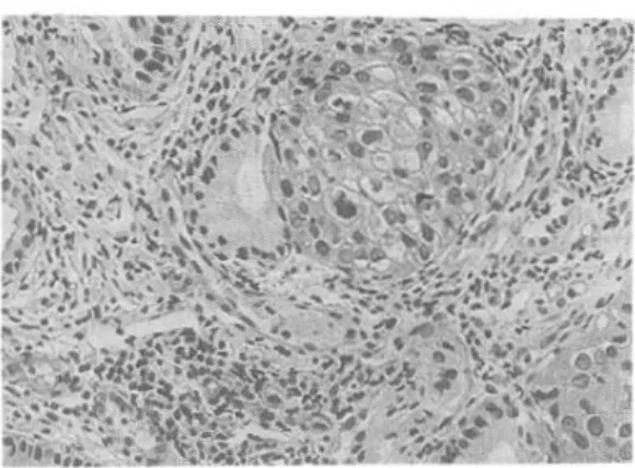
Ki-67, p53 및 p21 단백질들은 세포핵 내에서 특 이적으로 표출되었다. Ki-67은 특이적으로 세포 분 열이 진행되고 있는 세포에 표출되었으며, 염색체 분열상은 보이지 않으나, 분열주기에 있는 세포에 서도 표출되었다. p21 자궁경부 상피내종양 I 과 II 에서는 표출되지 않았으나 자궁경부 상피내 종양 III, 자궁경부 상피내암, 침윤성 편평상피암 및 자궁 경부 선암에서는 조직학적 등급이 높아질수록 이의 표출도 증가하였다.(Fig 1~6)
2. 연령에 따른 Ki- 67, p53 및 p21 wa f1/cip1의 발현
연령에 따른 Ki-67, p53 및 p21 의 발현은 분열기 세포를 표지하는 Ki-67인 경우 30세 이하의 연령군 에서는 positive index가 97±11.37로 30세 이상의 연 령군에 비하여 매우 높았다(P<0.005). 그러나 p53 의 발현은 각 연령군 사이에 특이한 통계적 차이가 없 었다. 반면, p21 waf1/cip1의 발현은 30세 이하의 연령 군에 비하여 50세 이상의 연령군에서 현저히 낮은 발현율을 보였다(P<0.005)(Table 2).
Fig 1. The s qua mous cell ca rcinoma in s itu with gla ndula r involve ment s hows s trong pos itive re action for Ki- 67(Pe rxida s e , DAB, ×200).
Fig 2. The s qua mous cell ca rcinoma in s itu with gla ndula r involve ment s hows pos itive re a ction for p53(P eroxida s e , DAB, ×200)
Fig 3. The s qua mous cell ca rcinoma in s itu with
gla ns ular involveme nt s hows pos itive rea ction for
p21waf1/cip1(pe roxida s e, DAB, ×200).
Table 2. Expres s ion of Ki- 67, p53 a nd p21waf1/cip1 a ccording to the age by pos itive Inde x
Age(years) Ki-67 p53 p21
20-29 97±11.37* 29±7.91 34.67±3.31 30-39 27.35±5.39 17.48±3.40 4.17±0.17 40-49 29.32±3.32 20.21±7.35 15.65±5.33 50-59 45.07±9.61 27.79±6.65 6.71±1.33 60-69 36.15±4.64 25.00±8.68 5.38±0.17
3. 조직학적 등급에 따른 Ki- 67, p53 및 p21 의 발현
Ki-67은 조직학적 등급이 낮은 자궁경부 상피내 종양 I에서는 9±1.76으로 자궁경부 상피내 종양 II 이상의 조직학적 등급과 비교할 때 통계적으로 유 의성 있게 낮은 발현율을 보였다.(P<0.005)
p53의 발현은 자궁경부 상피내 종양 I, II, III 및 침윤성 편평상피암에서 각각 12±1.98, 8.46±0.25, 12.07±4.26, 10.87±2.41으로 통계학적으로 유의성 이 없었다. 그러나 자궁경부 선암의 예에서는 22.34
±4.95으로 자궁경부 상피내 종양 I과 비교할 때 높 게 나타났으나 통계학적 유의성은 없었다.
p21 waf1/cip1의 발현은 자궁경부 상피내 종양 I과 II 에서는 발현이 관찰되지 않았으나, 자궁경부 상피 내 종양 III, 침윤성 편평상피암 및 자궁경부 선암에 서 각각 8.51±2.46, 24.75±6.57 및 25.75±14.75의 positive index를 보여 조직학적 등급이 높아질수록 발현율의 증가를 보였다
(Table 3).
Table 3. Expres s ions of Ki- 67, p53 a nd p21 a ccording to the his tologic types by pos itive Inde x
Histologic
diagnosis Ki-67 p53 p21
CIN I 9±1.76 12±1.98 -
CIN II 31.33±8.79 8.46±0.25 -
CIN III 31.57±8.34 12.07±4.26 8.51±2.46 Invasive ca. 36.88±12.56 10.87±2.41 24.75±6.57
Adeno ca. 34.33±9.59 22.34±4.95 25±14.75
* mean ± standard deviation
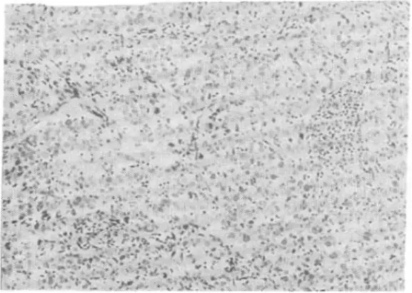
Fig 4. Invas ive s qa mous cell ca rcinoma ,
non- ke ra tinizing type , s hows s trong pos itive re action for Ki- 67, in s ome of the nuclei of the cells (Pe roxida s e, DAB, ×200).
Fig 5. Invas ive s qa mous cell ca rcinoma ,
non- ke ra tinizing type , s hows s trong pos itive re action for p53, in s ome of the nucle i of the cells (Pe roxida s e, DAB, ×200).
Fig 4. Invas ive s qa mous cell ca rcinoma ,
non- ke ra tinizing type , s hows s trong pos itive
re action for p21waf1/cip1, in s ome of the nucle i of
the ce lls (Pe roxida s e , DAB, ×200).
Ⅳ. 고 찰
p53 유전자는 염색체 17p13.1 위치에 존재하며 종양억제 역할을 하는 것으로 알려져 있으며9) 다른 세포 주기 조절 유전자의 전사(transcription)를 자극 하여 세포 증식을 조절하는데 관여한다10). 정상적인 wild type p53을 가진 세포는 비정상적인 DNA가 복 구되는 동안 세포주기 진행 중 G1기부터 S기까지의 진행을 지연시킬 수 있다. 그러나 불활성화 또는 돌 연변이 된 p53은 비정상적인 DNA의 복제를 방지하 지 못하여 암 발생을 가능케한다. p53 유전자의 변 이가 암 발생에 관여하는 흔한 단일 유전자 변이이 며11,12,13), 대부분의 변이는 p53을 안정화 및 축적시 키는 point mutation이다14). p53의 불활성화는 다음 3 가지 기전으로 발생한다고 추정되며 1) somatic point mutation15), 2) loss of heterozygosity16), 3) 인유 두종 바이러스 감염 등이다. 그러나 고위험군의 인 유두종 바이러스가 발견되는 자궁경부암에서는 E6 종양단백이 p53과 결합하여 ubiquitin mediated degradation을 통해서 불활성화되며 E7종양단백은 pRB와의 상호 작용으로 잠재적 억제 효과자 (suppresor effector)로 작용하여 p53을 불활성화 시키 는 것으로 추정된다. 그러므로, 인유두종 바이러스 를 포함하지 않는 세포들은 돌연변이 된 p53 유전 자를 갖게되어 p53 발현이 높은 반면 고위험군의 인 유두종 바이러스를 포함하고 있는 자궁경부암 세포 에서는 p53 단백 발현이 낮게 나타난다22). 자궁경부 암에서 p53 유전자의 변이는 0-8% 정도 매우 드문 일이라는 보고가 많으며17-21), Kurvien등은 전침윤 자 궁경부 병변(preinvasive cervical lesion)에서 아무런 p53 변이를 발견하지 못했다고 보고하였다17). 많은 면역조직화학적분석에 의한 연구에서 p53 표출율은 전침윤 병변에서는 0-83%, 침윤성암에서는 4-100%
로 큰 변이도를 보였는데23-28), 이는 사용한 항체, 고 정액, 면역조직화학분석방법 및 서로 다른 양성 역 가의 적용에 의한 것으로 보인다. 자궁경부암에서 는 다른 암에서와 다르게 진한 양성반응을 보이는 핵 염색이 드물다. 이와같이 자궁경부 이형성증이 나 침윤암에서 p53의 표출이 낮은 이유는 wild type p53이 아주 짧은 반감기를 갖기 때문이며8,29), 인유 두종 바이러스의 E6 종양단백의 발현이 p53 파괴를
가속화시키는 것으로 보인다.
분화의정도 또는 혈관 침습 등과 같은 조직학적 소견이 자궁경부암에서는 예후 인자로 인식되어 왔 으나 젊은 연령의 환자군에서는 반드시 일치하지 않을 수도 있다30,31,32). 젊은 연령군의 환자에서 침윤 성 자궁경부암의 발생빈도와 사망률이 증가하고 있 는 추세이나 생존율에 나이가 어떤 영향을 미치는 지는 논란이 많다33,34,35). 일부 보고에서는 국소적으 로 침윤된 젊은 연령의 환자군에서 5년 생존율이 50-70%로서 나쁜 예후를 보인다고 하며34,35,36), 또 다 른 보고에서는 연령에 따른 예후는 차이가 없다고 하였다32,37,38). 세포증식과 관련된 Ki-67 항원의 표출 에 대한 연구에서는 증식하고 있는 세포의 핵 내에 서 non-histone protein과 반응하는 항체인 MIB-1 단 일 클론 항체를 이용하여 면역조직화학적방법으로 연구할 결과 MIB-1 발현 정도와 종양의 성격 사이 에는 관련이 있다고 하였으며, 이는 MIB-1 index가 결합 조직질환, 임파 증-식 질환 및 자궁경부암을 포함한 일부 암에서 유용한 예후 인자의 하나가 될 수 있다는 것을 암시해준다고 하였다39,47). 젊은 연령 의 환자군에서는 높은 연령의 환자군에 비해서 더 높은 세포증식 지표(proliferative index)를 보이고 임 파절 전이도 더 흔히 발생하는 것으로 보아 세포증 식항원 Ki-67의 표출 정도가 생물학적 공격성 (biologic aggressiveness)을 암시해 준다고 하였다48).
cyclin-CDK inhibitory protein의 발견은 세포증식 을 조절하는 과정에서 새로운 물질로 등장했으며, DNA 손상에 대한 세포 반응, 분화, contact inhibiti- on, 노쇠, 성장억제신호에 대한 반응, 종양화의 억제 등 여러 생리적 과정에 관여한다고 한다. 이 CDK inhibitor는 cyclin-CDK 복합체의 kinase 활성을 억제 하여 세포주기 transition에 필요한 인산화(phospho- rylation)를 방해한다. 이들 negative regulatory protein 중 첫 번째로 확인된 것이 cyclin-CDK 복합체와 DNA 복제 억제 물질인 p21waf1/cip1 단백질이다49-53).
DNA 손상이 있은 후 p53 dependent p21 induction이 발생하여 세포주기를 정지시키나 p53과 무관한 p21induction 역시 세포 분화기에서 세포분열을 억제하 는 것으로 알려졌다54,55,56). 종양 억제 유전자 p53에 의한 직접적인 전사 유도(transcriptional induction) 외 에도 wild type p53 없이 여러 가지 물질에 의한 p21 induction이 보고되고 있으며, 이들 물질로서는
PDGF, FGF, okadaic acid, butyric acid, retionic acid, vitamine D3, TPA, G-CSF, IL-6, INF-γ 및 TGF-β 등이 있다57-62). 또 DNA 손상 후 wild type p53에 의 한 p21 활성화 외에도 MAP kinase signal transduction pathway 활성화에 의한 p53 independent p21 inducton 기전도 발표되었다63,64). p53 의존성 p21waf1/cip1 의 상 호 연관 표출이 유방암, astrocytoma, olfactory neuroblastoma, 갑상선암, 난소암, 대장암 등에서 나 타나기는 하지만 p53과는 무관하게 독럽적으로 p21waf1/cip1의 표출이 폐암, 췌장암, 일부 난소암, 대장 암 등에서 발견되었다65-71). 이는 종양의 종류에 따 라서 p21waf1/cip1의 활성화 기전이 다를 수 있다는 것 을 예측케 한다. p53의 불활성화로 p53의 발현이 감 소함에도 불구하고 편평세포암에서 p21waf1/cip1가 역 설적으로 증가(paradoxical increase)되는 현상은 말 단 분화 세포(terminally differentiated cell)의 존재에 기인한 것 같다72). 종양은 많은 세포군 즉, 증식 세 포군과 말단 분화 세포군을 갖고있기 때문에 증식 세포군과 말단 분화 세포군 모두가 증가하며 이 사 실은 종양에서 p21waf1/cip1과 Ki-67 항원의 상호 독립 적인 표출을 설명하고 있다. 대장암은 말단 분화 세 포군이 거의 없으므로 p21waf1/cip1의 표출이 적은 반 면 유방암에서는 말단 분화 세포가 많으므로 p21waf1/cip1 표출이 증가된다고 보고하였다65-71,73,74,75).
그러나, 현재까지 종양에서 p21waf1/cip1의 표출증가에 대한 설명이 만족스럽지는 못하므로 이 현상을 설 명하는데 더 많은 연구가 필요할 것으로 사료된다.
V. 결 론
1997년 7월부터 1998년 6월까지 순천향대학교 의 과대학 천안병원 산부인과에서 자궁경부 생검 또는 자궁적출술로 얻어진 자궁경부 상피내종양(I,II,III) 55예, 침윤성 편평 상피암 14예, 자궁경부 선암 3예 를 대상으로 연령, 조직학적 등급에 따른 면역조직 화학방법을 이용한 Ki-67과, p53 및 p21waf1/cip1 표출 에 대한 연구에서 다음과 같은 결과를 얻었다.
1. Ki-67은 특이적으로 세포분열이 진행되고있는 세
포에 표출되었으며, 세포분열상이 보이지 않더라 도 세포분열주기에 있는 세포에서도 표출되었다.
연령에 따라서는 30세 이하의 연령군에서는 positive index가 97±11.37로 30세 이상의 연령군 에 비하여 매우 높았다(p〈0.005). 또한 조직학적 등급에 따가서는 등급이 낮은 자궁경부 상피내 종양 I에서는 9±1.76으로 자궁경부 상피내 종양 II 이상의 등급에 비해 통계적으로 유의성 있게 낮은 발현율을 보였다(p<0.005).
2. 연령군에 따른 p53의 발현은 각 연령군 사이에 특이한 통계적 차이가 없었으며 조직학적 등급에 따라서도 자궁경부 상피내 종양 I, II, III 및 침윤 성 편평상피암에서 각각 12±1.98, 8.46±0.25, 12.07±4.26, 10.87±2.41으로 통계학적 유의성이 없었다.
3. p21waf1/cip1는 연령에 따라서는 30세 이하의 연령군 에 비하여 50세 이상의 연령군에서 현저히 낮은 발현율을 보였으며 조직학적 등급에 따라서는 자 궁경부 상피내종양 I과 II에서는 표출되지 않았으 나, 자궁경부 상피내 종양 III, 침윤성 편평상피암 및 자궁경부 선암에서 각각 8.51±2.46, 24.75±
6.57, 25.75±14.75의 positive index를 보여 조직학 적 등급이 높아질수록 발현율이 증가하였다.
결론적으로 세포증식항원 ki-67의 표출이 30세 이하의 연령군에서 30세 이상의 연령군에 비해 높 다는 것은 젊은 여성에서 자궁경부암의 예후가 더 불량할 것이라고 예측하게 하며 p53의 표출이 연령 및 조직학적 등급에 따라 유의한 차이가 없다는 것 은 p53의 변이가 자궁경부암의 발생과는 관련이 있 으나 암의 진행과는 관련이 없다는 것을 지적하고 있다. 또한 p21waf1/cip1의 표출이 p53의 발현과 무관하 게 조직학적 등급이 높아질수록 증가한다는 현상, 즉 p21waf1/cip1의 역설적 증가(paradoxical increase)는 말단 분화 세포 증식 또는 노화 세포의 증가에 의한 것으로 설명하고 있으나 앞으로 이 p21waf1/cip1의 역 설적증가에 대하여 더 많은 논의와 연구가 필요할 것이다.
-Reference-
1. Kurman RJ, Norris HJ, Wiikinson E. Tumors of the cervix. In: Rosai J, Sobin LH. editors. Tumors of the cervix, vagina, and vulva. Atlas of tumor pathology. 3rd series. Washington. D.C.: Armed Forces Institute of Pathology. 1992: 44.
2. Park T-W. Fufiwara H. Wright TC. Molecular biology of cervical cancer and its precursors. Cancer 1995;76:1902-13.
3. Kessis TD, Siebos RJ, Nelson WG, Kastan MB Plunkett BS. Han SM.
et al. Human papiilomavirus 16 E6 expression dirupts the p53-mediated cellular response to DNA damage. Prc. Natl. Acad. Sci USA 1993;90:3988-92.
4. Siebos JC, Lee MH, Plunkett BS, Kessis TD, Williams BO, Jacks T, Hedrick L, Kastan MB. Cho KR p53-dependent G1 arrest involves pRB-related proteins and is disrupted by the human papillomavirus 16 E, oncoprotein. Proc. Natl Acad SCiUSA 1994;91:5320-4.
5. Scheffener M, Werness BA, Hurtbregtse JM, Levine AJ. : Howley PM.
The E6 oncoprotein encoded by hyman papillomavirus type 16 and 18 promotes the degradation of p53. Cell 1990;63:1129-36.
6. Waldman T, Kinzler KW, Volgetein B. p21 is necessary for the p53-mediated G1 arrest in human cancer cells. Cancer Res 1995;55:5187-90.
7. Levine AJ, Chang A. Dittmer D, Notterman DA, Silver A,Thorn K, et al.
The p53 tumor suppressor gene. J Lab Clin Med 1994;123:817-23.
8. Iggo R, Gatter K, Bartek J, Lane D, Harris A. Increased expression of mutant forms of p53 oncogene in primary lung cancer. Lancet 1990;335:675-9.
9. Levine AJ, Momand J, Finlay CA. The p53 tumour suppressor gene.
Nature 1991;351:453-6.
10. Finlay CA, Hinds PN, Levine AJ. The p53 proto-oncogene can act as a suppressor of transformation. Cell 1989;57: 1083-93.
11. Hollstein M, Sidrensky D, Vogelstein B, Harris CC. p53 mutations in human cancers. Science 1991;253:49-53.
12. Bartek F, Barkova J, Vojtestek B, Stakove Z, Lukas J, Rejthar A, et al.
Aberrant expression of the p53 oncoprotein is a common feature of a wide spectrum of human malignancies. Oncogene 1991;6:1699-703.
13. Levine AJ, Perry ME, Chang A, Silver A, Dittmer D, Wu M, et al. The 1993 Walter Hubert Lecture: the role of the p53 tumour suppressor gene in tumorigenesis. Br J Cancer 1994;69:409-16.
14. Scheffner M, Munger K, Byrne JC, Howley PM. The state of the p53 and retinoblastoma genes in human cervical carcinoma cell lines. Proc Nat Acad Sci USA 1991;88:5523-7.
15. Crook T, Wrede D, Tidy JA, Mason WP, Evans DJ, Vousden KH.
Clonal p53 mutations in primary cervcal cancer: association with human papillomavirus negative tumours. Lacet 1992;339:1070-3.
16. Park SY, Kang YS, Kim BG, Lee SH, Lee ED, Lee KH, et al. Loss of heterozygosity on the short arm of chromosome 17 in uterine cervical carcinoma. Cancer Genet Cytogenet 1995; 79:74-8.
17. Kurvinen K, Tervagauta A, Syrjanen S, Chang F, Syrjnen K. The state of the p53 gene in human papillomavirus(HPV) positive and HPV-negative genital precancer lesions and carcinomas as determined by single-strand conformation polymorphism analysis and sequencing.
Anticancer Res. 1994;14:177-81.
18. Busby-Earle RM, Steel CM, Williams AR, Cohen B, Bird CC. p53 mutations in cervical carcinogenesis:low frequency and lack of cerrelation with human papillomavirus status. Br J Cancer 1994;69:732-7.
19. Helland A, Holm R, Kristensen G, Kaern J, Karlsen F, Trope C, et al.
Genetic alterations of the TP53 gene, p53 protein expression, and HPV infection in primary cervical carcinomas. J Pathol 1993;171:105-14.
20. Chen TM, Chen CA, Hsieh CY, Chang DY, Chen YH, Defendi V. The state of p53 in primary human cervical caircinomas and its effects in human papillomavirus-immortalized human cervical cells. Oncogene 1993;8:1511-8.
21. Tsuda H, Hirohashi S. Frequent occurrence of p53 gene mutations in uterine cancers at advanced clinical stage and with aggressive histological phenotypes. Jpn J Cancer Res 1992;83:1184-91.
22. Scheffner M, Munger K, Byrne JC, Howley PM. The state of the p53 and retinoblastoma genes in human cervical carcinoma cell lines. Proc Nat Acad Sci USA 1991;88:5523-7.
23. Mittal KR, Lin O, Chan W, Goswami S, Demopoulos RI. Cervical squamous dysplasias and carcinomas with immunodetectable p53 frequently contain HPV. Gynecol Oncol 1995;58:289-94.
24. Kainz C, Kohlberg P, Gitsch G, Sliutz G, Breitenecker G. Mutant p53 in patients with invasive cervical cancer stages IB to IIB. Gynecol Oncol 1995;57:212-4.
25. Akasofu M, Oda Y. Immunohistochemical detection of p53 in cervical epithelial lesions with or without infection of human papillomavirus types 16 and 18. Virch Archiv 1995; 425:593-602.
26. Vecchione A, Cermele G, Giovagnoli MR, Valli C, Alomandi M, Crico E, et al. p53 Ezpression and genetic evidence for vital infection in intraepithelial neoplasia of the uterine cervix. Gynecol Oncol 1994;55:343-8.
27. Tervahauta AI, Syrjanen SM, Mantyjarvi R, Syrjnen KJ. Detection of p53 protein and Ki-67 proliferation antigen in human papillomavirus (HPV)-positive and HPV-negative cervical lesions by immunohistochemical double-staining. Cytopathology 1994;5:282-93.
28. Inoue M, Fujita M, Enomoto T, Mormoto H, Monden T,Shimano T.
Immunohistochemical analysis of p53 in gynecologic tumors. Am J Clin Pathol 1994;172:13-8.
29. Finlay CA, Hinds PW, Tan T-H, Dliyahu D, Oren M, Levine AJ.
Activating mutations for transformation by p53 produce a gene product that forms an hsc-70-p53 complex with an altered half-life. Mol Cell Biol 1988;8:531-9.
30. Thomassen LV, Warshaw J, Lawhead RA, Unger ER:Invasive cervical cancer in young women. J Reprod Med 37:901-6, 1992
31. Serur E, Fruchter RG, Maiman M, McGuire F, Arrastia CD, Gibbon D:Age, substance abuse, and survical of patients with cervical carcinoma.
Cancer 75:2530-38, 199533. Devesa SS, Young JL, Brinton LA, Fraumen JF: Recent trends in cervical uteri cancer. Cancer 64:2184-90, 1989 32. Crowther ME: Is the nature of cervical carcinoma changing in young
women? Obstet Gynecol Surv 50:71-82. 1995
33. Devesa SS, Young JL, Brinton LA, Fraumen JF: Recent trends in cervical uteri cancer. Cancer 64:2184-90, 1989
34. Free K, Roberts S, Bourne R, Dickie G, Ward B, Wright G, Hill B:
Cancer of the cervix-Old and young. now and then. Gynecol Oncol 43:129-36, 1991
35. Saint Paul MT, Bremond A, Rochet Y: Cervical carcinoma before 35 years: epidemiologic and prognostic aspects. A retrospective study of 46 cases of cervix carcinoma before 35 years, of 449 new cases with carcinoma of cervix FIGO stage IA2-IV. J Gynecol Obstet Biol Reprod 22:737-42, 1993
36. Pfeiffer D, Schubert Frischle F, Meier W, Scheidel P, Hepp H:
Age-dependent defferences and changes in the clinical pattern and course of cervical carcinoma since 1973. Geburtshlfe Frauenheikd 52:81-7, 1992 37. Russell JM, Blair V, Hunter RD: Cervical carcinoma: prognosis in young
pateints. Br Med J 295:300-3, 1987
38. Poka R, Juhansz B, Lampe L: Cervical cancer in young women: a poorer prognosis? Int J Gynecol Obstet 46:33-7, 1994
39. Gerdes J, Schwab U, Lemake H, Stein: Production of a mouse monoclonal antibody reactive with a human nuclear antigen associated with cell proliferation. Int J Cancer 31:13-20, 1983
40. Grogan TM, Lippman SM, Spider CM, Slymen DJ, Rybski JA, Rangel CS, Richter LC, Miller TP: Independent prognostic significance of a nuclear proliferation antigen in diffuse large cell lymphomas as determined by the mooclocal antibody Ki 67. Blood 71:1157-60, 1988 41. Hall PA Richard MA, Gregory WM, d' ardenne AJ, Lister T, Stensfeld AG:The prognostic value of Ki67 immunostaining in non-Hodgkin' s lymphoma. J pathol 154:223-35,1988
42. Lelle RJ, Heidenreich W, Stauch Gerdes J: The correlation of growth fractions with histologic grading and lymph node status in human mammary carcinoma. Cancer 59:83-8. 1987
43. Weiss LM, Strickler JG, Medeiros LJ, Gerdes J, Stein H, Warnkle RA:
Proliferative rates of non-Hodgkin' s lymphomas as assessed by Ki-67 antibody. Hum Pathol 18:1155-9, 1987
44. Ueda T, Aozasa K, Tsujimoto M: Prognostic significance of Ki-67 reactivity in soft tissue sarcoma. Cancer 63:1607-11, 1989
45. Robertson MJ, Ritz J: Biology and clinical relevance of human natural killer cells. Blood 12:2421-38, 1990
46. Garzetti GG, Ciavattini A, Goteri G, De Nictolis M, Stramazzotti D, Lucarini G, Biagini G: Ki67 immunostaining (MIB 1 monoclonal antibody) in serous ovarin tumors: index of proliferative activity with prognostic significance. Gynecol Oncol 56:169-74, 1995
47. Garzetti GG, Ciavattini A, Lucarini G, Goteri G, De Nictolis M, Muzzioli M, Fabris N, Romanini C, Biagini G: MIB 1 immunostaining in stage I squamous cervical carcinoma: relationship with nature killer cell activity. Gynecol Oncol 58:28-33, 1995
48. G Gioele G, Andrea C, Guendalina L, Gaia G, Michele DN, Graziella B: MIB1 immunostaing in Cervical Carcinoma of Young Patients.
Gynecol Oncol 67:184-7, 1997
49. Xiong Y, Hannon GJ, Zhang H, Casso D, Kobayashi R and Beach D.
(1993). Nature, 366, 704-7.
50. Harper JW, Adami GR, Wei N, Keyomarsi K and Elledge SJ.(1993).
Cell, 75,805-6.
51. E1-Deiry WS, Harper JW, O' Connor PM, Velculescu VE, Canman CE,.
Jackman J, Pietenpol JA, Burrell M, Hill DE, Wang YS, Wiman KS, Mercer WE, Kastan MB, Kohn KW, Elledge SJ, Kinzler KW and Vogelstein B. (1994). Cancer Res., 54,1169-74.
52. Waga S, Hannon GJ, Beach D and Stillman B. (1994). Nature, 369, 574-8.
53. Dulic V, Kaufmann WK, Wilson SJ, T1sty TD, Lees Harper JW, Elledge SJ and Reed SI. (1994). Cell. 76, 1013-24.
54. Parker SB, Eichele G, Zhang P, Rawls A, Sands AT, Bradley A, Olson EN, Harper JW and Elledge SJ. (1995). Science, 267, 1024-7.
55. Zhang W, Grasso L, McClain CD, Gambel AM, Cha Y, Travali S, Deisseroth AB and Mercer WE. (1995). Cancer Res., 55, 668-74.
56. E1-Deiry WS, Tokino T, Waldman T, Oliner JD, Velculescu VE, Burrell M, Hill DE, Hearly E, Rees JL, Hamilton SR, Kinzler KW and Vogelstein B. (1995). Cancer Res., 55, 2910-9.
57. Michieli P, Chedid M, Lin D, Pierce JH, Mercer WE and Givol D.(1994). Cancer Res., 54,3391-5.
58. Steinman RA, Hoffman B, Iro A, Guillouf C, Lieberman DA and E1-Houseini ME. (1994). Oncogene, 9, 3389-96.
59. Sheikh MS, Li XS, Chen JC, Shao ZM, Ordonez JV and Fontana JA.
(1994). Oncogene, 9, 3407-15.
60. Jiang H, Lin J, Su Z, Collart FR, Huberman E and Fischer PB.(1994).
Oncogene, 9,3397-406.
61. Elbendary A, Berchuck A, Davis P, Havrilesky L, Bast Jr RC, Iglehart D and Marks JR. (1994). Cell Growth and Diff., 5,1301-7.
62. Datto MB, Li Y, Panus JF, Howe DJ, Xiong Y and Wang XF. (1995).
Proc. Natl. acad. Sci. USA, 92, 5524-49.
63. Missero C, Clautti E, Eckner R, chin J, Tsai LH, Livingston DM.
Involvement of the cell-cycle inhibitor cip1/waf1 and the E1A-associated p300 protein in terminal differentiation. Proc Natl Acad Sci U S A 1995;92:5451-5.
64. Zhnag W, Grasso L, McClain CD, Gambel AM, Cha Y, Travali S, et al. P53-independent induction of waf1/cip1 in human leukemia cells is correlated with growth arrest accompanying monocyte/macrophage differentiation. Cancer Res 1995;55:668-74.
65. el-Deiry WS, Tokino T, Waldman T. Oliner JD. Velculescu VE. Burrell M. et al. Topological control of p21 waf1/cip1 expression in normal and n대plastic tissues. Cancer Res 1995,53:2910-9.
66. Barbareschi M, Caffo O, Doglioni C,. Fina P, Marchetti A. Buttitta F, et al. p21 waf1 immunohistochemical expression in breast carcinoma:
correlations with clinicopathological data. cestrogen receptor status, and relapse-free survival. Br J Cancer 1996:74:208-15.
67. ZedeniusJ, Larsson C, Wallin G, Backdahl M<Aspenblad U. Hoog A, et al.. Alterations of p53 and expression of waf1/p21 in human thyroid tumors. Thyroid 1996;6:1-9.
68. Ozcelik H, Mousses S, Andrulis IL. Low levels of expression of an inhibitor of cyclin-dependent kinase(cip1/waf1) in primary breast carcinomas with p53 mutations. Clin Cancer Res 1995;1:907-12.
69. Jung JM, Bruner JM, Ruan S, Langford LA. Kyritsis AP, Kobayashi T, et al. Increased levels of p21 waf1/cip1 in human brain tumors.
Oncogene 1995;11:2021-8.
70. Papadaki H, Kounelis S, Kapadia SB, Bakker A, Swalsky PA, Finkelstein SD. Relationship of p53 gene alterations with tumor progression and recurrence in olfactory neuroblastoma. Am J Surg Pathol 1996;20:715-21.
71. Elbebdery AA, Crisano FD, Evans AL Ji, Davis PL, Iglehart JD, Marks JR. et al. Relationship between p21 expression and mutation of the p53 tumor suppressor gene in normal and malignant ovarian epithelial cell.
Clin Cancer Res 1996;2:1571-5.
72. Pierce GB, Wallace C. Differentiation of malignant to benign cells.
Cancer Res 1971;31:127-34.
73. Marchetti A, Doglioni C, Barbareschi M, Buttita F.Pellegrini S. Bertacca G. et. at. p21 RNA and protein expression in non-small cell lung carcinoma: evidence of p53-independent expression and association with tumoral differentiation. Oncogene 1996;12:1319-24.
74. DiGiuseppe JA, F\Redston MS, Yeo CJ, Kern SE, Hruban RH.
p53-independent expression of the cyclin-dependent kinase inhibitor p21 in pancreatic carcinoma. Am. J. Pathol 1995;147:884-8.
75. Werness BA. fobe JS. DiCioccio RA. Piver MS. Expression of p21 waf1/cip1 in malignant ovarian epithelial tumors and correlation with p53 and Ki-67 immunohistochemistry. Int. J Gynecol pathol 1997;16:149-53.