INTRODUCTION
The S100 protein family consists of over 20 family members of low molecular weight calcium binding proteins [1]. These proteins share common amino acid sequence domains and a functional EF-hand motif that plays a key role in calcium binding through a helix-loop-helix topology [2,3].
Hornerin is a member of the S100-fused protein family. It was first identified in the mouse embryo epidermis. It was also detected in the skin, tongue, esophagus, and proximal stomach of adult mouse tissues. Hornerin has a calcium bind- ing EF-hand structure at the N-terminus followed by a spacer
sequence and a large repetitive domain [4]. The hornerin pro- tein is similar to profilaggrin (which is involved in cornifica- tion of keratinocytes) in its structural features, expression pro- files, extensive posttranslational proteolytic processing, and tissue localization [5,6]. Hornerin has been thought to be in- volved in pre- and post-natal mammary development in breast tissue [7]. Although the physiological role of S100 pro- teins has not been clearly identified, recent experimental data have suggested that they are involved in physiological and pathological processes such as gene transcription, inflamma- tory and immune responses, regulation of protein phosphoryl- ation, transcription factors, antimicrobial responses, calcium homeostasis, dynamics of cytoskeleton constituents and, cell proliferation, differentiation, and death [8]. Differential ex- pression of the S100 family members in certain cancers has been reported [1]. Experiments focusing on their role in mammary carcinogenesis have demonstrated that the expres- sion levels of some S100 family proteins in basal-type breast cancers are higher than that in nonbasal types breast cancers [8,9]. Upregulated expression of hornerin in less aggressive breast cancers has been reported [8-10]. S100A8 and S100A9 are known to be associated with high grade and basal-type
Hornerin Is Involved in Breast Cancer Progression
Jinhyuk Choi*, Dong-Il Kim1,*, Jinkyoung Kim, Baek-Hui Kim, Aeree Kim
Department of Pathology, Korea University Guro Hospital, Seoul; 1Green Cross Reference Laboratory, Yongin, Korea ORIGINAL ARTICLE
Purpose: The S100 gene family, which comprises over 20 mem- bers, including S100A1, S100A2, S100A8, S100A9, profilaggrin, and hornerin encodes low molecular weight calcium-binding proteins with physiological and pathological roles in keratiniza- tion. Recent studies have suggested a link between S100 pro- teins and human cancer progression. The purpose of the present study was to determine the expression levels of hornerin, S100A8, and S100A9 and evaluate their roles in the progression of invasive ductal carcinoma (IDC). Methods: Seventy cases of ductal carcinoma in situ (DCIS), IDC, and metastatic carcinoma in lymph nodes (MCN) were included. Tissue microarrays were constructed from lesions of DCIS, IDC, and MCN from the same patients. Expression of hornerin, S100A8, and S100A9 was anal- yzed using immunohistochemistry. Results: The expression of hornerin was associated with the estrogen receptor-negative (p=0.003) and the human epidermal growth factor receptor
2-positive (p=0.002) groups. The expression of S100A8 was as- sociated with a higher pT stage (p=0.017). A significant (p<0.001) correlation between the expression of S100A9 and S100A8 was also found. The mean percentages of hornerin- positive tumor cells in DCIS, IDC, and MCN were 1.0%±3.3%
(mean ±standard deviation), 12.0% ±24.0%, and 75.3% ± 27.6%, respectively. The expression of hornerin significantly (p<0.001) increased with the progression of carcinoma. The mean levels of S100A8 and S100A9 in DCIS, IDC, and MCN were not significantly (p>0.050) different. The expression of hornerin increased in a stepwise manner (DCIS<IDC<MCN).
Conclusion: Our data suggest that hornerin is involved in breast cancer progression and malignant transformation from preinva- sive lesions.
Key Words: Breast neoplasms, Hornerin protein, S100 proteins
Correspondence to: Aeree Kim
Department of Pathology, Korea University Guro Hospital, 148 Gurodong-ro, Guro-gu, Seoul 08308, Korea
Tel: +82-2-2626-1472, Fax: +82-2-2626-1486 E-mail: [email protected]
*These authors contributed equally to this work.
This research was supported by a grant (A120392) of the Korea Health Industry Development Institute (KHIDI) funded by the Ministry of Health &
Welfare (MOHW), Republic of Korea.
Received: September 23, 2015 Accepted: March 23, 2016
Cancer
breast cancer [8-10]. It is generally accepted that breast can- cers arise by a multistep process in which normal epithelial cells transform into invasive cancer via atypical ductal hyper- plasia and in situ carcinoma, though it is not necessarily a lin- ear process [11,12]. However, the roles of hornerin, S100A8, and S100A9 in the progression of mammary carcinogenesis have not been fully evaluated. Therefore, the objective of this study was to determine their expression levels in ductal carci- noma in situ (DCIS), invasive ductal carcinoma (IDC), and metastatic carcinoma in the same patient to clarify their roles in cancer progression.
METHODS
Tissue specimens
A total of 94 cases of surgically resected IDC at Korea Uni- versity Guro Hospital during 2007 to 2011 were included in this study with approval from the Institutional Review Board of the hospital (IRB number: KUGH 12149). All subjects had invasive carcinoma, adjacent DCIS component, and lymph node metastasis. Hematoxylin and eosin-stained slides for each case were reviewed for tumor subtype, histologic grade, nuclear grade, and lymph node status. The medical records of all subjects were reviewed. Nottingham’s histologic grade and nuclear pleomorphism score were analyzed in this study.
Clinicopathologic information was obtained by reviewing medical records, pathology reports, and hematoxylin and eo- sin-stained sections. The following histopathologic variables were determined in IDCs: tumor subtype, pT stage, pN stage, Nottingham combined histologic grade [13], estrogen recep- tor (ER), and human epidermal growth factor receptor 2 (HER2). Tissue microarrays (TMAs) were constructed using two representative cores (2.0 mm in diameter) of primary IDCs, adjacent DCISs, or metastatic carcinomas from the same case.
Immunohistochemical analysis and silver in situ hybridization Immunohistochemical (IHC) analyses of hornerin, S100A8, and S100A9 were performed using the Bond-Max system (Leica Biosystems, Wetzlar, Germany). Antigens were re- trieved according to the Bond Max ER1 antigen retrieval pro- tocol. Antibodies used in this study included those against hornerin (rabbit polyclonal anti-human antibody, dilution 1/200; Novus Biologicals, Littleton, USA), S100A8 (mouse anti-human antibody, 1/800; Lifespan Bioscience, Seattle, USA), and S100A9 (goat polyclonal anti-human antibody, 1/400; Santa Cruz Biotechnology, Santa Cruz, USA).
The percentage of tumor cells exhibiting intense staining for hornerin, S100A8, and S100A9 were determined in 10 high-
power fields. Cases were considered positive when more than 10.0% of tumor cells were stained or negative when 10.0% or less were stained [14]. As tumor heterogeneity can exist, any expression of protein at more than 10.0% in two TMA cores was interpreted as positive.
IHC analyses of ERs (Ventana Medical Systems, Tucson, USA) and HER2s (Ventana Medical Systems) were performed using the Ventana BenchMark automatic staining system (Ventana Medical Systems). Cancer cells with ER staining in the nucleus were considered immunoreactive and scored. The evaluation of hormone receptor expression was based on the Allred scoring method and the American Society of Clinical Oncology/College of American Pathologists (ASCO/CAP) guidelines [15]. For HER2, membranous staining was also evaluated according to the guidelines of ASCO/CAP. Cases with a score of 3 were considered HER2-positive, whereas those with a score of 2 were evaluated for HER2 gene amplifi- cation according to ASCO/CAP guidelines. Silver in situ hy- bridization (SISH) was performed with a Ventana BenchMark automated instrument (Ventana Medical Systems) according to the manufacturer’s protocols using INFORM HER2DNA probe (Ventana Medical Systems) or chromosome 17 probes (Ventana Medical Systems). These probes were labeled with dinitrophenol (DNP) and visualized using rabbit anti-DNP primary antibody and the ultraView SISH Detection Kit.
Briefly, the HER2 DNA probe was denatured at 95°C for 4 minutes and hybridized at 52°C for 2 hours. The chromosome 17 probe was denatured at 95°C for 4 minutes and hybridized at 44°C for 2 hours. The final reaction was driven by the se- quential addition of silver acetate, hydroquinone, and hydro- gen peroxidase to the peroxidase-conjugated goat anti-rabbit antibody in the detection kit to produce a silver precipitate, which was deposited into the HER2 genes. Red centromeric signals in chromosome 17 were seen as red dots. For SISH test, we defined HER2 positivity as HER2 gene amplification by SISH with a gene copy ratio of HER2:chromosome 17 centromere ≥2.0 as described previously [16].
Statistical analysis
Statistical analyses were performed using the SPSS version 12.0 for Windows (SPSS Inc., Chicago, USA). Pearson chi- square test (or Fisher exact test when appropriate) was used to compare the binary categories of hornerin, S100A8, and S100A9 expression between groups. Paired t-tests were per- formed to determine whether there were significant differences between the mean percentages of hornerin, S100A8, and S100A9 expression in DCIS, IDC, and metastatic carcinoma in lymph node (MCN). McNemar test was used to assess the cor- relation between S100A8 and S100A9. Data were considered
statistically significant when the p-value was less than 0.050.
RESULTS
Demographic and pathologic data (clinicopathologic data) From the 94 cases, the data that we required were available for 70 cases. The median age of the 70 patients was 51 years (range, 33−83 years). Clinicopathologic features are shown in Table 1. According to the seventh edition of the American Joint Committee on Cancer Staging Manual [17], the primary tumor pT1 occurred in 40.0%, pT2 in 55.7%, pT3 in 4.3%, pN1 in 55.7%, pN2 in 34.3%, pN3 in 10.0%, TNM stage 0/IA/
IB in 0.0%, IIA in 22.9%, IIB in 31.4%, IIIA in 35.7%, IIIB in 0.0%, and IIIC in 10.0% of cases. Based on the Nottingham histologic grade, the distribution of grades was as follows:
grade I in 21.4%, grade II in 50.0%, and grade III in 28.6% of cases. Based on the nuclear grade of the primary tumor, the distribution of grades was as follows: grade I in 2.9%, grade II in 70.0%, and grade III in 27.1% of cases.
Expression of hornerin, S100A8, and S100A9 in breast cancer Hornerin, S100A8, and S100A9 were easily detectable in the cytoplasm and focally positive in the nucleus of tumors (Figure 1). Hornerin was positive in 15 of 70 IDCs (21.4%).
S100A8 was positive in 22 of 70 IDCs (31.4%), and S100A9 was positive in 39 of 70 IDCs (55.7%).
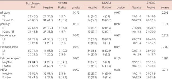
Hornerin positivity in IDC was significantly higher in ER- negative (p=0.003) and HER2-positive groups (p=0.002).
However, hornerin expression in IDC did not show any sig- nificant relationship with stages or grades. Expression of S100A8 was associated with a higher pT stage (p=0.017);
however, it was not correlated with other prognostic factors.
There was no significant relationship between S100A9 expres- sion and prognostic markers (Table 2). A significant (p<
0.001) correlation was found between S100A9 and S100A8 expression (Table 3). The number of S100A9-positive IDC cases was higher than that of S100A8-positive IDC cases. In- terestingly, S100A8 was not expressed in the absence of S100A9 expression. Hornerin showed no significant (p>
0.050) correlation with S100A8 or S100A9 expression (data not shown).
Hornerin expression is increased during breast cancer progression
Next, we investigated whether changes in the expression of hornerin, S100A8, and S100A9 were correlated with can- Table 1. Clinicopathologic features
Variable No. (%)
Age (yr)* 51 (33–83)
T stage
T1 28 (40.0)
T2 39 (55.7)
T3 3 (4.3)
N stage
N1 39 (55.7)
N2 24 (34.3)
N3 7 (10.0)
TNM stage
0/IA/IB 0
IIA 16 (22.6)
IIB 22 (31.4)
IIIA 25 (35.7)
IIIB 0
IIIC 7 (10.0)
Nuclear grade
I 2 (2.9)
II 49 (70.0)
III 19 (27.1)
Histologic grade
I 15 (21.4)
II 35 (50.0)
III 20 (28.6)
*Median (range).
Figure 1. Immunohistochemical staining for hornerin, S100A8, and S100A9 in ductal carcinoma in situ (DCIS), invasive ductal carcinoma (IDC), and metastatic carcinoma in lymph nodes (MCN). Hornerin, S100A8, and S100A9 were easily detectable in the cytoplasm and fo- cally positive in the nucleus of tumor cells. The top, middle, and bottom rows show the expression of hornerin, S100A8, and S100A9, respec- tively. The column on the left, middle, and right shows DCIS, IDC; and MCN, respectively (magnification, ×200).
DCIS IDC MCN
Hornerin
S100A8
S100A9
cer progression. The mean percentages of hornerin-positive tumor cells in DCIS, IDC, and MCN were 1.0% ±3.3%
(mean±standard deviation), 12.0%±24.0%, and 75.3%±
27.6%, respectively. Hornerin expression was significantly (p<0.001) higher in the MCN group compared to that of the DCIS or IDC group (Figure 2). Expression of hornerin ap- peared to increase in a stepwise manner (DCIS <IDC <
MCN). The mean levels of S100A8 and S100A9 expression in DCIS, IDC, and MCN were not significantly (p>0.050) dif- ferent (Figure 2).
DISCUSSION
Previous studies on “fused gene”-type cornified envelop precursor proteins have mainly focused on their physiological and pathologic roles in keratinization [4,6,18,19]. However, recent studies have reported that “fused gene”-type S100 pro- teins may also be involved in the pathogenesis of neoplasia [10,20]. The objective of this study was to determine the ex- pression levels of hornerin, S100A8, and S100A9 in DCIS,
IDC, and metastatic carcinoma in the same patient to clarify their roles in cancer progression. Our results revealed a posi- tive correlation between hornerin expression and ER-negativ- ity with HER2-amplification, in agreement with the results of an in vitro study that used the MCF10A cancer progression model to show that hornerin mRNA expression increased as tumorigenicity progressed [10]. However, in this study, we confirmed our results with IHC staining using formalin fixed paraffin embedded tissue and found that hornerin expression was correlated with the lobular carcinoma or node-negative group. To demonstrate the changes in expression, we specially constructed TMAs from DCIS, IDC, and metastatic lesion in Table 2. Expression of hornerin, S100A8, and S100A9 in invasive breast cancer and their correlation with clinicopathologic prognostic factors
No. of cases (%)
Hornerin S100A8 S100A9
Negative Positive p-value Negative Positive p-value Negative Positive p-value
pT stage 0.373 0.017 0.202
T1 28 (40.0) 24 (34.3) 4 (5.7) 24 (34.3) 4 (5.7) 15 (21.4) 13 (18.6)
T2 and T3 42 (60.0) 31 (44.3) 11 (15.7) 24 (34.3) 18 (25.7) 16 (22.9) 26 (37.1)
pN stage 0.150 0.242 0.071
N1 39 (55.7) 28 (40.0) 11 (15.7) 29 (41.4) 10 (14.3) 21 (30.0) 18 (25.7)
N2 and N3 31 (44.3) 27 (38.6) 4 (5.7) 19 (27.1) 12 (17.1) 10 (14.3) 21 (30.0)
Nuclear grade 0.543 0.987 0.823
I, II 51 (72.9) 41 (58.6) 10 (14.3) 35 (50.0) 16 (22.9) 23 (32.9) 28 (40.0)
III 19 (27.1) 14 (20.0) 5 (7.1) 13 (18.6) 6 (8.6) 8 (11.4) 11 (15.7)
Histologic grade 0.269 0.871 0.939
I, II 50 (71.4) 41 (58.6) 9 (12.9) 34 (48.6) 16 (22.9) 22 (31.4) 28 (40.0)
III 20 (28.6) 14 (20.0) 6 (8.6) 14 (20.0) 6 (8.6) 9 (12.9) 11 (15.7)
Estrogen receptor* 0.003 0.168 0.487
Negative 24 (34.3) 14 (20.0) 10 (14.3) 19 (27.1) 5 (7.1) 12 (17.1) 12 (17.1)
Positive 46 (65.7) 41 (58.6) 5 (7.1) 29 (41.4) 17 (24.3) 19 (27.1) 27 (38.6)
HER2† 0.002 0.336 0.271
Negative 39 (56.7) 36 (51.4) 3 (4.3) 25 (35.7) 14 (20.0) 15 (21.4) 24 (34.3)
Positive 31 (44.3) 19 (27.1) 12 (17.1) 23 (32.9) 8 (11.4) 16 (22.9) 15 (21.4)
HER2=human epidermal growth factor receptor 2.
*Evaluated in invasive ductal carcinoma; †Determined by the guidelines of the American Society of Clinical Oncology/College of American Pathologists.
Table 3. Correlation of S100A8 and S100A9 expression with invasive ductal carcinoma
S100A9 S100A8
p-value Negative Positive
Negative 31 0 <0.001
Positive 17 22
Figure 2. Expression patterns of hornerin, S100A8, and S100A9 in ductal carcinoma in situ (DCIS), invasive ductal carcinoma (IDC), and metastatic carcinoma in lymph nodes (MCN). The expression of horn- erin was increased in a stepwise manner (DCIS<IDC<MCN).
80 70 60 50 40 30 20 10 0
DCIS IDC MCN Site of expression
Mean percentage of positive cells (%)
p<0.001 Hornerin
S100A8 S100A9
p>0.050 p>0.050
lymph nodes of the same patients. IHC analyses of TMAs showed that hornerin expression differed in breast tissue ac- cording to the progression stages in the same patient, includ- ing preinvasive in situ, invasive, and metastatic. In addition, the expression of hornerin dramatically increased in a step- wise manner as breast cancer progressed from DCIS to MCN.
These findings strongly suggest that hornerin is involved in breast cancer progression, specifically in the transformation from preinvasive carcinoma to invasive carcinoma. Further- more, it was remarkable to find that the expression levels of hornerin in IDC were much higher than those in DCIS. Re- cently, it has been proposed that exosomes are involved in the formation of organ-specific metastatic niches [21]. Therefore, as an exosome protein, hornerin may play an essential role in the invasive process of mammary cancer progression, sug- gesting that it might be used as a potential marker to predict recurrence or potential metastasis. In this study, a survival analysis for hornerin in breast cancer was not performed.
Therefore, further study with survival data is necessary.
S100 proteins are low molecular weight proteins ranging in size from 9 to 13 kDa. Most S100 proteins can form hetero- and homo-dimers that might be essential for the generation of their active form [22]. S100A8 and S100A9 proteins usually form a heterodimer called calprotectin that is present in high levels in endothelium, macrophages, and neutrophils [23-25].
Our study showed a significant correlation between S100A8 and S100A9 expression. This finding is in accordance with a previous study [8]. Since S100A8 and S100A9 proteins have been known to exist as heterodimeric molecules [26], it would not be surprising if their expression was coordinately con- trolled. In the present study, a positive correlation between S100A8 expression and higher pT stage was demonstrated.
However, no association between S100A9 and clinicopathol- ogic parameters with prognostic impact was observed. Some studies have indicated that overexpression of S100A8 and S100A9 is associated with poor prognosis of IDC and lung adenocarcinoma [9,27]. Another study has suggested an asso- ciation between hornerin expression and ER-negative or high grade tumors [8].
There are some differences between our study and another study with regard to how protein expression was interpreted [10]. We interpreted it to be positive when we observed more than 10.0% of protein expressed in two TMA cores because any resultant subclone could have a specific biologic behavior due to evolutionary changes among cancer cells. As we used TMA with a unique design, including in situ, invasive, and metastatic lesions from each patient, we only enrolled a small number of cases in this study. Furthermore, we only evaluated 70 of the 94 cases recruited for this study because not all the
cores fulfilled the required data.
In this study, the expression of hornerin steadily increased during breast cancer progression from preinvasive lesions to metastatic carcinoma. In addition, hornerin was expressed at higher levels in ER-negative but HER2-positive groups. These findings suggest that hornerin may be involved in mammary cancer progression. Therefore, it might be used as potential marker for poor prognosis in breast cancer management. Re- cently, it has been proposed that exosome proteins, such as S-100 family members, are involved in the formation premeta- static niches at metastatic sites [21]. The relationship between exosomes and S100 family members in cancer progression is starting to emerge. To clarify the roles of S-100 family mem- bers including hornerin in cancer progression, further studies with a large number of cases along with survival data are needed.
CONFLICT OF INTEREST
The authors declare that they have no competing interests.
REFERENCES
1. Salama I, Malone PS, Mihaimeed F, Jones JL. A review of the S100 pro- teins in cancer. Eur J Surg Oncol 2008;34:357-64.
2. Leclerc E, Heizmann CW. The importance of Ca2+/Zn2+ signaling S100 proteins and RAGE in translational medicine. Front Biosci (Schol Ed) 2011;3:1232-62.
3. Carafoli E. Calcium signaling: a tale for all seasons. Proc Natl Acad Sci U S A 2002;99:1115-22.
4. Makino T, Takaishi M, Morohashi M, Huh NH. Hornerin, a novel pro- filaggrin-like protein and differentiation-specific marker isolated from mouse skin. J Biol Chem 2001;276:47445-52.
5. Henry J, Hsu CY, Haftek M, Nachat R, de Koning HD, Gardinal-Galera I, et al. Hornerin is a component of the epidermal cornified cell enve- lopes. FASEB J 2011;25:1567-76.
6. Wu Z, Meyer-Hoffert U, Reithmayer K, Paus R, Hansmann B, He Y, et al. Highly complex peptide aggregates of the S100 fused-type protein hornerin are present in human skin. J Invest Dermatol 2009;129:1446- 58.
7. Hovey RC, Trott JF, Vonderhaar BK. Establishing a framework for the functional mammary gland: from endocrinology to morphology. J Mammary Gland Biol Neoplasia 2002;7:17-38.
8. McKiernan E, McDermott EW, Evoy D, Crown J, Duffy MJ. The role of S100 genes in breast cancer progression. Tumour Biol 2011;32:441-50.
9. Arai K, Takano S, Teratani T, Ito Y, Yamada T, Nozawa R. S100A8 and S100A9 overexpression is associated with poor pathological parameters in invasive ductal carcinoma of the breast. Curr Cancer Drug Targets 2008;8:243-52.
10. Fleming JM, Ginsburg E, Oliver SD, Goldsmith P, Vonderhaar BK.
Hornerin, an S100 family protein, is functional in breast cells and aber- rantly expressed in breast cancer. BMC Cancer 2012;12:266.
11. Dupont WD, Page DL. Risk factors for breast cancer in women with proliferative breast disease. N Engl J Med 1985;312:146-51.
12. Beckmann MW, Niederacher D, Schnürch HG, Gusterson BA, Bender HG. Multistep carcinogenesis of breast cancer and tumour heterogene- ity. J Mol Med (Berl) 1997;75:429-39.
13. Elston CW, Ellis IO. Pathological prognostic factors in breast cancer. I.
The value of histological grade in breast cancer: experience from a large study with long-term follow-up. Histopathology 1991;19:403-10.
14. Lee HJ, Kim DI, Kwak C, Ku JH, Moon KC. Expression of CD24 in clear cell renal cell carcinoma and its prognostic significance. Urology 2008;72:603-7.
15. Hammond ME, Hayes DF, Dowsett M, Allred DC, Hagerty KL, Badve S, et al. American Society of Clinical Oncology/College of American Pa- thologists guideline recommendations for immunohistochemical test- ing of estrogen and progesterone receptors in breast cancer (unabridged version). Arch Pathol Lab Med 2010;134:e48-72.
16. Wolff AC, Hammond ME, Schwartz JN, Hagerty KL, Allred DC, Cote RJ, et al. American Society of Clinical Oncology/College of American Pathologists guideline recommendations for human epidermal growth factor receptor 2 testing in breast cancer. Arch Pathol Lab Med 2007;
131:18-43.
17. Edge SB, Compton CC. The American Joint Committee on Cancer: the 7th edition of the AJCC cancer staging manual and the future of TNM.
Ann Surg Oncol 2010;17:1471-4.
18. Eckert RL, Broome AM, Ruse M, Robinson N, Ryan D, Lee K. S100 proteins in the epidermis. J Invest Dermatol 2004;123:23-33.
19. Takaishi M, Makino T, Morohashi M, Huh NH. Identification of hu- man hornerin and its expression in regenerating and psoriatic skin. J Biol Chem 2005;280:4696-703.
20. Wang L, Wang YY, Cao Q, Chen Z, Chen SJ. Hornerin gene was in- volved in a case of acute myeloid leukemia transformed from myelo- dysplastic syndrome with t(1;2)(q21;q37). Leukemia 2006;20:2184-7.
21. Liu Y, Cao X. Organotropic metastasis: role of tumor exosomes. Cell Res 2016;26:149-50.
22. Donato R. S100: a multigenic family of calcium-modulated proteins of the EF-hand type with intracellular and extracellular functional roles.
Int J Biochem Cell Biol 2001;33:637-68.
23. Passey RJ, Xu K, Hume DA, Geczy CL. S100A8: emerging functions and regulation. J Leukoc Biol 1999;66:549-56.
24. Fagerhol MK. Calprotectin, a faecal marker of organic gastrointestinal abnormality. Lancet 2000;356:1783-4.
25. Yui S, Nakatani Y, Mikami M. Calprotectin (S100A8/S100A9), an in- flammatory protein complex from neutrophils with a broad apoptosis- inducing activity. Biol Pharm Bull 2003;26:753-60.
26. Vogl T, Leukert N, Barczyk K, Strupat K, Roth J. Biophysical character- ization of S100A8 and S100A9 in the absence and presence of bivalent cations. Biochim Biophys Acta 2006;1763:1298-306.
27. Su YJ, Xu F, Yu JP, Yue DS, Ren XB, Wang CL. Up-regulation of the ex- pression of S100A8 and S100A9 in lung adenocarcinoma and its corre- lation with inflammation and other clinical features. Chin Med J (Engl) 2010;123:2215-20.