Biomedical Science Letters 2018, 24(3): 221~229 https://doi.org/10.15616/BSL.2018.24.3.221 eISSN : 2288-7415
Genetic Variations of Trichophyton rubrum Clinical Isolates from Korea
Nam-Sup Yoon1,2,§,*, Hyunjung Kim3,**, Sung-Bae Park4,5,***, Min Park6,****, Sunghyun Kim4,5,†,**** and Young-Kwon Kim2,†,****
1Department of Laboratory Medicine, Asan Medical Center, Seoul 05505, Korea
2Department of Biomedical Laboratory Science, College of Medical Sciences, Konyang University, Daejeon 35365, Korea
3QuantaMatrix, Inc., Seoul 03082, Korea
4Department of Clinical Laboratory Science, College of Health Sciences, Catholic University of Pusan, Busan 46252, Korea
5Clinical Trial Specialist Program for in vitro Diagnostics, Brain Busan 21 Plus Program, Graduate School, Catholic University of Pusan, Busan 46252, Korea
6Department of Biomedical Laboratory Science, Daekyung University, Gyeongsan 38547, Korea
Trichophyton rubrum is one of the well-known pathogenic fungi and causes dermatophytosis and cutaneous mycosis in human world widely. However, there are not an available sequence type (ST) classification methods and previous studies for T. rubrum until now. Therefore, currently, molecular biological tools using their DNA sequences are used for genotype identification and classification. In the present study, in order to characterize the genetic diversity and the phylogenetic relation of T. rubrum clinical isolates, five different housekeeping genes, such as actin (ACT), calmodulin (CAL), RNA polymerase II (RPB2), superoxide dismutase 2 (SOD2), and β-tubulin (BT2) were analyzed using by multi- locus sequence typing (MLST). Also, DNA sequence analysis was performed to examine the differences between the sequences of Trichophyton strains and the identified genetic variations sequence. As a result, most of the sequences were shown to have highly matched rates in their housekeeping genes. However, genetic variations were found on three different positions of β-tubulin gene and were shown to have changed from C→G (1766), G→T (1876), and C→A (1886). To confirm the association with T. rubrum inheritance, a phylogenetic tree analysis was performed. It was classified as four clusters, but there was little significant correlation. Even so, MLST analysis is believed to be helpful for determining the genetic variations of T. rubrum in cases where there is more large-scale data accumulation. In conclusion, the present study demonstrated the first MLST analysis of T. rubrum in Korea and explored the possibility that MLST could be a useful tool for studying the epidemiology and evolution of T. rubrum through further studies.
Key Words: Trichophyton rubrum, Multi-locus sequence typing, Genetic variations, Clinical isolates
Original Article
Received: August 17, 2018 / Revised: September 13, 2018 / Accepted: September 13, 2018
*Medical technologist, **Research scientist, ***Graduate student, ****Professor.
§Equal contributors.
†Corresponding author: Sunghyun Kim. Department of Clinical Laboratory Science, College of Health Sciences, Catholic University of Pusan, Busan 46252, Korea.
Tel: +82-51-510-0560, Fax: +82-51-510-0568, e-mail: [email protected]
†Corresponding author: Young-Kwon Kim. Department of Biomedical Laboratory Science, College of Medical Sciences, Konyang University, Daejeon 35365, Korea.
Tel: +82-42-660-6371, Fax: +82-42-543-6370, e-mail: [email protected]
○CThe Korean Society for Biomedical Laboratory Sciences. All rights reserved.
○CCThis is an Open Access article distributed under the terms of the Creative Commons Attribution Non-Commercial License (http://creativecommons.org/licenses/by-nc/3.0/) which permits unrestricted non-commercial use, distribution, and reproduction in any medium, provided the original work is properly cited.
서 론
피부 진균 감염증은 피부, 머리카락, 손톱, 발톱의 각질 을 침습하는 피부사상균에 의해 일어나는 주요 진균 감 염이다(Kwon-Chung and Bennett, 1992). 감염 시 원형으로 확산되는 임상증상으로 인해 'Ringworm'으로도 알려져 있 으며, 최근 'Tinea'로 통일하여 사용하고 있다(Lee et al., 1995). Epidermophyton, Microsporum, Trichophyton의 세 가지 속으로 대표되는 피부사상균(Dermatophyte)은 피부 진균증 (Cutaneous mycosis)을 나타내며, 비감염성 조건뿐만 아니 라 심각한 감염을 제어하기 위한 면역 억제제의 과다 사 용 때문에 피부사상균에 의한 감염의 중요성이 과거보다 더 강조되고 있는 상황이다(Atef et al., 2008; Kim and Kim, 2016). 이 진균들은 각질(Keratin)을 분해하는 각질 분해효 소(Keratinase)를 생성하면서 표면피부조직을 침습하여 인 간과 동물의 피부 각화조직에 감염되어 Dermatophytosis, Tinea와 Trichophytosis라 불리는 피부 진균증의 원인이 되 는 것으로 보고되어 있다(Atef et al., 2008; Vikesh and Prakash, 2014).
인체 친화성 피부사상균인 Trichophyton rubrum은 족부 백선에서 감염이 시작하여 수부 백선, 서혜부 백선, 손톱 과 발톱(조갑 백선) 등의 부위로 퍼지며, 가벼운 증상이지 만 만성으로 진행하여 치료가 어려운 경향이 있는 것으로 알려져 있다(Oh and Ahn, 2009). 전 세계적으로 가장 빈도 가 높은 피부 진균 종은 T. rubrum으로, 1910년 Castellani에 의해 처음으로 명명되었으며(Yang, 1949), 약 30년 전에는 English 등이 T. rubrum 감염으로 인한 백선증의 높은 유병 률을 보고했다(Judy et al., 1998).
현재 피부 진균 감염증은 인체 감염 부위 또는 증상에 따라 분류 및 진단 후 항진균제의 처방이 이루어 지고 있 으며, 원인 진균의 일반적인 동정은 형태학적, 생화학적 특성을 기반으로 하고 있다(Kim et al., 2011). Trichophyton 속의 경우 잘 발달된 대분생자(Macroconidia)와 소분생자 (Microconidia)를 생성하며 약 20여 종이 알려져 있으나 대, 소분생자를 모두 생성하는 균종과 분생자를 생성하지 않 는 균종 등 다양한 특성을 가지고 있어, 실험실 내에서의 형태학적 진단이 어려운 것으로 알려져 있다(Weitzman and Summerbell, 1995). 또한 균종 간 다양성이 적거나 동 일한 부분이 많아 정확한 동정을 위해 형태학적 진단 외 다른 동정법이 요구되고 있다. 최근 다면 젤 전기영동법 (Pulsed field gel electrophoresis, PFGE), Random amplified
polymorphic DNA analysis (RAPD), NTS와 Internal transcribed spacer (ITS) primer를 이용한 중합효소 연쇄반응법(Poly- merase chain reaction, PCR), Nested-PCR, PCR을 이용한 제 한효소 단편 다형성 분석법(PCR-restriction fragment length polymorphism, PCR-RFLP), Arbitrary primer PCR, ITS region sequence analysis를 포함한 분자유전학적 기법들이 피부사 상균 종 및 균주의 동정을 위해 사용되어 왔다(Anderson et al., 1996; Semighini et al., 2001; Dodgson et al., 2003; Balajee et al., 2006; Litvintseva et al., 2006; Anne et al., 2010; Kim et al., 2011; Jin et al., 2014).
이러한 분자생물학적 분석방법 중 진균의 유전자에 존 재하는 매우 가변적인 Microsatellite인 단순 반복 뉴클레 오티드(GACA)를 이용한 유전자 증폭법은 피부 백선 감염 과 병원성 Candida 종의 효율적인 동정을 비롯한 인체 병 원성 진균 분류와 동정을 위해 이용되고 있으며(Atef et al., 2008; Guofang et al., 2014), 또 다른 분자생물학적 분석 기 법인 Multi-locus sequence typing (MLST)은 세균 및 진균의 동정을 위해 몇 개의 유전자 염기서열을 이용하여 염기 서열 분석을 통해 얻어진 정보를 통해 종(Species) 단계의 진균 동정은 물론 단일 클론인지, 또는 혼합된 클론인지 를 확인할 수 있어 정확한 동정을 할 수 있는 장점이 있 는 것으로 보고되어 있다(Bongnoux et al., 2003; John and Matthew, 2003; Anne et al., 2010). MLST에서 주로 사용되는 유전자는 유전자 다형성(Polymorphism)과 microsatellite가 존재하는 유전자들로 약 10개 정도의 Housekeeping 유전 자를 대상으로 분석하며, 해당 유전자 부위 약 200~500 bp 정도 영역을 Typing에 이용하고 있다. 기존에 많이 사 용되었던 Multi-locus enzyme electrophoresis (MLEE)는 분석 에 많은 수의 균주가 필요하고, 숨겨진 변이(Variation)를 분석할 수 없었던 점과 Phenotyping이나 Mating test를 통 해서 동정하지 못했던 부분까지도 MLST는 분석이 가능 한 장점이 있는 것으로 알려져 있다.
MLST 분석을 통한 진균 분석에 있어서 최초로 Candida albicans 동정에 이용된 후 Coccidioides immitis, Histoplasma capsulatum, Cryptococcus neoformans, Fusarium oxysporum 과 같은 전신성 진균증 및 식물 병원성 진균 등에 적용 되어 이미 그 결과가 보고되어 있다(Dodgson et al., 2003;
Bougnoux et al., 2003; Litvintseva et al., 2006).
백선증(Dermatophytosis, tinea, ringworm, trichophytosis)은 수의학 및 공중 보건과 관련성을 가지고 있고, 지리적 분 포와 기타 역학적 인자들인 연령, 성별과 계절 등에 의해 영향을 받는 것으로 보고되어 있다(Lee et al., 1995; Kim et
al., 2001). 이러한 이유로 MLST 분석법은 동일 균주 간에 도 유전학적 및 형태학적 특성이 다른 양상으로 분석될 수 있어, 균주 간 구분을 통해 균의 감염 경로 추적이 가 능하여 재감염, 재발 및 새로운 변이 균주의 감염이 만성 감염에 의한 것인지 분석이 가능한 것으로 보고되어 있다 (Bernhardt et al., 2013).
아직까지 가장 흔하게 나타나는 인체 진균 감염증의 원 인체인 T. rubrum을 대상으로 MLST를 수행한 연구는 보 고된 바 없었다. 따라서, 본 연구에서는 임상 검체에서 분 리된 T. rubrum 균주들에 대해 형태학적 분석과 분자유전 학적 분석인 MLST를 실시하여, MLST 기법이 T. rubrum 의 분자유전학적 특성 분석 및 역학적 조사를 실시하는 데 활용될 수 있을 지와 이를 이용해 백선증 원인균의 정 확한 동정을 위한 적용 가능성을 분석해 보고자 하였다.
재료 및 방법 연구 균주
국내 3차 의료기관의 내원 환자로부터 분리, 배양 후 형태학적 동정 및 분자생물학적 동정이 완료되어 한국의 진균자원은행(Korean Collection of Medical Fungi, KCMF)에 기탁되어 보관중인 의진균자원 중 T. rubrum 30주를 분양 받아 본 연구에 사용하였다. 분양 받은 임상분리 균주 간 의 임상 역학적 특성을 분석하기 위해 환자의 성별, 연령, 분리된 검체의 종류 및 주 질환 등의 일반적인 환자의 임 상정보 또한 KCMF로부터 제공받아 사용하였다(Table 1).
Genomic DNA (gDNA) 추출
20~30 mL의 Sabouraud dextrose 액체 배지를 이용하여 25℃에서 3~4일간 120 RPM으로 교반하면서 배양을 실시 하였다. 균사체가 충분히 자란 후 멸균된 거즈로 걸러내 고 멸균 증류수로 3회 세척하였다. 균사체는 수분을 완전 히 제거하고, 1.5 mL tube로 옮긴 후 Deep freezer에서 급속 동결시킨 후 동결 건조기를 이용하여 균사체는 5 mmTorr 이하의 압력에서 완전히 동결 건조시켰다. 건조된 균사체 20~300 mg을 Sea sand (15~20 mesh, Sigma, USA) 소량을 첨가한 후 액체질소를 이용해 빠르고 강하게 분쇄하였다.
균사체 분말의 적당량을 새 1.5 mL tube로 옮긴 후 CTAB (Cetyltrimethyllammonium bromide) 방법을 기준으로 Geno- mic DNA (gDNA)를 추출하였다. 400 μL의 Lysis buffer (200 mM Tris-Cl, pH 8.0), 200 mM NaCl, 30 mM EDTA, 0.5%
SDS, Proteinase K 5 μL를 첨가하여 37℃에서 1시간 Incu-
bation한 후 400 μL 2 × CTAB solution (2% CTAB, pH 8.0 1 mM EDTA)을 첨가하고 Inverting하여 천천히 섞어주었다.
이후 700 μL의 Chloroform : Isoamylalcohol (24:1)을 첨가하 고, 12,000 x g에서 10분간 원심분리하고 상층액 600 μL를 새로운 1.5 mL tube로 옮긴 후 0.7 × Volume (420 μL)의 Isopropanol을 첨가하고 -20℃ 냉동고에서 30분간 방치한 후 다시 12,000 x g로 원심분리하였다. 상층액을 제거한 후 남아 있는 DNA pellet은 70% EtOH 500 μL로 세척한 뒤 Vacuum pump를 이용해 15분간 건조시킨 후 Tris-EDTA buffer에 DNA를 녹였다. 마지막으로 RNase (10 mg/mL) 2 μL를 첨가하여 RNA를 제거한 후 사용 전까지 냉동보관 하였다.
T. rubrum 임상분리 균주의 Multi locus sequence typing (MLST) 분석
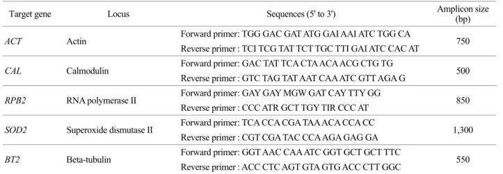
T. rubrum 균주를 대상으로 MLST 분석을 통해 Sequence type (ST) 분석을 실시한 기존 연구가 없었기 때문에, 피부 사상균과 특성이 유사한 사상균에 속하는 Scedosporium apiospermum을 기준으로 하여 ST 분석에 사용되고 있는 Housekeeping gene 다섯 종류를 MLST 분석을 위한 타겟으 로 설정하였고, National Center for Biotechnology Information (NCBI) GenBank의 유전자 Database에서 T. rubrum의 다섯 가지 유전자의 gDNA 염기서열을 참고로 하여 Oligonu- cleotide primer set을 디자인 및 제작하였다(Table 2).
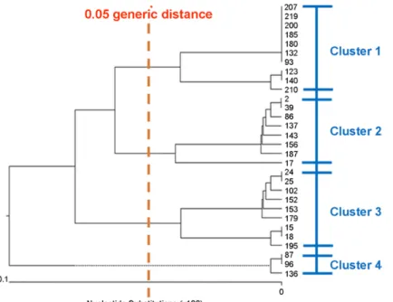
분석 대상 유전자의 염기서열 분석이 완료된 후 유전자 의 염기서열 차이를 분석하기 위해 유전자의 일부 염기서 열을 따로 분리하여 여러 개의 유전자를 하나의 파일로 결합하였고, 결합된 염기서열 정보를 이용하여 염기서열 의 차이를 분석하였다(Fig. 1). T. rubrum의 경우 Allele type 이나 ST이 아직 정의되어 있지 않아 ST 분석은 생략하였 다. T. rubrum 임상분리 균주들의 유전학적 연관성 분석을 위해 Phylogenetic tree 분석을 추가적으로 수행하였다. 다 섯 가지 표적 유전자 각각의 염기서열을 Molecular Evo- lutionary Genetics Analysis (MEGA) v. 7.0 프로그램을 통해 하나의 파일로 결합하였고, 결합된 염기서열 정보를 이용 해 1,000 bootstrap replication 조건으로 unweighted pair group method using arithmetic average (UPGMA) dendrogram을 생성 하였고, 유전적 Cluster 및 연관성을 구분 짓기 위해 Cut- off limit을 0.05 Generic distance로 설정하여 분석하였다.
결 과 주 질환별 환자 및 검체별 분석
주 질환에 따른 환자 분포에서는 조갑 진균증(Tinea unguium)이 12건(40%)으로 가장 많은 비중을 차지했고, 다음으로는 피부염(Dermatitis)이 4건(13.3%)이었으며, T.
rubrum이 분리된 임상 검체의 종류는 손톱, 발톱 검체가 17건(56.7%)으로 가장 많았고, 다음으로 피부 및 조직 검 체가 13건(43.3%)으로 많았다.
MLST 분석을 위한 유전자 증폭 및 Nucleotide substitution 분석
T. rubrum 임상분리 균주 30주에 대한 MLST 분석을 위 해 Actin (ACT, 750 bp), Calmodulin (CAL, 500 bp), RNA polymerase II (RPB2, 850 bp), Superoxide dismutase 2 (SOD2, 1,300 bp), β-tubulin (BT2, 550 bp)을 포함한 5개의 House- keeping gene을 대상으로 PCR을 수행하였다. 증폭된 PCR 산물을 대상으로 염기서열 분석을 실시한 결과, T. rubrum Table 1. Fungal strains and clinical data for this study
Strain No. Clinical data
Specimen Sex Age Diagnosis
1 Skin swab M 60 Seborrhoeic keratosis
2 Forearm (Rt.) F 70 Tinea corporis
3 Tissue M 60 Nummular dermatitis
4 Others M 30 Tinea corporis
5 Toe nail (Lt. 2nd) F 52 Tinea unguium
6 Toe nail (Lt. 1st) M 63 Tinea unguium
7 Toe nail (Lt. 2nd) M 58 Tinea unguium
8 Others M 40 Cellulitis
9 Toe nail (Rt. 5th) F 67 Other rosacea
10 Finger nail (Lt. 1st) F 64 Other prurigo
11 Tissue M 62 Irritant contact dermatitis
12 Toe nail (Lt. 4th) M 59 Onychomycosis
13 Toe nail (Lt. 4th) M 54 Tinea unguium
14 Others M 76 Atopic dermatitis
15 Toe nail (Lt. 1st) F 10 Tinea unguium
16 Tissue F 65 Onychomycosis
17 Others F 85 Tinea unguium
18 Toe nail (Rt. 4th) M 85 Contact dermatitis
19 Toe nail (Lt. 1st) M 19 Vitiligo
20 Toe nail (Rt. 1st) F 5 Tinea unguium
21 Groin M 35 Tinea corporis
22 Toe nail (Rt. 1st) M 30 Tinea unguium
23 Toe nail (Rt. 1st) M 59 Tinea unguium
24 Tissue F 63 Tinea corporis
25 Finger nail M 53 Tinea unguium
26 Toe nail (Rt. 1st) M 82 Tinea unguium
27 Others M 59 Tinea unguium
28 Toe nail (Lt. 2nd) M 54 Tinea cruris
29 Foot F 77 Tinea pedis
30 Arm M 56 Psoriasis (unspecified)
임상분리 균주들의 대부분의 Housekeeping 유전자에서는 염기서열이 모두 일치하는 결과를 보였다. 그러나, β-tubulin 유전자(BT2)에서 3개의 위치에서 염기서열의 변이가 다양 하게 나타남을 확인하였다(Fig. 1). 변이가 나타난 세 염기 는 주로 C→G (1766), G→T (1876), 그리고 C→A (1886)로 치환이 발생한 것을 확인하였다.
Phylogenetic tree 상관성 분석
T. rubrum 임상분리 균주들의 분자유전학적 연관성을 파악하기 위해 Phylogenetic tree 분석을 수행하였다. Cluster 분류를 위한 Cut-off limit을 0.05 Generic distance로 설정하 였고, 그 결과 Cluster 1부터 Cluster 4까지 총 4개의 Cluster Table 2. Specific oligonucleotide primer sets for T. rubrum MLST analysis
Target gene Locus Sequences (5' to 3') Amplicon size
(bp)
ACT Actin Forward primer : TGG GAC GAT ATG GAI AAI ATC TGG CA
Reverse primer : TCI TCG TAT TCT TGC TTI GAI ATC CAC AT 750
CAL Calmodulin Forward primer : GAC TAT TCA CTA ACA ACG CTG TG
Reverse primer : GTC TAG TAT AAT CAA ATC GTT AGA G 500
RPB2 RNA polymerase II Forward primer : GAY GAY MGW GAT CAY TTY GG Reverse primer : CCC ATR GCT TGY TIR CCC AT 850
SOD2 Superoxide dismutase II Forward primer : TCA CCA CGA TAA ACA CCA CC
1,300 Reverse primer : CGT CGA TAC CCA AGA GAG GA
BT2 Beta-tubulin Forward primer : GGT AAC CAA ATC GGT GCT GCT TTC
Reverse primer : ACC CTC AGT GTA GTG ACC CTT GGC 550
*I=, Y=T or C, M=A or C, W=A or T, R=G or A.
Fig. 1. Nucleotide substitutions in BT2 gene of T. rubrum clinical isolates. Genetic variations of T. rubrum clinical isolates were found on three different positions of β-tubulin gene and were shown to have changed from C→G (1766), G→T (1876), and C→A (1886).
로 구분되었다(Fig. 2). T. rubrum 임상분리 균주 간 분자유 전학적 연관성 차이를 분석한 결과, Cluster 1의 경우 Tinea unguium 환자가 4명으로 가장 많은 분포를 보였고(Table
3), Cluster 2의 경우 질병에서의 차이는 없었으나 대부분이 남자 환자에서 많이 나타남을 확인할 수 있었다. 하지만, 균이 분리된 환자 성별 간의 통계학적인 유의성은 나타내 Table 3. Type of dermatophytosis and clinical specimen sources according to the molecular genetic cluster of T. rubrum clinical isolates
Clusters Diagnosis No. of cases Specimens No. of cases Sex (M/F)
I (N=10)
Tinea unguium 4 Nail 4
6/4
Others 3 Others 4
Cruris 1 Skin 2
Pedis 1
Corporis 1
II (N=8)
Tinea unguium 2 Nail 3
7/1
Dermatitis 2 Tissue 3
Cellulitis 1 Skin 2
Tinea corporis 1
Seborrhoeic keratosis 1
Onychomycosis 1
III (N=9)
Tinea unguium 5 Nail 7
Tinea corporis 2 Arm 1 6/3
Vitiligo 1 Others 1
Onychomycosis 1
IV (N=3)
Tinea unguium 1 Nail 2
1/2
Dermatitis 1 Tissue 1
Rosacea 1
Fig. 2. Dendrogram of a total of 30 T. rubrum clinical isolates determined by MLST with five housekeeping genes. It was classified as four clus- ters, however there was little significant correlation.
지 않았다. Cluster 3의 경우는 주 질환이 Tinea unguium인 환자가 5명이었으며, 대부분 손, 발톱에서 분리된 균주들 로 확인되었다. Cluster 4에서는 균주 간의 뚜렷한 차이를 보이지 않았다.
고 찰
2006년부터 2010년까지 국내 진균 감염증 건강보험 청 구 자료를 분석한 결과에 따르면, 연평균 인구의 9.4%가 진균증으로 치료를 받고 있으며, 이 중 백선증에 의한 감 염이 6.5%를 차지하였다. 2010년 한 해 동안 백선증에 의 한 요양급여 비용은 3,205억을 사용하여 매우 높은 것으 로 알려져 있다(Yoon et al., 2014). 최근 보고에 의하면, 현 재 한국의 경우 약 40여 종의 피부사상균이 동정되었으 며, 주로 T. rubrum, T. mentagrophytes, M. canis, E. floccosum 등이 대표 균종이다. 하지만 예전에 주로 동정되던 M.
ferrugineum, T. schoenleinii 등은 현재 거의 사라진 것으로 보고되고 있다. 현재 국내에서 가장 높은 빈도로 발생하고 있는 균종 역시 T. rubrum으로 보고자에 따라 다양한 수 치를 보이나 대개 80~92% 정도의 발생 빈도를 보이고 있 으며, T. mentagrophytes와 M. canis 등은 10%대 이하로 감 소하는 추세를 보이고 있다. 1979년에서 2013년까지 국내 의 T. rubrum에 의한 진균 감염 환자 수는 매년 1,436명에 서 5,565명 수준으로 평균 3,310명이 발생하였으며, 1979년 이후 꾸준한 증가 추세를 보이고 있다(Lee et al., 2015).
최근 진균 종의 정확한 동정과 유전자형 분석 및 분자 유전학적 변이 유형을 연구하기 위해 소개되고 있는 다양 한 분자생물학적 분석법들은 특정 표적 유전자를 증폭하 여 분석하는 방법을 기본적으로 사용하고 있으며, 대표적 으로 교잡법(Hybridization), PFGE, RFLP, AFLP, RAPD, 염기 서열 분석법 등이 존재한다(Anderson et al., 1996; Semighini et al., 2001; Dodgson et al., 2003; Balajee et al., 2006; Litvintseva et al., 2006; Anne et al., 2010; Kim et al., 2011; Jin et al., 2014).
하지만, 유전적 변이가 많이 발생하는 진균 종의 경우 분 석에 한계가 있으며, PCR 증폭물의 밴드 패턴 분석(Poly- morphism)에 있어서 연구자 또는 실험실마다 결과가 일정 치 않아 사용에 제한적이며, 표준화가 어려운 단점이 있 는 것으로 보고된 바 있다(Bidet et al., 2000). 특히, PFGE 법은 분석시간이 4~5일로 노동 집약적이며, 결과 분석에 자의적인 견해가 개입될 수 있는 단점이 있는 것으로 알 려져 있다. 최근 이용이 되고 있는 MLST 기법은 진균의 유전자 중 4~10개의 Housekeeping gene을 선정하여 염기
서열 분석을 통해 균종, 균주 별 또는 유전자형 구별이 가 능한 동시에 유전자 변이 분석이 가능하여 전 세계적으로 진균의 분포, 호발 원인균 등 분자역학적 분석이 가능하 다는 장점이 있는 것으로 보고되고 있다(Byun et al., 2012).
진균을 대상으로 한 MLST 분석을 이용한 역학적 연구 는 최초로 Candida albicans의 유전자형 분석에 이용된 이 후, Coccidioides immitis, Histoplasma capsulatum, Cryptococcus neoformans, Fusarium oxysporum과 같은 인체 전신 감염성 진균 및 식물 병원성 진균 등에 적용되어 이미 그 결과가 보고되어 있다(Dodgson et al., 2003; Bougnoux et al., 2003;
Litvintseva et al., 2006). 동일 균주 간에도 유전학적 및 형 태학적 특성이 서로 다른 양상으로 분석될 수 있기 때문 에, 균주 간 구분을 통해 진균의 감염 경로 추적이 가능 하여 재감염, 재발 및 새로운 변이 균주의 감염이 만성 감 염에 의한 것인지 분석이 가능한 것으로 보고되어 있다 (John and Matthew, 2003). 이는 현재 광범위 항진균제의 처 방보다 적절한 항균제의 처방 및 치료에 도움을 줄 수 있 고 항진균제 내성균의 출현을 감시할 수 있으며 더불어 지리적, 시간적인 역학 조사의 방법으로 적용될 수 있을 것으로 여겨지고 있다.
그러나 백선증의 원인 진균으로서 T. rubrum에 대한 MLST를 이용한 분석은 아직 보고된 바 없어 본 연구에서 는 MLST 기법이 T. rubrum의 분석에 이용될 수 있는 지 와 이를 이용하여 백선증의 원인균 동정에 관한 가능성을 분석하였다. MLST 분석에 있어서 T. rubrum 30검체에 대 해 actin (ACT), calmodulin (CAL), RNA polymerase II (RPB2), superoxide dusmutase 2 (SOD2) 및 β-tubulin (BT2)를 포함한 5개의 Housekeeping gene을 대상으로 염기서열에서의 변 화를 분석한 결과, 대부분의 유전자에서 원인균 동정 결 과가 T. rubrum으로 모두 일치하는 결과를 보였다. 그러나, β-tubulin 유전자(BT2)에서 3개의 염기 위치에서 변이가 다 양하게 나타남을 확인하였으며 변이가 나타난 세 염기는 주로 C→G (1766), G→T (1876), 그리고 C→A (1886)로 치 환을 보였다.
T. rubrum의 분자유전학적 연관성 분석을 위해 Phylo- genetic tree 분석을 수행한 결과, 크게 4개의 Cluster로 구 분되었으나 각 Cluster 마다의 연관성은 서로 크지 않은 것으로 나타났다. 본 연구에서 MLST 분석의 표적으로 사용된 5가지 종류의 Housekeeping gene 중 BT2에서만 Nucleotide substitution이 확인되었기 때문에, BT2에서의 유전적 변이가 4가지 Cluster로의 구분에 큰 영향을 준 것 으로 사료되며, 이후 T. rubrum의 ST 분석을 위한 하나의
지표로 활용이 가능할 것으로 사료된다. 하지만, 추후 BT2 에 의해 구분된 Cluster 간의 표현형으로 나타나는 특성 차이를 규명하기 위해서는 추가적인 연구가 필요할 것으 로 사료된다. T. rubrum 균주 간 임상적 연관성 분석에서 는 Cluster 1의 경우 Tinea unguium 환자가 4명으로 가장 많은 분포를 보였고, Cluster 2의 경우 질병에서의 차이는 없었으나 대부분이 남자 환자였다. Cluster 3의 경우는 주 질환이 Tinea unguium인 환자가 5명이였으며, 대부분 손, 발톱에서 분리된 균주였다. Cluster 4에서는 뚜렷한 차이를 보이지 않았다.
본 연구에서 사용한 검체의 수가 제한적이긴 하나, 지 금까지 진균 종에서의 MLST 분석은 Coccidioides immitis, Histoplasma capsulatum, Fusarium oxysporum 등 특정 사상 균에 대해서만 한정적으로 이루어 졌지만, 본 연구에서 T.
rubrum를 대상으로 유전적 변이 및 분자역학적 분석을 위 해 MLST법의 적용 가능성을 탐색하였다는 점에서 의미 가 있다고 할 수 있다. 향후 추가적인 연구를 통해 전국 단위의 분리 균주를 대상으로 한 연구 결과를 데이터베이 스화 한다면 T. rubrum의 검체 별 유전적 변이 패턴, 항진 균제 내성 패턴, 지역적, 시간적 변이 등에 대한 국내 피 부 진균증 대표 원인균에 관한 정확한 분자역학적 분석이 가능할 것으로 생각된다.
본 연구를 통해 T. rubrum에 대한 MLST 분석의 가능성 을 확인함으로써 이를 기반으로 한 전 세계의 균주 간 분 자유전학적 다양성이나 분자역학적 특성 파악이 가능할 수 있을 것으로 사료되며, 정확한 데이터베이스가 축적된 다면 임상적으로 다방면에서 이용될 수 있을 것으로 예상 된다. 또한, 국내뿐만 아니라 세계적으로도 피부 진균증의 유병률이 전 세계 인구의 약 20%에 달하고 있고, 그 중 피부 진균증의 80~90%가 T. rubrum이 원인이 되고 있기 때문에, T. rubrum에 대한 정확한 진단과 분자역학적 분석 에 관한 결과들이 이후 감염증의 조기 치료와 감염 전파 예방에 도움을 줄 뿐만 아니라 역학적 감시체계를 구축하 는데 큰 기여를 할 수 있을 것으로 사료된다.
ACKNOWLEDGEMENTS None.
CONFLICT OF INTEREST
No conflict of interests exists for any of the authors.
REFERENCES
Anderson MJ, Gull K, Denning DW. Molecular typing by random amplification of polymorphic DNA and M13 Southern hy- bridization of related paired isolates of Aspergillus fumigatus.
Journal of Clinical Microbiology. 1996. 34: 87-93.
Anne D, Cecile G, Christophe H, Nelly CA, Sybren de H, Marie M. Development of a new MLST scheme for differentiation of Fusarium solani species complex (FSSC) isolates. Journal of Microbiological Methods. 2010. 82: 319-323.
Atef SS, Pranab KM, Hassan NA, Atef I, El A, Said HA, Mahmoud AG. Single-Step PCR Using (GACA) 4 Primer: Utility for Rapid Identification of Dermatophyte Species and Strains.
Journal of Clinical Microbiology. 2008. 46: 2641-2645.
Balajee SA, Nickle D, Varga J, Marr KA. Molecular studies reveal frequent misidentification of Aspergillus fumigatus by mor- photyping. Eukaryotic Cell. 2006. 5: 1705-1712.
Bernhardt A, Sedlacek L, Wagner S, Schwarz C, Würstl B, Tintelnot K. Multi locus sequence typing of Scedosporium apiospermum and Pseudallescheria boydii isolates from cystic fibrosis patients. Journal of Cystic Fibrosis. 2013. 12: 592-598.
Bidet P, Lalande V, Salauze B, Burghoffer B, Avesani V, Delmée M, et al. Comparison of PCR-ribotyping, arbitrarily primed PCR, and pulsed-field gel electrophoresis for typing Clostridium difficile. Journal of Clinical Microbiology. 2000. 38: 2484 -2487.
Bougnoux, ME, Tavanti A, Bouchier C, Gow NAR, Magnier A, Davidson AD, et al. Collaborative consensus for optimized multilocus sequence typing of Candida albicans. Journal of Clinical Microbiology. 2003. 41: 5265-5266.
Byun JH, Yoo JH, Park CM, Lee Dg, Park SH, Choi SM. Mole- cular epidemiologic analysis of community-onset extended spectrum beta-lactamase (ESBL) producing Escherichia coli using infrequent-restriction site polymerase chain reaction (IRS-PCR) with comparison by pulsed-field gel electropho- resis (PFGE). Infect Chemotherapy. 2012. 44: 5-10.
Dodgson AR, Pujol C, Denning DW, Soll DR, Fox AJ. Multilocus sequence typing of Candida glabrata reveals geographically enriched clades. Journal of Clinical Microbiology. 2003. 41:
5709-5717.
Guofang L, Chenghua HE, Haibin Z. Identification and character- ization of dermatophyte species and strains with PCR amplifi- cation. Experimental and Therrapeutic Medicine. 2014. 8: 545
-550.
Jin H, Kim H, Kim S, Choi Y, Bang H, Park S, Wang H, Lee JH, Jang IH, Kim YK, Lee H. Evaluation of a PCR-Reverse Blot Hybridization Assay to Identify Six Dermatophytes Predo- minant in the Republic of Korea. Biomedical Science Letters.
2014. 20: 139-146.
John WT, Matthew CF. Fungal multilocus sequence typing-it's not just for bacteria. Current Opinion Microbiology. 2003. 6: 351 -356.
Judy RR, Robert WP, Rana AH, Mary EB, Arthur LR. The epi- demiological features of invasive mycotic infections in the San Francisco bay area, 1992-1993: Results of population-based laboratory activity surveillance. Clinical Infectious Diseases.
1998. 27: 1138-1147.
Kim KH, Jun JB, Yoo HJ. Superficial and cutaneous mycoses.
Dermatology revised vol. 4. Seoul: Ryo Moon Gak. 2001. 310 -340.
Kim H, Jin H, Kim S, Wang H, Choi Y, Bang H, Park JS, Lee JH, Won YH, Ahn KJ, Kim YK, Lee H. PCR-reverse Blot Hybri- dization Assay for Species Identification of Dermatophytes.
Korean Journal of Medical Mycology. 2011. 16: 86-98.
Kim SJ, Kim SJ. A Distribution of Keratinophilic Fungi Isolated from the Soil of Haeundae Beach in Korea, Korean Journal of Clinical Laboratory Science. 2016. 48: 343-347.
Kwon-Chung KJ, Bennett JE. Philadelphia: Lea & Febigger.
Medical mycology. 1992. 105-161.
Lee SK, Choi JS, Kim KH. A clinical and mycological Study of Tinea Pedis. Annals of Dermatology. 1995. 33: 1029-1037.
Lee WJ, Kim SL, Jang YH, Lee SJ, Kim DW, Bang YJ, et al. In- creasing Prevalence of Trichophyton rubrum Identified through an Analysis of 115,846 Cases over the Last 37 Years. Journal
of Korean Medical Science. 2015. 30: 639-643.
Litvintseva AP, Thakur R, Vilgalys R, Mitchell TG. Multilocus sequence typing reveals three genetic subpopulations of Cry- ptococcus neoformans var. grubii (serotype A), including a unique population in Botswana. Genetics. 2006. 172: 2223 -2238.
Oh BH, Ahn KJ. Drug Therapy of Dermatophytosis. Journal of the Korean Medical Association. 2009. 52: 1109-1114.
Semighini CP, Delmas G, Park S, Amstrong D, Perlin D, Goldman GH. New restriction fragment length polymorphism (RFLP) markers for Aspergillus fumigatus. FEMS Immunology and Medical Microbiology. 2001. 31: 15-19.
Vikesh KB, Prakash CS. Epidemiological studies on Dermatophy- tosis in human patients in Himachal Pradesh. India: Springer- Plus. 2014. 134.
Weitzman I, Summerbell RC. The dermatophytes. Clinical Micro- biology Reviews. 1995. 8: 240-259.
Yang JH. Statistical observation of skin fungal disease for last 10 years. Korean Dermatology & Urology Society Newsletter.
1949. 1: 10-78.
Yoon HJ, Choi HY, Kim YK, Song YJ, Ki Moran. Prevalence of fungal infections using National Health Insurance data from 2009-2013. Epidemiology and Health. 2014. 36.
https://doi.org/10.15616/BSL.2018.24.3.221
Cite this article as: Yoon NS, Kim H, Park SB, Park M, Kim S, Kim YK. Genetic Variations of Trichophyton rubrum Clinical Isolates from Korea. Biomedical Science Letters. 2018. 24: 221-229.