• 검색 결과가 없습니다.

Genetic Variations of Candida glabrata Clinical Isolates from Korea using Multi-locus Sequence Typing

N/A
N/A
Protected

Academic year: 2021

Share "Genetic Variations of Candida glabrata Clinical Isolates from Korea using Multi-locus Sequence Typing"

Copied!
7
0
0

로드 중.... (전체 텍스트 보기)

전체 글

(1)

Genetic Variations of Candida glabrata Clinical Isolates from Korea using Multi-locus Sequence Typing

Min Ji Kang

1

, Kyung Eun Lee

2

and Hyunwoo Jin

2

*

1

Department of Microbiology, College of Medicine, Chungnam National University, Daejeon 34134, Korea

2

Department of Clinical Laboratory Science, College of Health Science, Catholic University of Pusan, Busan 46252, Korea Received October 23, 2019 /Revised January 17, 2020 /Accepted January 31, 2020

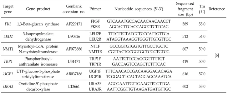
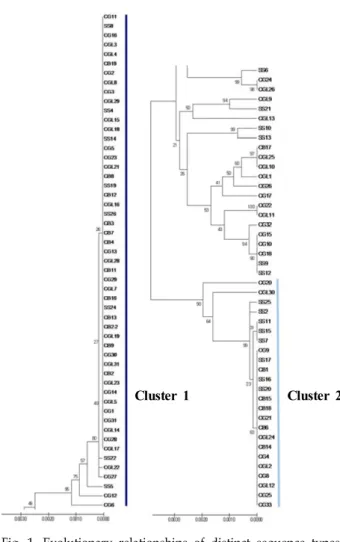
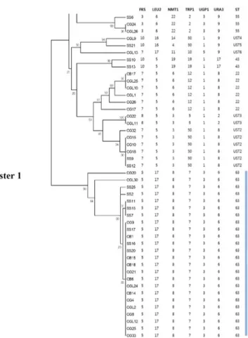
Although Candida albicans is the major fungal pathogen of candidemia, severe infections by non-albi- cans Candida (NAC) spp. have been increasing in recent years. Among NAC spp., C. glabrata has emerged as the second most common pathogen. However, few studies have been conducted to inves- tigate its structure, epidemiology, and basic biology. In the present study, multi-locus sequence typing (MLST) was performed with a total of 102 C. glabrata clinical isolates that were isolated from various types of clinical specimen. For MLST, six housekeeping genes—FKS, LEU2, NMT1, TRP1, UGP1, and URA3—were amplified and sequenced. The results were analyzed using the C. glabrata database. Out of a total of 3,345 base-pair DNA sequences, 49 variable nucleotide sites were found, and the results showed that 12 different sequence types (ST) were identified from the 102 clinical isolates. The data also demonstrated that the undetermined ST1 was the most predominant ST in Korea. Further, seven undetermined STs (UST) containing UST2-8 were classified at specific loci. The data from this study may provide a fundamental database for further studies on C. glabrata, including its epidemiology and evolution. The data may also contribute to the development of novel antifungal agents and diagnostic tests.

Key words : Candida glabrata, non-albicans Candida, candidiasis, genetic variations, multi-locus sequence typing

*Corresponding author

*Tel : +82-51-510-0567, Fax : +82-51-510-0568

*E-mail : jjjnhw@cup.ac.kr

This is an Open-Access article distributed under the terms of the Creative Commons Attribution Non-Commercial License (http://creativecommons.org/licenses/by-nc/3.0) which permits unrestricted non-commercial use, distribution, and reproduction in any medium, provided the original work is properly cited.

Journal of Life Science 2020 Vol. 30. No. 2. 122~128 DOI : https://doi.org/10.5352/JLS.2020.30.2.122

Introduction

Candida spp. belong to the normal flora of the vaginal tract, the gastrointestinal tract, and the oral cavity in human [8, 18]. However, rarely, serious infections, ranging from mucosal infections to systemic infection have been caused by Candida spp. [8, 18]. Fungal infections caused by Candida spp. have increased significantly, especially in acquired im- mune deficiency syndrome (AIDS) and immunocompro- mised individuals, including intensive care and, elderly pa- tients [3, 8]. Also, candidemia is associated with a high mor- tality rate approximately 30 to 40% in hospitalized patients and is difficult to treat, thus increasing the cost of medical care [3, 8].

C. glabrata has emerged as the second or third most com- mon Candida pathogen after C. albicans in the United States,

depending on the site [3, 7, 21]. Despite its increased preva- lence, there have been relatively few studies on the pop- ulation structure, epidemiology, and basic biology of C. glab- rata compared to those conducted on other Candida spp.

[6-8].

As mentioned above, C. albicans has considered to be the major fungal pathogen of candidemia in the past [6, 23].

However, as the number of severe infections caused by non-albicans Candida spp. (NAC) have increased, studies have shifted from C. albicans to NAC such as C. glabrata in recent years [11, 21]. Furthermore, since C. glabrata infections are often resistant to azole antifungal drugs, especially fluco- nazole, it is important to distinguish NAC from C. albicans to ensure the appropriate antifungal therapy and clinical management [8, 20, 21]. Thus, the discrimination of subtypes in these species are required for investigating their epidemi- ology and evolutionary biology [15, 23, 24].

In recent years, there has been substantial progress in the

development of several molecular methods for typing sub-

species and strains of fungi [26]. For instance, pulsed-field

gel electrophoresis (PFGE) compares total DNA band pat-

terns with or without restriction enzyme digestion, while

multilocus variable-number tandem-repeat (VNTR) analysis

(2)

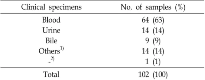
Table 1. Details of diverse yeast isolates used in this study Clinical specimens No. of samples (%)

Blood Urine Bile Others

1)

-

2)

64 (63) 14 (14) 9 (9) 14 (14) 1 (1)

Total 102 (100)

1)

Others: ascitic fluid, joint fluid, pleural fluid, tissue etc.

2)

No clinical information.

Table 2. Sequences of primer pairs for fungal species identification

Target region Primer Nucleotide sequences (5' to 3') Amplicon size (bp) Tm (℃) Reference

ITS pITS-F

pITS-R

GTCCTAACAAGGTTAACCTGCGG

TCCTCCGCTTATTGATATGC 970-980 62.4

55.3 [16]

examines length variations in six to nine PCR-amplified loci that contain polymorphic tandem repeats. Further, the ran- dom amplification of polymorphic DNA compares banding patterns following PCR with a nonspecific primer. Finally, multilocus enzyme electrophoresis, studies the different elec- trophoretic mobility of multiple core metabolic enzymes.

These four approaches have some limitations, such as a low- er reproducibility and portability [10, 22], and the results obtained in different laboratories are difficult to compare [2, 13].

Among these genotyping methods, multilocus sequence typing (MLST) is a useful tool to assign single nucleotide polymorphisms as allele numbers, which are stored in a da- tabase on line (PubMLST) and determine the differences from between closely related isolates by their geographical origins, sources, and other properties [23]. Also, it is possible that database are accessed by laboratories worldwide [13, 24].

In the present study, MLST was performed with a total of 102 C. glabrata clinical isolates from various clinical speci- mens such as blood, urine, and other body fluids in Korea.

The results were analyzed by using the C. glabrata MLST database (http://pubmlst.org/cglabrata/). The aim of the study is to discriminate STs in the same C. glabrata spp. by using common MLST and investigate the most prevalent ST from the C. glabrata in Korea.

Materials and Methods

Clinical strains

A total of 102 C. glabrata clinical isolates were provided

from Korean Culture Collection of Medical Fungi (KCMF) and those isolates were collected from tertiary hospitals in Korea. Clinical isolates were isolated from a wide variety of clinical samples, including blood, catheterized urine, bile and other body fluids (Table 1).

Genomic DNA extraction from fungal isolates Genomic DNA of C. glabrata clinical isolates was extracted using a I-genomic BYF DNA Extraction Mini Kit (iNtRON Inc., Seongnam, Korea) according to the manufacturer's in- structions [5]. The concentration and purity of the genomic DNA were checked by 260/280 optical density using a Nanodrop 2000 Spectrophotometer (Thermo Scientific, Wil- mington, DE, USA). The extracted genomic DNA was stored at 4℃ until use.

Polymerase chain reaction and sequence analysis of the fungal ITS region for precise identification of C. glabrata clinical isolates

The fungal internal transcribed spacer (ITS) region, the conserved region between the 18S and 28S ribosomal RNA (rRNA) was amplified and sequenced using each primer pairs (Table 2). Target amplification was carried out in 20 μl reaction mixture containing 10 μl Prime Taq Primix (Genet Bio Inc., Daejeon, Korea), 5 μl of distilled ultra-pure water, 1 μl of each primer (10 pmol/μl), and 3 μl of genomic DNA template. The PCR condition was: an initial denaturation at 94℃ for 1 min, 30 cycles including subsequent denaturation at 94℃ for 30 sec, annealing at 57℃ for 30 sec and extension at 72℃ for 45 sec followed by final extension at 72℃ for 7 min and holding at 4℃. The amplified products were vi- sualized by gel electrophoresis to confirm the presence of desired product.

The resulting amplicon was purified and sequenced by

Macrogen Inc. (Daejeon, Korea). All sequences with low-

quality bases in the chromatogram were re-sequenced for

the high-quality results. The obtained sequences were aligned

with reference sequences in the Genebank database using

the basic local alignment search tool (BLAST) at the National

Center for Biotechnology Information (NCBI), and percent

homology scores were generated to precise identification of

(3)

Table 3. List of gene fragments and primer sequences for C. glabrata MLST analysis Target

gene Gene product GenBank

accession no. Primer Nucleotide sequences (5'-3')

Sequenced fragment size (bp)

Tm

(℃) Reference

FKS 1,3-Beta-glucan synthase AF229171 FKSF FKSR

GTCAAATGCCACAACAACAACCT

AGCACTTCAGCAGCGTCTTCAG 589 55.0

[6]

LEU2 3-Isopropylmalate

dehydrogenase U90626 LEU2F

LEU2R

TTTCTTGTATCCTCCCATTGTTCA

ATAGGTAAAGGTGGGTTGTGTTGC 512 54.0

NMT1 Myristoyl-CoA, protein

N-myristoyltransferase AF073886 NT1F NMT1R

GCCGGTGTGGTGTTGCCTGCTC

CGTTACTGCGGTGCTCGGTGTCG 607 59.0

TRP1 Phosphoribosyl-

anthranilate isomerase U31471 TRP1F TRP1R

AATTGTTCCAGCGTTTTTGT

GACCAGTCCAGCTCTTTCAC 419 50.0

UGP1 UTP-glucose-1-phosphate

uridylyltransferase AB037186 UGP1F UGP1R

TTTCAACACCGACAAGGACACAGA

TCGGACTTCACTAGCAGCAAATCA 616 57.0

URA3 Orotidine-5'-phosphate

decarboxylase L13661 URA3F

URA3R

AGCGAATTGTTGAAGTTGGTTGA

AATTCGGTTGTAAGATGATGTTGC 602 53.0

C. glabrata clinical isolates.

MLST analysis for identifying sequence type of C.

glabrata clinical isolates

Table 3 shows primers for the amplification and sequence analysis of C. glabrata six housekeeping gene fragments in- cluding FKS, LEU2, NMT1, TRP1, UGP1, and URA3. For the PCR amplification, 20 μl of final mixture contained 10 μl of Prime Taq Premix, 5 μl of distilled ultra-pure water, 1 μl of each forward and reverse primer (10 pmol/μl), and 3 μl of genomic DNA template. To amplify each six gene, the PCR reaction conditions were as follows: 7 min at 94℃, 30 cycles of 1 min at the relevant annealing temperature (Table 3), and 1 min at 74℃, followed by 10 min at 74℃.

The resulting sample was analyzed by gel electrophoresis.

The PCR product of all loci were purified and sequenced using reverse sequence primer at Macrogen Inc.. The ob- tained sequences were analyzed by using the C. glabrata MLST database (http://pubmlst.org/cglabrata/). Each unique sequence at a locus defined an allele number, and unique combinations of alleles assigned as a ST.

Data analysis

The alignment of combined six target gene sequences and loci (3,345 bp) was performed using the Molecular Evolutio- nary Genetics Analysis (MEGA) v. 7.0 software [12]. For re- latedness of the same species, the phylogenetic tree was drawn with the Unweighted Pair Group Method using Arithmetic algorithm (UPGMA) with randomized 1,000 bootstrapping. And then the eBURST package (http://eburst.

mlst.net/) was used to determine that all related isolates were grouped into clonal complexes.

Results

PCR and sequence analysis of the fungal ITS region for species identification

1.5% TBE agarose gel DNA electrophoresis data showed that the size of amplified fungal ITS region was 978 bp, and amplicons have shown one clear band (data not shown). As a analysis result of comparison by Genebank BLAST tool for verifying the amplified PCR products, all clinical isolates used in this study were identified as C. glabrata with high concordance rate (97.8%±2.9).

PCR, sequence analysis, obtaining allele number of six housekeeping genes for the MLST analysis

In order to perform the MLST analysis, six housekeeping genes of 102 C. glabrata clinical isolates were amplified by PCR. The size of amplified fragments of FKS, LEU2, NMT1, TRP1, UGP1, and URA3 were 589 bp, 512 bp, 607 bp, 419 bp, 616 bp, and 602 bp, respectively as the expected size and they represented clear band (data not shown).

Amplified six housekeeping gene fragments were se-

quenced and then sequenced data was trimmed as each

length manually by using Chromas software. Trimmed se-

quence of six housekeeping gene fragments was analyzed

at the C. glabrata PubMLST database then allele number of

each gene was obtained. Obtained allele number of FKS gene

were 3, 5, 7, 8, and 10. Those of LEU2 gene were 5, 6, 16,

(4)

Table 4. The number of polymorphic site and different alleles, and sequence variable position of six housekeeping gene fragments in 102 C. glabrata clinical isolates

Locus No. of polymorphic sites (%)

No. of alleles

defined Variable positions

FKS LEU2 NMT1 TRP1 UGP1 URA3

5 (0.9) 6 (1.2) 12 (2.0) 13 (3.1) 4 (0.7) 9 (1.5)

5 4 8 8 4 7

403, 352, 154, 118, 43 392, 384, 336, 290, 111, 54

575, 551, 512, 480, 434, 396, 341, 328, 305, 301, 243, 155 387, 378, 357, 352, 333, 309, 272, 268, 255, 229, 176, 162, 158 585, 435, 413, 195, 99

581, 574, 556, 549, 440, 380, 257, 164, 44

Total 49

Table 5. Unique sequence types determined with a combination of six loci

C. glabrata sequence types no. of isolates (%) ST63

ST22 ST55 ST43 undetermined ST1 undetermined ST2 undetermined ST3 undetermined ST4 undetermined ST5 undetermined ST6 undetermined ST7 undetermined ST8

24 (23.5) 6 (5.9) 3 (2.9) 2 (1.2) 54 (52.9)

6 (59) 2 (1.2) 1 (1) 1 (1) 1 (1) 1 (1) 1 (1)

Total 102 (100)

Cluster 1 Cluster 2

Fig. 1. Evolutionary relationships of distinct sequence types.

UPGMA dendrogram used to determine the pairwise differences of the concatenated sequence of C. glabrata clinical isolates.

and 17. Those of NMT1 gene were 3, 4, 6, 8, 11, 14, 19, and 22. Those of TRP1 gene were 2, 3, 5, 7, 10, 12, 19, and 50.

Those of UGP1 gene were 1, 3, 5, and 51. Those of URA3 gene were 2, 4, 6, 8, 9, 17, and 20 (Table 4). A total of 49 (1.5%) polymorphic sites were found among six house- keeping gene fragments. The number of polymorphic site in each gene fragment were as follows: FKS (5, 0.9%); LEU2 (4, 1.2%); NMT1 (8, 2.0%); TRP1 (8, 3.1%); UGP1 (4, 0.7%);

and URA3 (7, 1.5%), as shown in Table 4. Additionally, in- sertions, deletions, or heterozygosity were only detected in the NMT1 fragments.

Sequence type and cluster of C. glabrata clinical isolates

The MLST scheme revealed a high diversity of C. glabrata isolates with a total of 12 STs, 8 of which were identified as undetermined STs (USTs) that were not discovered in the previous studies.

The data demonstrates that the UST1 was the most pre- dominant ST in this study as a total of 54 clinical isolates

(52.9%) were contained in this ST, and the following most

predominant ST was the ST63 as a total of 24 clinical isolates

(23.5%) were contained in this ST. In addition, this study

obtained the ST55, ST22, and ST43 were as a total of 3 (2.9%),

6 (5.9%), and 2 (1.2%) clinical isolates were contained in re-

spective ST and the UST2 was identified in 6 isolates (5.9%).

(5)

Fig. 2. Phylogenetic tree generated from each isolate. The MLST allele profile, sequence type (ST), and eBURST group (Cluster 1) are listed.

Fig. 3. Phylogenetic tree generated from each isolate. The MLST allele profile, sequence type (ST), and eBURST group (Cluster 2) are listed.

The UST3 was identified in 2 isolates (1.2%). The remaining 5 USTs (UST4, -5, -6, -7, and -8) were classified only once each (1%) (Table 5).

Combined sequence (3,345 bp) of six housekeeping genes was used for the phylogenetic tree analysis. Apart from 3 outliers, the isolates were divided into 2 major clusters: clus- ter 1 and cluster 2 (Fig. 1). Cluster 1 consisted of the UST1 and cluster 2 consisted of the ST63 (Fig. 2, Fig. 3).

Discussion

C. glabrata is a highly opportunistic pathogen of the ur- ogenital tract and the bloodstream in humans [17]. It is espe- cially prevalent in the elderly and within the human im- munodeficiency virus positive population [19]. Although candidiasis is frequently treated with azole antifungal agents, treatment failure has become a serious concern with azole-resistant clinical isolates due to widespread and long-term use of these agents. Nevertheless, few studies

have been conducted on the structure, epidemiology, and basic biology of C. glabrata.

Healthcare-associated infections may be endogenous in origin or nosocomially transmitted, and the only way to dis- tinguish them is through strain typing. Recently, MLST di- rectly investigated the DNA sequence variations in a set of housekeeping genes and characterized the strains by their unique allelic profiles. The principle of MLST is simple, in- volving PCR amplification followed by DNA sequence analysis. Nucleotide differences between strains can be veri- fied at a variable number of genes depending on the desired degree of discrimination. MLST schemes now exist for sev- eral important bacterial pathogens including Neisseria menin- gitidis, Streptococcus pneumoniae, Staphylococcus aureus, Strep- tococcus pyogenes, and Campylobacter jejuni. The technique has also been used to assess genetic relatedness among strains of Candida spp. including C. glabrata, C. albicans, C. tropicalis, and C. krusei. However, MLST scheme for C. glabrata has only been used by Dodgson, A. R. (2003) and Katiyar, S.

(2016).

(6)

Hence, in this study, the first MLST analysis with the yeast pathogen C. glabrata was performed and evaluated in Korea. 6 loci were selected for this study, as recommended by previous studies. While a ST3 was defined as prevalent ST in Dodgson, A. R. (2003), the data in this study demon- strates that the UST1 was the most predominant ST.

Additionally, the data defined a total of 12 STs among the 102 clinical isolates, and found 8 USTs as a result.

In conclusion, prevalent and novel C. glabrata STs were found in the present study. The data might provide a funda- mental database for further studies on C. glabrata, including its epidemiology and evolution. Furthermore, these data might also contribute to the development of novel antifungal agents and diagnostic tests. It might even be possible to dis- cover the virulence factors associated with disease, which population genetic studies currently struggle to monitor.

Acknowledgements

This study was supported by Basic Science Research Program through the National Research Foundation of Korea (NRF) funded by the Ministry of Education (NRF- 2017R1C1B5076822) and Brain Busan 21 Plus project.

The Conflict of Interest Statement

The authors declare that they have no conflicts of interest with the contents of this article.

References

1. Berila, N. and Subik, J. 2010. Molecular analysis of Candida glabrata clinical isolates. Mycopathologia 170, 99-105.

2. Bidet, P., Lalande, V., Salauze, B., Burghoffer, B., Avesani, V., Delmée, M., Rossier, A., Barbut, F. and Petit, J. C. 2000.

Comparison of PCR-ribotyping, arbitrarily primed PCR, and pulsed-field gel electrophoresis for typing Clostridium difficile. J. Clin. Microbiol. 38, 2484-2487.

3. Cheng, M. F., Yang, Y. L., Yao, T. J., Lin, C. Y., Liu, J. S., Tang, R. B., Yu, K. W., Fan, Y. H., Hsieh, K. S., Ho, M.

and Lo, H. J. 2005. Risk factors for fatal candidemia caused by Candida albicans and non-albicans Candida species. BMC Infect. Dis. 5, 22-26.

4. Da Matta, D. A., Melo, A. S., Guimarães, T., Frade, J. P., Lott, T. J. and Colombo, A. L. 2010. Multilocus sequence typing of sequential Candida albicans isolates from patients with persistent or recurrent fungemia. Med. Mycol. 48, 757- 762.

5. Da Silva-Rocha, W. P., Lemos, V. L., Svidizisnki, T. I., Milan, E. P. and Chaves, G. M. 2014. Candida species dis-

tribution, genotyping and virulence factors of Candida albi- cans isolated from the oral cavity of kidney transplant recip- ients of two geographic regions of Brazil. BMC Oral Health 14, 20-28.

6. Dodgson, A. R., Pujol, C., Denning, D. W., Soll, D. R. and Fox, A. J. 2003. Multilocus sequence typing of Candida glab- rata reveals geographically enriched clades. J. Clin. Microbiol.

41, 5709-5717.

7. Dodgson, A. R., Pujol, C., Pfaller, M. A., Denning, D. W.

and Soll, D. R. 2005. Evidence for recombination in Candida glabrata. Fungal Genet. Biol. 42, 233-243.

8. Fidel, P. L. Jr., Vazquez, J. A. and Sobel, J. D. 1999. Candida glabrata: review of epidemiology, pathogenesis, and clinical disease with comparison to C. albicans. Clin. Microbiol. Rev.

12, 80-96.

9. Hesham, A. E. L., Wambui, V., JO, H. O. and Maina, J.

M. 2014. Phylogenetic analysis of isolated biofuel yeasts based on 5.8 S-ITS rDNA and D1/D2 26S rDNA sequences.

J. Genet. Engineer. Biotechnol. 12, 37-43.

10. Katiyar, S., Shiffrin, E., Shelton, C., Healey, K., Vermitsky, J. P. and Edlind, T. 2016. Evaluation of polymorphic locus sequence typing for Candida glabrata epidemiology. J. Clin.

Microbiol. 54, 1042-1050.

11. Krcmery, V. and Barnes, A. J. 2002. Non-albicans Candida spp. causing fungaemia: pathogenicity and antifungal re- sistance. J. Hosp. Infect. 50, 243-260.

12. Kumar, S., Stecher, G. and Tamura, K. 2016. MEGA7:

Molecular evolutionary genetics analysis version 7.0 for big- ger datasets. Mol. Biol. Evol. 33, 1870-1874.

13. Maiden, M. C., Bygraves, J. A., Feil, E., Morelli, G., Russell, J. E., Urwin, R., Zhang, Q., Zhou, J., Zurth, K., Caugant, D. A., Feavers, I. M., Achtman, M. and Spratt, B. G. 1998.

Multilocus sequence typing: a portable approach to the identification of clones within populations of pathogenic microorganisms. Proc. Natl. Acad. Sci. USA. 95, 3140-3145.

14. Meats, E., Feil, E. J., Stringer, S., Cody, A. J., Goldstein, R., Kroll, J. S., Popovic, T. and Spratt, B. G. 2003. Characteriza- tion of encapsulated and noncapsulated Haemophilus influ- enzae and determination of phylogenetic relationships by multilocus sequence typing. J. Clin. Microbiol. 41, 1623-1636.

15. Odds, F. C. and Jacobsen, M. D. 2008. Multilocus sequence typing of pathogenic Candida species. Eukaryot. Cell 7, 1075-1084.

16. Pryce, T. M., Palladino, S., Kay, I. D. and Coombs, G. W.

2003. Rapid identification of fungi by sequencing the ITS1 and ITS2 regions using an automated capillary electro- phoresis system. Med. Mycol. 41, 369-381.

17. Rahimkhani, M., Saberian, M., Mordadi, A., Varmazyar, S.

and Tavakoli, A. 2015. Urinary tract infection with Candida glabrata in a patient with spinal cord injury. Acta. Med. Iran.

53, 516-517.

18. Rodrigues, C. F., Silva, S. and Henriques, M. 2013. Candida glabrata: a review of its features and resistance. Eur. J. Clin.

Microbiol. Infect. Dis. 33, 673-688.

19. Sanson, G. F. and Briones, M. R. 2000. Typing of Candida

glabrata in clinical isolates by comparative sequence analysis

(7)

초록:Multi-locus sequence typing을 이용한 한국에서 분리한 Candida glabrata 임상균주의 유전자 유형 분석

강민지

1

․이경은

2

․진현우

2

*

(

1

충남대학교 의과대학 미생물학교실,

2

부산가톨릭대학교 보건과학대학 임상병리학과)

Candida albicans가 칸디다 혈증의 주요 원인 균으로 알려져 있지만, 최근에는 non-albicans Candida (NAC)에 의한 심각한 감염이 증가하고 있다. NAC 중에서 C. glabrata는 두 번째로 병원성을 나타내는 원인 균이지만 C.

glabrata의 구조, 역학 등의 기본적인 연구는 거의 없는 실정이다. 따라서 본 연구에서는 다양한 유형의 임상 표본 으로부터 분리된 총 102개의 C. glabrata균주로 multi-locus sequence typing (MLST)을 수행하였다. FKS, LEU2, NMT1, TRP1, UGP1 및 URA3을 포함한 6개의 하우스키핑 유전자를 증폭하여 염기서열을 분석하였다. 총 3,345 개의 DNA 염기서열 중 49개의 가변 염기서열 부위가 발견되었으며, 그 결과 102개의 균주에서 총 12개의 상이한 서열 유형을 확인하였고, 알려지지 않은 서열 유형(USTs) 중 UST1이 가장 많이 나타났다. 이 연구의 결과는 국내 C. glabrata 항생제 처방에 도움이 될 것으로 사료되며, 한국에서 유행하는 C. glabrata에 대한 추가 연구를 위한 기본 역학자료로 사용될 것으로 기대한다.

of the cytochrome c oxidase subunit 2 gene distinguishes two clusters of strains associated with geographical se- quence polymorphisms. J. Clin. Microbiol. 38, 227-235.

20. Silva, S., Negri, M., Henriques, M., Oliveira, R., Williams, D. W. and Azeredo, J. 2012. Candida glabrata, Candida para- psilosis and Candida tropicalis: biology, epidemiology, pathoge- nicity and antifungal resistance. FEMS Microbiol. Rev. 36, 288-305.

21. Spampinato, C. and Leonardi, D. 2013. Molecular finger- prints to identify Candida species. Biomed. Res. Int. 2013, 923742.

22. Tatay-Dualde, J., Prats-van der Ham, M., de la Fe, C., Gómez-Martín, Á., Paterna, A., Corrales, J. C., Contreras, A. and Sánchez, A. 2016. Multilocus sequence typing of Mycoplasma mycoides subsp. capri to assess its genetic varia- bility in a contagious agalactia endemic area. Vet. Microbiol.

191, 60-64.

23. Tavanti, A., Davidson, A. D., Johnson, E. M., Maiden, M.

C., Shaw, D. J., Gow, N. A. and Odds, F. C. 2005. Multilocus sequence typing for differentiation of strains of Candida tropicalis. J. Clin. Microbiol. 43, 5593-5600.

24. Urwin, R. and Maiden, M. C. 2003. Multi-locus sequence typing: a tool for global epidemiology. Trends Microbiol. 11, 479-487.

25. Wang, Y., Shi, C., Liu, J. Y., Li, W. J., Zhao, Y. and Xiang, M. J. 2016. Multilocus sequence typing of Candida tropicalis shows clonal cluster enrichment in azole-resistant isolates from patients in Shanghai, China. Infect. Genet. Evol. 44, 418- 424.

26. Xu, J. 2006. Fundamentals of fungal molecular population

genetic analyses. Curr. Issues Mol. Biol. 8, 75-89.

수치

Table  1.  Details  of  diverse  yeast  isolates  used  in  this  study Clinical  specimens No
Table  3.  List  of  gene  fragments  and  primer  sequences  for  C.  glabrata  MLST  analysis Target
Table  5.  Unique  sequence  types  determined  with  a  combination  of  six  loci
Fig.  2.  Phylogenetic  tree  generated  from  each  isolate.  The  MLST  allele  profile,  sequence  type  (ST),  and  eBURST  group  (Cluster  1)  are  listed.

참조

관련 문서

We compared the distribution of Acinetobacter species in 95 clinical isolates which were determined by rpoB gene analysis, 16S rRNA gene analysis, and Vitek 2 system..

Anti-fungal effect of isolated bactrial strains was tested in vitro by incubating in potato dextrose agar with isolates of four fungal plant pathogens

Phylogenetic analysis of Orientia tsutsugamushi strains based on the sequence homologies of 56-kDa type-specific antigen genes. FEMS

We determined the nucleotide sequences of the mitochondrial DNA (mtDNA) control region using cloning and sequencing, and obtained the complete sequence from the cattle bones

Full genome cloning and nucleotide sequence analysis of hepatitis C virus from sera of chronic hepatitis patients in Korea. Detection of hepatitis C virus RNA

양성 환자에서 CNS (Coagulase-Negative Staphylococcus)균을 제외하고, Type1 환자 5명에서 Staphylococcus aureus, Escherichia coli, Acinetobacter Baumannii,

 Manipulation of protein’s amino acid sequence to change its function or properties.

최 지 애.. The distribution of specimens from which 100 clinical isolates of A.. Antimicrobial Susceptibility Pattern of 100 clinical isolates of A. Relative