pISSN: 0378-6471⋅eISSN: 2092-9374
http://dx.doi.org/10.3341/jkos.2014.55.7.1030
Original Article
개방각녹내장에서 맥락막분수계역, 야간 혈압하강 및 녹내장성 시야결손의 상관관계
Relationship among Water-Shed Zone, Nocturnal Dip and Visual Field Progression in Open Angle Glaucoma
서홍융1⋅김남영2⋅노세현2
Hong Ryung Seo, MD, PhD1, Nam Yeong Kim, MD2, Sae Heun Rho, MD, PhD2 왈레스기념 침례병원 안과1, 동아대학교 의과대학 안과학교실2
Department of Ophthalmology, Wallace Memorial Baptist Hospital1, Busan, Korea Department of Ophthalmology, Dong-A University College of Medicine2, Busan, Korea
Purpose: To investigate the influence of water-shed zone (WSZ) and nocturnal dip (ND) on the progression of the glaucoma- tous visual field (V/F) defects in open-angle glaucoma (OAG) patients when the intraocular pressure (IOP) was maintained un- der the target pressure.
Methods: We performed fluorescence angiography (FAG), 24-hour ambulatory blood pressure monitoring (24-hr ABPM), and V/F tests. We examined the relationships among WSZ in early-FAG, ND over 10% (dip), and the progression of the glaucoma- tous V/F defects using chi-square, Fisher’s exact, and multivariate logistic regression tests. A p-value < 0.05 was considered statistically significant.
Results: When considering the correlation between WSZ and dip, statistical significance was found in OAG (p = 0.024, odds ra- tio (OR) = 3.308) and normal tension glaucoma (NTG) (p = 0.029, OR = 4.364) patients. In patients with dip, glaucomatous V/F defects significantly progressed (OAG: p = 0.003, OR = 5.938, NTG: p = 0.005, OR = 13.929). In patients with WSZ, the glau- comatous V/F defects progressed in all groups (OAG: p = 0.002, OR = 5.156, NTG: p = 0.024, OR = 4.750, primary open angle glaucoma (POAG): p = 0.021, OR = 8.750). In the patients with WSZ involving optic nerve head, the glaucomatous V/F defects had progressed in OAG (p = 0.004, OR = 5.958) and NTG (p = 0.009, OR = 8.333) groups. Based on binary logistic regression analysis, dip (p = 0.010, OR = 6.227) significantly affected V/F progression only in OAG patients.
Conclusions: In the OAG and NTG groups, ND over 10% influenced the progression of the glaucomatous V/F defects. The pa- tients with WSZ tended to have ND over 10% in OAG and NTG groups and glaucomatous V/F defects progressed in all patients.
Therefore, performing early FAG and 24-hr ambulatory blood pressure monitoring may be helpful for glaucoma patients with progressing glaucomatous V/F defects even when the IOP was maintained under the target pressure.
J Korean Ophthalmol Soc 2014;55(7):1030-1038
Key Words: Nocturnal dip, Progression of the glaucomatous visual field defects, Twenty-four-hour blood pressure monitoring, Vascular dysregulation, Water-shed zone
■Received: 2013. 9. 13. ■ Revised: 2014. 1. 18.
■Accepted: 2014. 6. 10.
■Address reprint requests to Sae Heun Rh, MD, PhD Department of Ophthalmology, Dong-A University Hospital,
#26 Daesingongwon-ro, Seo-gu, Busan 602-715, Korea Tel: 82-51-240-5222, Fax: 82-51-254-1987
E-mail: [email protected]
* This study was presented as an e-poster at the 108th Annual Meeting of the Korean Ophthalmological Society 2012.
ⓒ2014 The Korean Ophthalmological Society
This is an Open Access article distributed under the terms of the Creative Commons Attribution Non-Commercial License (http://creativecommons.org/licenses/by-nc/3.0/) which permits unrestricted non-commercial use, distribution, and reproduction in any medium, provided the original work is properly cited.
녹내장의 병인은 아직 명확하게 밝혀져 있지 않으나, 안압 상승이 녹내장의 중요한 위험인자로 알려졌다.1 그러나 안압 이 정상 범위이면서도 녹내장성 시신경 손상과 시야결손을 보이는 정상 안압녹내장(normal tension glaucoma, NTG)과1-3 안압을 목표안압 이하로 유지하였음에도 불구하고 녹내장 성 시신경 손상과 시야결손이 진행한다는 사실은4,5 녹내장 의 발생과 진행에 안압 이외의 다른 위험 인자가 있음을 알 수 있다.
정상안압녹내장은 아시아, 특히 일본과 한국에서 높은 유병률을 보이는 녹내장으로 한국의 유병률은 2.7%이며 개 방각녹내장(open angle glaucoma, OAG)의 77%을 차지한 다.6 원발성 개방각녹내장(primary open angle glaucoma, POAG)은 주로 상승된 안압이 점진적으로 시신경 손상을 야기하는데 반해,7-9 정상안압녹내장에서는 안압 이외의 비 안압성 인자들에 더 많이 영향을 받는 것으로 알려졌고,10-16 특히 녹내장성 시신경 손상보다 먼저 나타나는 안혈류량의 감소가 정상안압녹내장의 발생과 진행에 관여할 것으로 생 각한다.17 안혈류량이 감소하면 안관류압(ocular perfusion pressure)이 줄어들고 혈관의 자동조절기능(vascular autor- egulation)의 이상을 보이는 혈관성 자율조절장애(vascular dysregulation)가 나타나 녹내장성 시신경 손상 및 시야결손 의 진행을 야기한다.17 손발 저림 및 차가움, 어지러움, 기립 성 저혈압, 편두통 등의 증상이 안혈류량이 감소된 녹내장 환자에서 동반될 수 있고 이를 원발성혈관성자율조절장애 (primary vascular dysregulation, PVD)라고 한다.18 당뇨병, 고혈압, 저혈압, 편두통, 말초 혈액 순환장애 등이 있는 환 자에서 녹내장의 발생과 진행이 많은 것도 혈관성 자율조 절장애가 원인으로 생각한다.19-24
24시간 활동혈압검사(24 hours ambulatory blood pressure monitoring, 24 hrs ABPM)는 일상 생활 중의 수축기 혈압 (systolic blood pressure, SBP), 이완기 혈압(diastolic blood pressure, DBP), 심박수(heart rate, HR), 평균동맥압(mean arterial pressure, MAP) 및 야간 혈압하강(nocturnal dip, ND)을 살펴볼 수 있다. 24 hrs ABPM에 이상 소견을 보이 는 경우는 전신적인 요인으로는 심혈관계 질환, 혈압약제, 교감 신경 장애 등이 있고25,26 국소적인 요인으로는 동맥경 화, 모세혈관 장애, 혈관 경련, 자율 신경 장애 등이 있
다.27-31 24 hrs ABPM을 이용한 연구들에서 녹내장 환자와
허혈성 시신경병증 환자는 정상인보다 낮은 평균 혈압과 평균 동맥압을 보이고 10-15% 이상의 야간혈압하강이 나 타나 녹내장성 시야결손의 진행을 야기하였다.32-40 본 저자 들의 이전 연구에서도 목표안압 이하로 안압을 유지함에도 불구하고 야간 혈압하강이 10% 이상인 개방각녹내장 및 정상안압녹내장 환자군에서 녹내장성 시야결손이 진행되 었고, 고혈압 치료를 받는 환자에서는 10% 이상의 ND을 보이는 환자가 더 많았으며, 10% 이상의 야간 혈압하강 녹 내장성 시야결손의 진행과도 관련이 있었다.41
맥락막 순환의 동맥계는 주로 안동맥에서 분지된 2개 내 지 3개의 후모양체동맥과 이들에서 분지된 10개 내지 20개 의 단후모양동맥으로 이루어지며, 일부는 2개의 장후모양 동맥과 전모양동맥 및 대홍체동맥륜에서 나오는 회귀분지 등으로 이루어져 있다.42 후모양체동맥에 의한 시신경 유두
(optic nerve head; ONH)의 혈액공급 형태는 이러한 혈관들 의 수나 모양에 따라 결정되며, 개인마다 차이를 보이고43,44 맥락막 순환에서 맥락막분수계역(watershed zone)은 혈관 분포 영역 사이의 경계 구역을 의미하며, 후모양체동맥과 이들의 분지가 생리학적으로 서로 문합하지 않는 종동맥 (end artery)이기 때문에 나타나게 된다.42-45 시신경유두 주 위 맥락막과 단후모양체동맥의 공급을 받는 시신경 앞부분 에서는 혈관분포가 적기 때문에 맥락막분수계역이 나타나 게 되며, 안관류압이 감소할 경우 허혈성 손상이 발생하기 쉬운 영역이다.45 Hayreh는 정상안압녹내장과 허혈성 시신 경 병증에서 시신경유두 주위 맥락막이 다른 부위보다 혈 류공급이 감소되기 때문에 맥락막분수계역이 나타나고, 맥 락막분수계역이 녹내장성 시신경 손상과 관련이 있다고 하 였다.42,43,46
이에 저자들은 목표안압 이하로 안압을 유지하는 개방각 녹내장 환자에서 초기 형광안저촬영을 통해 맥락막분수계 역을 관찰하고, 10% 이상의 야간 혈압하강 및 녹내장성 시 야결손의 진행 사이의 상관관계에 대해 알아보고자 하였다.
대상과 방법
2008년 1월부터 2012년 8월까지 본원에서 개방각녹내장 을 진단받고 경과 관찰 중인 녹내장 환자의 의무 기록을 후 향적으로 조사하여 이 중 추적 관찰 기간이 2년 이상이면 서 목표안압 이하로 안압을 유지하고, 최소 4회 이상 시야 검사를 실시하였으며, 24 hrs ABPM과 초진 시 형광 안저 검사(fluorescence angiography, FAG)를 시행한 환자 36명 (68안)을 대상으로 하였다.
정상안압녹내장은 안압이 <21 mmHg이면서 전방각경검 사상 전방각이 개방된 상태이고 특징적인 시신경 유두의 형태적 변화, 망막신경다발층의 결손과 녹내장성 시야결손 을 보이는 환자로 정의하였으며 원발성개방각녹내장 환자 는 안압이 ≥22 mmHg이면서 마찬가지로 전방각경검사상 전방각이 개방된 상태이고 특징적인 시신경 유두의 형태적 변화, 망막신경다발층의 결손과 녹내장성 시야결손을 보이 는 환자로 정의하였다.47
대상 환자 중 개방각녹내장 이외의 형태의 녹내장을 진 단 받은 환자, 수정체 및 각막 혼탁 같은 시력이나 시야에 영향을 끼칠 수 있는 상태의 환자, 시력저하를 일으키는 안 과 질환을 이미 진단 받았거나 경과 관찰 중 진단받은 환 자, 녹내장 수술 및 백내장 수술, 유리체 절제술 등 눈 수술 의 병력이 있거나 경과 관찰 중 수술을 시행한 환자, 경과 관찰 기간 동안 안압 측정 시간이 제대로 기록되어 있지 않 은 환자, 시야 검사 및 FAG 촬영 과정에서 협조가 이루어
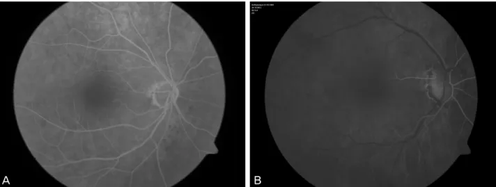
Figure 1. The pattern of watershed zone in fluorescence angiography. (A) 65-year-old female NTG patient shows that watershed
zone is not including the optic nerve head. (B) 72-year-old female POAG patient shows that watershed zone including the optic nerve head. NTG = normal tension glaucoma; POAG = primary open-angle glaucoma.지지 않은 환자, FAG 검사상 결과가 부정확한 환자, 시야 검사 결과 신뢰도가 떨어지는 환자, 정기적으로 외래를 방 문하지 못한 환자는 대상에서 제외하였다.
안압 검사는 낮 시간에 한 명의 동일 검사자가 골드만 압 평 안압계를 이용하여 3회 측정한 안압의 평균치를 사용하 였고, 환자의 안압은 항녹내장 점안제만으로 치료하였으며, 정상안압녹내장에서는 기저안압(Baseline)에 비해 20% 이 상 감소된 경우로, 원발개방각녹내장에서는 30% 이상 감소 된 안압을 목표안압으로 정의하였다.47,48
시야검사는 자동시야검사(Humphrey field analyzer, 24-2 Swedish Interactive Thresholding Algorithm (SITA) stand- ard program, Carl-Zeiss Meditec, Dublin, CA, USA)를 이용 하여 적어도 2년 이상 경과 관찰한 환자 중 최소 6개월 간 격으로 4회 이상 시행하였다. 시야결손의 진행은 6개월 전 후의 시야검사에서 the Advanced Glaucoma Intervention Study (AGIS) method를 이용하여 AGIS score상 적어도 2 unit 이상의 감소가 있거나 mean deviation (MD)이 2 dB 이 상의 감소가 있는 경우로 정의하였다.49,50 또한 거짓양성반 응(false positive response)이 20%, 거짓음성반응(false neg- ative response)이 33%를 넘거나 주시 실패(fixation loss)가 20% 이상일 때에는 검사의 신뢰도가 낮다고 판단하여 결 과분석에서 제외하였다.51
24 hrs ABPM은 전자혈압계인 24시간 활동혈압측정기 (TONOPORT V. GM Medical System, Germany)를 이용하 여 비우세완(non-dominant arm)에서 측정하였다. 주간 혈압 (오전 7시-오후 10시)은 30분 간격으로, 야간 혈압(오후 10 시-오전 7시)은 1시간 간격으로 측정하였다. SBP, DBP, HR 를 측정하여 최고치, 최저치, 평균치를 조사하였고, MAP
및 ND는 아래의 공식으로 구할 수 있다.52
×
ND는 주간 혈압의 평균치에 대한 야간 혈압의 평균치의 감소 비율로 구하며,53-55 야간 혈압하강이 10% 미만인 경우 를 non-dip, 10% 이상인 경우를 dip로 정의하였다.56,57
형광안저촬영은 초진 시 FF 450 plus IR (Carl Zeiss Meditec, Germany)을 이용하여 한 명의 숙련된 촬영자에 의해 시행되었다. 촬영 결과는 2명의 안과 전문의에 의해 형광조영 초기 맥락막 순환기와 초기 동맥기에 나타나는 저형광대를 관찰해 맥락막분수계역의 유무를 판정하였다 (Fig. 1).
위의 검사 내용을 근거로 목표안압 이하로 안압을 유지 하는 개방각녹내장환자를 대상으로 dip의 유무, 맥락막분 수계역의 유무가 녹내장성 시야결손 진행에 미치는 영향을 Fisher’s exact test 및 Binary logistic regression test를 통해 분석하였다(SPSS ver.12.0, SPSS Inc. Chicago, USA).
결 과
개방각녹내장 환자 36명(68안) 중 정상안압녹내장 환자 는 23명(43안)이고 원발개방각녹내장 환자는 13명(25안)이 었다. 개방각녹내장 환자군의 평균 연령은 62.07세이고 정 상안압녹내장 환자군의 평균 연령는 58.35세이며 원발개방 각녹내장 환자군의 평균 연령 68.48세이었다(Table 1).
목표안압을 유지하기 위해 항녹내장 약물을 단일 또는
A B
Table 1. Clinical and demographic data of the patients with OAG
OAG NTG POAG p-value
M:F (eye) 22:46 11:32 11:14 0.194*
Mean age (years) 62.07 ± 11.32 58.35 ± 12.44 69.48 ± 9.72 0.351†
Mean baseline IOP (mm Hg) 19.64 ± 3.68 17.57 ± 3.14 23.78 ± 4.56 0.062†
Mean IOP (mm Hg) 13.04 ± 2.64 12.89 ± 2.18 14.64 ± 3.73 0.072†
Mean Δ IOP decrease (mm Hg) 6.64 ± 2.98 4.68 ± 1.59 9.14 ± 3.74 0.068†
Mean Δ IOP decrease (%) 33.60 ± 3.46 26.64 ± 2.57 42.65 ± 3.41 0.057†
Mean deviation of visual field (dB) -5.85 ± 4.84 -5.59 ± 4.21 -6.82 ± 6.33 0.083†
Pattern standard deviation of visual field (dB) -3.89 ± 3.05 -3.73 ± 3.25 -4.29 ± 2.96 0.682† Mean deviation change of visual field (dB) -3.24 ± 2.86 -3.26 ± 0.45 -3.42 ± 2.64 0.068†
Mean AGIS score change (point) -3.25 ± 2.13 -3.15 ± 3.16 -3.49 ± 1.56 0.105†
Mean nocturnal dip of SBP (%) 7.31 ± 9.16 7.26 ± 9.81 7.44 ± 8.8 0.947†
Mean nocturnal dip of DBP (%) 8.84 ± 8.72 9.20 ± 8.67 8.63 ± 8.94 0.817†
Incidence of watershed zone (%) 39.71 (27 eyes) 46.51 (20 eyes) 28.00 (7 eyes) 0.135* Incidence of watershed zone with ONH involvement (%) 25.00 (17 eyes) 27.90 (12 eyes) 20.00 (5 eyes) 0.179* Values are presented as mean ± SD.
OAG = open angle glaucoma; IOP = intraocular pressure; AGIS = advanced glaucoma intervention study; SBP = systolic blood pressure;
DBP = diastolic blood pressure; ONH = optic nerve head; NTG = normal tension glaucoma; POAG = primary open-angle glaucoma.
*Fisher’s exact test, statistical significance: p < 0.05; †Student t-test, statistical significance: p < 0.05.
Table 2. Anti-glaucoma eyedrops to obtain target intraocular pressure
Groups Anti-glaucoma eyedrops Eyes
NTG
PG 21
PG + CAI + β-blocker 12
PG + CAI + β-blocker + α-agonist 10
POAG CAI + β-blocker + α-agonist 12
PG + CAI + β-blocker + α-agonist 13
NTG = normal tension glaucoma; POAG = primary open-angle glaucoma; PG = prostaglandin; CAI = carbonic anhydrase inhibitor; β-blocker
= beta-adrenergic blocker; α-agonist = alpha-adrenergic agonist.
Table 3. The relationship between watershed zone and nocturnal dip in OAG
p-value* Odds ratio
OAG Watershed zone 0.024 3.308
ONH involvement with watershed zone 0.633 1.600
NTG Watershed zone 0.029 4.364
ONH involvement with watershed zone 1.000 1.444
POAG Watershed zone 0.568 1.667
ONH involvement with watershed zone 1.000 1.500
OAG = open-angle glaucoma; NTG = normal tension glaucoma; POAG = primary open-angle glaucoma; ONH = optic nerve head.
*Fisher's exact test, statistical significance: p < 0.05.
병용 사용하였다(Table 2). 치료 후 안압은 기저 안압에 비 해 정상안압녹내장 환자군은 26.64%, 원발개방각녹내장 환 자군은 42.65%로 기대했던 목표안압 이하의 수준으로 유 지되었다(Table 1).
시야검사 결과에서 정상안압녹내장환자 43안 중 14안 (32.6%), 원발개방각녹내장환자 25안 중 9안(36%)에서 녹 내장성 시야결손의 진행이 나타났다. 시야검사상 녹내장성 진행이 나타난 환자들 중 정상안압녹내장환자는 평균 -3.15 unit의 AGIS score 감소 및 -3.26 dB의 MD 변화를 보였고 원발개방각녹내장환자는 AGIS score -3.49 unit 및 MD -3.42
dB의 변화를 보였다. 그 외 24 hrs ABPM 결과 및 맥락막분 수계역은 각 군 간 의미 있는 차이는 없었다(Table 1).
맥락막분수계역은 68안 중 27안(39.7%)에서 발견하였으 며 이 중 17안(25%)에서 시신경유두를 침범하였다. 정상안 압녹내장환자 43안 중에서는 20안(46.51%)에서 맥락막분 수계역이 관찰되었으며 이 중 12안(27.90%)에서 시신경 유 두를 침범하였다. 원발개방각녹내장환자에서는 25안 중 7안 (28%)에서 맥락막분수계역이 관찰되었으며 이 중 5안(20%) 에서 시신경 유두 침범이 나타났다(Table 1).
맥락막분수계역의 유무와 dip 유무와의 상관관계에서도
Table 4. The risk factors of glaucomatous visual field progression in OAG
Fisher's exact test* Binary logistic regression test†
p-value Odds ratio p-value Odds ratio
OAG Sex 0.931 0.1058 0.982 0.985
Age over 60-year-old 0.189 0.966 0.060 3.672
Nocturnal dip over 10% 0.003 5.938 0.010 6.227
Watershed zone 0.002 5.156 0.210 2.841
Watershed zone with ONH involvement 0.004 5.958 0.430 2.034
NTG Sex 0.827 1.246 0.954 1.065
Age over 60-year-old 0.579 0.984 0.418 2.007
Nocturnal dip over 10% 0.005 13.929 0.060 11.335
Watershed zone 0.024 4.750 0.836 1.246
Watershed zone with ONH involvement 0.009 8.333 0.209 3.947
POAG Sex 0.757 0.713 0.189 0.152
Age over 60-year-old 0.198 0.900 0.999 7.629
Nocturnal dip over 10% 0.226 3.333 0.081 13.605
Watershed zone 0.021 8.750 0.999 2.630
Watershed zone with ONH involvement 1.000 1.500 0.999 0.000
OAG = open-angle glaucoma; ONH = optic nerve head; NTG = normal tension glaucoma; POAG = primary open-angle glaucoma.
*Fisher's exact test, statistical, significance: p < 0.05, confidence interval (CI) was calculated at 95 percentile; †Binary logistic regression test, significance: p < 0.05, confidence interval (CI) was calculated at 95 percentile.
개방각녹내장 환자군과, 정상안압녹내장 환자군에서 통계 학적으로 유의한 상관관계를 보였으며(OAG: p=0.024, NTG: p=0.029), 각각의 OR은 OAG 환자군에서 OR=3.308, 정상안압녹내장 환자군에서 OR=4.364로 나타났다. 하지만 맥락막분수계역을 보이는 환자들 중 시신경유두침범여부 와 dip 사이에는 관련성을 찾을 수 없었다(Table 3).
녹내장성 시야결손의 진행에 대한 성별 및 60세 이상의 고령은 모든 군에서 상관관계를 찾을 수 없었다. dip의 유 무에 따른 녹내장성 시야결손의 진행 사이의 상관 분석에 서는, 개방각녹내장환자군과 정상안압녹내장환자군에서 통계 학적으로 유의한 상관관계를 보였고(OAG: p=0.003, NTG:
p=0.005), 각각의 odds ratio (OR)는 개방각녹내장환자군에 서 OR=5.938, 정상안압녹내장 환자군에서 OR=13.929로 나타났다(Table 4).
맥락막분수계역의 유무와 녹내장성 시야결손의 진행과 의 상관 분석은 모든 군에서 유의한 상관관계를 보였다 (OAG: p=0.002, NTG: p=0.023, POAG: p=0.021). 각각의 OR은 개방각녹내장환자군에서 OR=5.156, 정상안압녹내장 환자군에서 OR=4.750, 원발개방각녹내장 환자군은 OR=8.750 로 나타났다(Table 4). 그리고 맥락막분수계역의 시신경유 두 침범 유무와 녹내장성 시야결손 진행의 유무와의 상관 분석에서는 개방각녹내장 환자군과 정상안압녹내장 환자 군에서 통계학적으로 유의한 상관관계를 보였고(OAG:
p=0.004, NTG: p=0.009), 각각의 odds ratio (OR)는 개방각 녹내장환자군에서 OR=5.958, 정상안압녹내장 환자군에서 OR=8.333으로 나타났다(Table 4).
녹내장성 시야결손에 대한 Binary logistic regression test
에선 10% 이상의 야간 혈압하강이 가장 큰 위험요인으로 나타났지만 개방각녹내장환자군에서만 통계적으로 유의성 을 나타내었다(p=0.010, OR=6.227) (Table 4).
고 찰
녹내장 환자의 시신경 및 시야결손의 녹내장성 변화에는 안압 상승으로 인한 기계적 요인과 안혈류량의 감소로 인 한 허혈성 요인이 중요하다.1 목표안압 이하로 안압이 일정 하게 유지됨에도 불구하고 시신경의 녹내장성 변화와 녹내 장성 시야결손이 진행되는 것은5 안압 이외의 다른 여러 요 인들이 녹내장의 발생 및 진행에 영향을 주고 있음을 알 수 있다. 개방각녹내장환자, 특히 정상안압녹내장에서 안혈류 량의 감소가 녹내장의 발생과 진행에 영향을 주고 있어, 안 관류압의 개선을 통해 녹내장의 발생과 진행을 억제할 수 있을 것으로 생각한다.57
시신경유두의 안관류압은 평균동맥압과 연관되어 있고, 안압의 변화와는 관계없이 혈압의 감소만으로도 평균동맥 압이 감소하여 안관류압이 부족해질 수 있다.58-61 안관류압 과 안혈류량을 살펴보는 다른 검사에 비해 비교적 간단하 게 시행 가능한 24 hrs ABPM은 녹내장 환자들의 야간 혈 압하강, 평균동맥압를 알 수 있고, 평균동맥압과 야간 혈압 하강의 감소가 심한 녹내장 환자들은 시신경유두 및 시야 의 녹내장성 진행과 연관이 있다.32,40 저자들이 보고한 이전 연구에서 목표안압 이하로 안압을 유지함에도 불구하고 야 간 혈압하강이 10% 이상인 개방각녹내장 및 정상안압녹내 장 환자군에서 녹내장성 시야결손이 진행되고, 특히 고혈
압 치료를 받는 환자에서 10% 이상의 야간혈압하강이 많 이 나타났고, 녹내장성 시야결손의 진행과도 관련이 있었 다고 보고하였다.41 따라서 목표안압 이하로 안압을 유지시 켜 시신경유두에 가해지는 기계적 손상을 억제하더라도 야 간 혈압하강과 평균동맥압의 감소로 인한 안관류압의 하강 만으로도 시신경유두와 시야의 녹내장성 변화와 진행을 야 기할 수 있다.32,40,41
맥락막분수계역은 망막의 종동맥들의 분포 영역 사이의 경계구역으로 형광 조영 초기 맥락막 순환기와 초기 동맥 기에 저형광대로 관찰되는데, 망막 질환, 허혈성 안질환, 녹 내장 등에서 빈번하게 발생한다.44 혈관성 자율조절장애가 생길 수 있는 모든 경우에서 안관류압이 감소하여 맥락막 분수계역이 생길 위험이 높다.42,56 이러한 맥락막분수계역 의 위치와 범위는 혈관의 분포와 위치에 따라 개인마다 차 이가 있다. Hayreh42-45 맥락막분수계역의 위치에 대하여 60%에서 시신경유두의 이측에 존재하였고, 16%의 환자에 서 시신경유두 전체를 포함하였으며, 10%의 환자에서 시신 경유두의 비측에 존재한다고 보고하였다. 이번 연구에서는 68안 중 27안(39.7%)에서 맥락막분수계역을 발견하였으며 이 중 17안(25%)에서 시신경유두를 침범하였다.62 이 중 13 안(76.5%)은 시신경 유두에서 이측부위에 존재하였다. 정 상안압녹내장환자 43안 중에서는 20안(46.5%)에서 맥락막 분수계역이 관찰되었으며 이 중 12안(60%)에서 시신경 유 두를 침범하였다.63 이는 Xu et al64의 연구와 비교할 때 맥 락막분수계역의 관찰 비율과 시신경유두침범의 비율 모두 낮았다.
Singh and Dass65,66는 개방각녹내장 환자에서 안관류압의 감소가 발생할 경우 맥락막분수계역이 상대적으로 허혈성 손상에 취약하여 녹내장성 시신경 변화를 유발할 수 있다 고 보고하였다. 하지만 녹내장이 없는 정상인의 대부분에 서 시신경유두를 포함하는 맥락막분수계역이 나타나며 녹 내장 환자와 정상인 사이에 맥락막분수계역의 형태는 큰 차이가 없다는 보고도 있다.65,66 맥락막분수계역의 형태나 위치는 단후모양체동맥의 위치를 반영할 뿐이고, 안혈류량 을 나타내는 것은 아니며, 맥락막분수계역의 형태 및 위치 와 시신경유두의 녹내장성 손상 사이에 연관성은 없었다는 연구 결과도 있지만67-69 반대로 맥락막분수계역을 보이는 환자에서 시신경유두를 더 많이 포함할수록 시야검사상 시 야결손이 더 크다는 연구도 있다.70
본 연구에서는 개방각녹내장 및 정상안압녹내장 환자군 에서 맥락막분수계역과 dip 사이에서는 유의한 상관관계를 보였다. 해부학적 형태를 반영하는 맥락막분수계역과 전신 적인 자율신경반응을 나타내는 야간혈압저하 사이는 개별 적인 것으로 보이지만 각각 녹내장환자에서 위험요인이며
본 연구에서 통계적으로 유의한 상관관계를 보였다는 것은 녹내장 환자에서 해부학적인 차이와 생리적인 요인의 결합 이 녹내장의 발생에 관여한다는 측면에서 의미가 있다고 생각한다.
모든 군에서 맥락막분수계역과 녹내장성 시야결손의 진 행 사이에 유의한 상관관계가 있었다. 이는 해부학적으로 허혈성 손상에 취약한 맥락막분수계역을 보이는 환자에서 야간 혈압하강과 같은 전신 혈압저하가 있을 경우 녹내장 성 시야결손을 진행시켰을 것이라 생각한다. 추가적으로 시행한 맥락막분수계역의 시신경유두에 대한 침범 여부와 dip의 유무 및 녹내장성 시야결손의 진행 유무 사이의 상관 분석에서는, 통계학적으로 유의성을 찾을 수 없었다. 맥락 막분수계역을 보이는 환자에서 시신경유두를 침범한 형태 를 보일 때 안관류압이 저하된 환경에서 시신경에 직접적 인 손상의 가능성이 더 클 것으로 생각하나 이번 연구에서 는 대상 환자 수가 부족하여 통계적으로 의미있는 차이를 보이지 못하였다고 생각하며, 좀 더 많은 환자를 대상으로 조사하면 통계적으로 유의성을 찾을 수 있을 것으로 생각 한다. 또 안관류압에 영향을 줄 수 있는 다른 요인들에 대 한 분석은 추가적인 연구가 필요하다.
이항로지스틱회귀분석을 통해 녹내장성 시야결손의 진 행에 대한 성별, 60세 이상의 고령, 야간혈압저하, 맥락막분 수계역의 존재 및 시신경유두에 침범여부의 영향은 야간혈 압저하가 가장 큰 위험요인으로 나타났으며 그 외 60세 이 상의 고령, 맥락막분수계역 등이 위험요인으로 생각하였다 (Table 4).
따라서 목표안압 이하로 안압이 유지됨에도 불구하고 녹 내장성 시야결손이 진행되는 개방각녹내장 환자에서 24 hrs ABPM을 통해 야간혈압저하를 평가하고 초기 형광안 저촬영을 통해 맥락막분수계역의 유무만을 평가하기보다 시신경유두에 대한 침범까지 평가하는 것이 녹내장의 진행 을 예측하는 데 도움이 되리라 생각한다. 목표안압 이하로 안압을 조절한 상태에서 여러 가지 전신적 및 국소적인 허 혈성 위험 인자들을 확인하고 이들을 가능한 방법을 사용 하여 치료할 때에 녹내장으로 인한 실명의 위험을 최대한 예방할 수 있을 것으로 생각한다.
REFERENCES
1) Armaly MF. Ocular pressure and visual fields. A ten-year fol- low-up study. Arch Ophthalmol 1969;81:25-40.
2) Levens RZ. Low tension glaucoma: a critical review and new material. Surv Ophthalmol 1980;24:621-64.
3) Perkins ES. The Bedford glaucoma survey. I. Long-term follow-up of boarderline cases. Br J Ophthalmol 1973;57:179-85.
4) Van Buskirk EM, Cioffi GA. Glaucomatous optic neuropathy. Am
J Ophthalmol 1992;113:447-52.
5) Brubaker RF. Delayed functional loss in glaucoma. LII Edward Jackson Memorial Lecture. Am J Ophthalmol 1996;121:473-83.
6) Kim CS, Seong GJ, Lee NH, Song KC. Prevalence of primary open-angle glaucoma in central South Korea the Namil study.
Ophthalmology 2011;118:1024-30.
7) Detry M, Boschi A, Ellinghaus G, de Plaen JF. Simultaneous 24-h monitoring of intraocular pressure and arterial blood pressure in patients with progressive and non-progressive primary open-angle glaucoma. Eur J Ophthalmol 1996;6:273-8.
8) Fechtner RD, Weinreb RN. Mechanisms of optice nerve damage in primary open angle glaucoma. Surv Ophthalmol 1994;39:23-42.
9) Juronen E, Tasa G, Veromann S, et al. Polymorphic glutathione S-transferase M1 is a risk factor of primary open-angle glaucoma among Estonians. Exp Eye Res 2000;71:447-52.
10) Araie M, Sekine M, Suzuki Y, Koseki N. Factors contributing to the progression of visual field damage in eyes with normal-tension glaucoma. Ophthalmology 1994;101:1440-4.
11) Beano F, Org ul S, Stumpfig D, et al. An evaluation of the effect of unoprostone isopropyl 0.15% on ocular hemodynamics in normal- tension glaucoma patients. Graefe’s Arch Clin Exp Ophthalmol 2001;239:81-6.
12) Buckley C, Hadoke PWF, Henry E, O’Brien C. Systemic vascular endothelial cell dysfunction in normal pressure glaucoma. Br J Ophthalmol 2002;86:227-32.
13) Chung HS, Harris A, Kagemann L, Martin B. Peripapillary retinal blood flow in normal tension glaucoma. Br J Ophthalmol 1999;
83:466-9.
14) The effectiveness of intraocular pressure reduction in the treatment of normal-tension glaucoma. Collaborative Normal-Tension Glaucoma Study Group. Am J Ophthalmol 1998;126:498-505.
15) Drance SM, Douglas GR, Wijsman K, et al. Response of blood flow to warm and cold in normal and low-tension glaucoma patients. Am J Ophthalmol 1988;105:35-9.
16) Drance S, Anderson DR, Schulzer M. Risk factors for progression of visual field abnormalities in normal-tension glaucoma. Am J Ophthalmol 2001;131:699-708.
17) Flammer J, Orgül S, Costa VP, et al. The impact of ocular blood flow in glaucoma. Pro Retin Eye Res 2002;21:359-93.
18) Flammer J, Pache M, Resink T. Vasospasm, its role in the patho- genesis of disease with particular reference to eye. Pro Retin Eye Res 2001;20:319-49.
19) Becker B. Diabetes mellitus and primary open-angle glaucoma.
The XXVII Edward Jackson Memorial Lecture. Am J Ophthalmol 1971;71:1-16.
20) Mcleod SD, West SK, Quigley HA, Fozard JL. A longitudinal study of the relationship between intraocular and blood pressures.
Invest Ophthalmol Vis Sci 1990;31:2361-6.
21) Armstrong JR, Daily RK, Dobson HL, Girard LJ. The incidence of glaucoma in diabetes mellitus. A comparison with the incidence of glaucoma in the general population. Am J Ophthalmol 1960;
50:55-63.
22) Mitchell P, Lee AJ, Rochtchina E, Wang JJ. Open-angle glaucoma and systemic hypertension: the blue mountains eye study. J Glaucoma 2004;13:319-26.
23) Pradalier A, Hamard P, Sellem E, Bringer L. Migraine and glauco- ma: an epidemiologic survey of French ophthalmologists. Cephalalgia 1998;18:74-6.
24) Broadway DC, Drance SM. Glaucoma and vasospasm. Br J Ophthalmol 1998;82:862-70.
25) Millar-Craig MW, Bishop CN, Raftery EB. Circadian variation of blood pressure. Lancet 1978;1:795-7.
26) Pickering T. Recommendations for the use of home (self) and am- bulatory blood pressure monitoring. American Society of Hyper- tension Ad Hoc Panel. Am J Hypertens 1996;9:1-11.
27) Arend O, Remky A, Cantor LB, Harris A. Altitudinal visual field asymmetry is coupled with altered retinal circulation in patients with normal pressure glaucoma. Br J Ophthalmol 2000;84:1008-12.
28) Arend O, Remky A, Redbrake C, et al. [Retinal hemodynamics in patients with normal pressure glaucoma. Quantification with digi- tal laser scanning fluorescein angiography]. Ophthalmologe 1999;
96:24-9.
29) Flammer J, Orgül S. Optic nerve blood-flow abnormalities in glaucoma. Progr Ret Eye Res 1998;17:267-89.
30) Hayreh SS. The 1994 Von Sallman Lecture. The optic nerve head circulation in health and disease. Exp Eye Res 1995;61:259-72.
31) Plange N, Remky A, Arend O. [Absolute filling defects of the optic disc in fluorescein angiograms in glaucoma--a retrospective clin- ical study]. Klein Monatsbl Augenheilkd 2001;218:214-21.
32) Be´chetoille A, Bresson-Dumont H. Diurnal and nocturnal blood pressure drops in patients with focal ischemic glaucoma. Graefe’s Arch Clin Exp Ophthalmol 1994;232:675-9.
33) Chumbley LC, Brubaker RF. Low-tension glaucoma. Am J Ophthalmol 1976;81:761-7.
34) Dielemans I, Vingerling JR, Algra D, et al. Primary open-angle glaucoma, intraocular pressure, and systemic blood pressure in the general elderly population. The Rotterdam Study. Ophthalmology 1995;102:54-60.
35) Drance SM, Wheeler C, Pattullo M. Uniocular open-angle glaucoma. Am J Ophthalmol 1968;65:891-902.
36) Francois J, Neetens A. The deterioration of the visual field in glau- coma and the blood pressure. Doc Ophthalmol 1970;28:70-132.
37) Geijssen HC. Systemic Vascular Risc Factors. In: Geijssen HC , ed.
Studies on Normal Pressure Glaucoma. New York: Kugler Publications,1991;chap. 6.
38) Hayreh SS, Podhajsky P, Zimmermann MB. Role of nocturnal arte- rial hypotension in optic nerve head ischemic disorders. Ophthal- mologica 1999;213:76-96.
39) Jampol LM, Board RJ, Maumenee AE. Systemic hypotension and glaucomatous changes. Am J Ophthalmol 1978;85:154-9.
40) Hayreh SS, Zimmerman MB, Podhajsky P, Alward WL. Nocturnal arterial hypotension and its role in optic nerve head and ocular is- chemic disorders. Am J Ophthalmol 1994;117:603-24.
41) Seo HR, Ryu WY, Rho SH. Corrlelation between nocturnal dip and progression of glaucoma. J Korean Ophthalmol Soc 2010;51:
1471-8.
42) Hayreh SS. In vivo choroidal circulation and its watershed zones.
Eye (Lond) 1990;4:273-89.
43) Hayreh SS. Segmental nature of the choroidal vasculature. Br J Ophthalmol 1975;59:631-48.
44) Hayreh SS. Inter-individual variation in blood supply of the optic nerve head. Its importance in various ischemic disorders of the op- tic nerve head, and glaucoma, low-tension glaucoma and allied disorders. Doc Ophthalmol 1985;59:217-46.
45) Hayreh SS. Posterior ciliary artery circulation in health and dis- ease: the Weisenfeld lecture. Invest Ophthalmol Vis Sci 2004;
45:749-57.
46) Hayreh SS, Ritch R, Shield MB, Krupin T. Blood supply of the an- terior optic nerve. The glaucomas. St. Louis: C.V. Mosby, 1989;
133-61.
47) Huang P, Shi Y, Wang X, et al. Interocular Asymmetry of the Visual Field Defects in Newly Diagnosed Normal-tension Glaucoma, Primary Open-angle Glaucoma, and Chronic Angle-clo- sure Glaucoma. J Glaucoma 2013 Apr 29 [Epub ahead of print].
48) Aoyama A, Ishida K, Sawada A, Yamamoto T. Target intraocular pressure for stability of visual field loss progression in nor- mal-tension glaucoma. Jpn J Ophthalmol 2010;54:117-23.
49) The effectiveness of intraocular pressure reduction in the treatment of normal-tension glaucoma. Collaborative Normal-Tension Glaucoma Study Group. Am J ophthalmol 1998;126:498-505.
50) Advanced Glaucoma Intervention Study. 2. Visual field test scor- ing and reliability. Ophthalmology 1994;101:1445-55.
51) Kim J, Dally LG, Ederer F, et al. The Advanced Glaucoma Intervention Study (AGIS): 14. Distinguishing progression of glaucoma from visual field fluctuations. Ophthalmology 2004; 111:2109-16.
52) Wilkins MR, Fitzke FW, Khaw PT. Pointwise linear progression criteria and the detection of visual field change in a glaucoma trial.
Eye (Lond) 2006;20:98-106.
53) Zheng L, Sun Z, Li J, et al. Pulse pressure and mean arterial pres- sure in relation to ischemic stroke among patients with uncon- trolled hypertension in rural areas of China. Stroke 2008;39:
1932-7.
54) Plange N, Kaup M, Daneljan L, et al. 24-h blood pressure monitor- ing in normal tension glaucoma: night-time blood pressure variability. J Hum Hypertens 2006;20:137-42.
55) Graham SL, Drance SM, Wijsman K, et al. Ambulatory blood pres- sure monitoring in glaucoma. The nocturnal dip. Ophthalmology 1995;102:61-9.
56) Hirotsu C, Ohta E, Hirose N, Shimizu K. Profile analysis of 24-hours measurements of blood pressure. Biometrics 2003;59:
907-15.
57) Choi J, Jeong J, Cho H, Kook MS. Effect of nocturnal blood pres- sure reduction on circadian fluctuation of mean ocular perfusion pressure: a risk factor for normal tension glaucoma. Invest Ophthalmol Vis Sci 2006;47:831-6.
58) Collignon N, Dewe W, Guillaume S, Collignon-Brach J. Ambulatory blood pressure monitoring in glaucoma patients. The nocturnal systolic dip and its relationship with disease progression. Int Ophthalmol 1998;22:19-25.
59) Hayreh SS. The blood supply of the optic nerve head and the evalu- ation of it - myth and reality. Prog Retin Eye Res 2001;20:563-93.
60) Gherghel D, Orgul S, Gugleta K, et al. Relationship between ocular perfusion pressure and retrobulbar blood flow in patients with glaucoma with progressive damage. Am J Ophthalmol 2000;130:
597-605.
61) Sehi M, Flanagan JG, Zeng L, et al. Relative change in diurnal mean ocular perfusion pressure: a risk factor for the diagnosis of primary open-angle glaucoma. Invest Ophthalmol Vis Sci 2005;46:
561-7.
62) Hayreh SS. Duke-elder lecture. Systemic arterial blood pressure and the eye. Eye (Lond) 1996;10:5-28.
63) Harris A, Rechtman E, Siesky B, et al. The role of optic nerve blood flow in the pathogenesis of glaucoma. Ophthalmol Clin North Am 2005;18:345-53.
64) Xu W, Grunwald JE, Metelitsina TI, et al. Association of risk fac- tors for choroidal neovascularization in age-related macular degen- eration with decreased foveolar choroidal circulation. Am J Ophthalmol 2010;150:40-7.
65) Singh S, Dass R. The central artery of the retina. I. Origin and course. Br J Ophthalmol 1960;44:193-212.
66) Singh S, Dass R. The central artery of the retina. II. A study of its distribution and anastomosis. Br J Ophthalmol 1960;44:280-99.
67) Giuffre G. Main posterior watershed zone of the choroid.
Variations of its position in normal subjects. Doc Ophthalmol 1989;72:175-80.
68) Onda E, Sato Y, Takahashi S, et al. Angiographic evaluation of wa- tershed zone in the peripapillary choroid in glaucoma and non-glaucoma eyes. Invest Ophthalmol Vis Sci 1996;37:267.
69) Onda E, Cioffi GA, Bacon DR, van Buskirk EM. Microvasculature of the human optic nerve. Am J Ophthalmol 1995;120:92-102.
70) Sato Y1, Tomita G, Onda E, et al. Association between watershed zone and visual field defect in normal tension glaucoma. Jpn J Ophthalmol 2000;44:39-45.
= 국문초록 =
개방각녹내장에서 맥락막분수계역, 야간 혈압하강 및 녹내장성 시야결손의 상관관계
목적: 맥락막분수계역(water-shed zone, WSZ)과 야간 혈압하강(nocturnal dip, ND)이 녹내장성 시야결손 진행에 미치는 영향에 대해 알아보고자 하였다.
대상과 방법: 목표안압 이하로 안압이 유지되는 OAG환자를 대상으로 형광안저촬영, 24시간활동혈압검사 및 시야검사를 시행하여 WSZ, 10% 이상의 ND (dip) 및 녹내장성 시야결손 진행과의 관계를 분석하였다.
결과: WSZ과 dip의 상관관계는 OAG군(p=0.024) 및 NTG군(p=0.029)에서 유의하였다. dip와 시야결손 진행과의 상관관계는 OAG군 (p=0.003)과 NTG군(p=0.005)에서 의미를 보였다. WSZ과 시야결손 진행의 관계는 모든 군에서 유의하였다(OAG: p=0.002, NTG:
p=0.023, POAG: p=0.021). WSZ의 시신경유두 침범과 시야결손의 진행 사시에는 OAG군(p=0.004)과 NTG군(p=0.009)에서 유의했 다. 시야결손의 진행에 대한 이항로지스틱회귀분석에선 OAG군에서 dip (p=0.010, OR=6.227)만이 통계적으로 유의한 위험요인으로 나타났다.
결론: OAG군 및 NTG군에서 WSZ는 dip와 상관관계를 보였고 dip는 녹내장성 시야결손의 진행에 영향을 주었다. WSZ는 모든 군에서 시야결손 진행에 영향을 주었고 시신경유두 침범여부가 중요하였다. 목표안압 이하로 안압이 유지됨에도 불구하고 녹내장성 시야결 손 진행을 보이는 OAG환자에서 WSZ와 시신경유두의 침범여부, dip를 확인하는 것이 도움이 될 것으로 생각한다.
<대한안과학회지 2014;55(7):1030-1038>