Risk factors for pancreatogenic diabetes after pancreaticoduodenectomy
Hyoung Min Oh, Yoo-Seok Yoon, Ho-Seong Han, Ji Hoon Kim, Jai Young Cho, and Dae-Wook Hwang
Department of Surgery, Seoul National University Bundang Hospital, Seoul National University College of Medicine, Seongnam, Korea
Backgrounds/Aims: Postoperative diabetes mellitus (DM) after pancreaticoduodenectomy (PD) may compromise the long-term quality of life in survivors after the operative procedure due to the treatment difficulty and its related complications. The aim of this study is to determine the incidence of new-onset pancreatogenic DM after PD and investigate the risk factors for this complication. Methods: Among 170 patients who had undergone PD between November 2003 and September 2009, 98 patients were selected for this study. The selected patients were non-diabetic prior to the operation and had undergone follow-up tests for glucose metabolism and an abdominal computed tomog- raphy (CT) scan 1 year after the operation. The clinical data of these patients were retrospectively analyzed by review- ing the medical records, radiologic images, and pathologic reports. Results: Postoperative pathology confirmed malig- nant tumors in 91 patients, borderline malignancy in 5, and benign tumor in 2. The tumor locations included the pancre- atic head (n=30), the common bile duct (CBD) (n=30), ampulla of Vater (n=30), and the duodenum (n=8). New-onset DM occurred in 17 (17.4%) of the 98 patients during the first year after the operation. The comparative analysis be- tween postoperative DM (+) and DM (−) groups revealed that the atrophy of the remaining pancreas was the only significant risk factor for development of postoperative DM after PD. Conclusions: This study suggests that the atrophy of the remaining pancreas increases the risk of pancreatogenic DM after PD, and efforts to prevent pancreatic atrophy are needed to decrease this complication. (Korean J Hepatobiliary Pancreat Surg 2012;16:167-171)
Key Words: Pancreaticoduodenectomy; Pancreatogenic diatetes mellitus; Pancreatic atrophy
Received: August 14, 2012; Revised: September 15, 2012; Accepted: October 15, 2012 Corresponding author: Yoo-Seok Yoon
Department of Surgery, Seoul National University Bundang Hospital, Seoul National University College of Medicine, Gumi-dong, Bundang-gu, Seongnam 463-707, Korea
Tel: +82-31-787-7096, Fax: +82-31-787-4055, E-mail: [email protected]
Copyright Ⓒ 2012 by The Korean Association of Hepato-Biliary-Pancreatic Surgery Korean Journal of Hepato-Biliary-Pancreatic Surgery ∙ pISSN: 1738-6349
INTRODUCTION
There is limited data on the long-term quality of life of the patients following the pancreaticoduodenectomy (PD), mainly due to the poor prognosis of the malignant disease constituting the major indications for this oper- ative procedure, along with the high postoperative mor- bidity and mortality rates. However, the long-term surviv- al rate has recently increased due to the refined surgical techniques, improvement in perioperative management, development of adjuvant treatments, and more frequent use of PD in benign and borderline malignant tumors.1 The improved survival after PD has led to an increased concern regarding the long-term metabolic consequen- ces.2,3
The loss of pancreatic parenchyma resulting from pan-
createctomy can lead to the abnormality in the glucose ho- meostasis known as pancreatogenic diabetes mellitus (DM). Impairment of the glucose metabolism after PD is difficult to predict and can range from minimal to major metabolic derangements. Although pancreatic insuffici- ency is generally known to occur when more than 80%
of the pancreas is resected, new-onset DM has been re- ported to develop in 20-50% of the patients even after PD involving the removal of approximately 40-50% of pan- creatic volume.1,4 Pancreatogenic DM can compromise the long-term quality of life of the patients, because it is fre- quently associated with iatrogenic hypoglycemia resulting from increased peripheral sensitivity to insulin, unlike oth- er types of DM; hence, it is difficult to control with medication. Long-term inadequate glycemic control can also cause nephropathy, neuropathy, and retinopathy.
Therefore, it is important to evaluate the incidence of pan- creatogenic DM after PD, and identify the risk factors of developing this complication. Previous studies on pan- creatogenic DM mainly examined the changes in hormone secretion or glucose metabolism after pancreatectomy.2,5 There have been few clinical studies which have aimed to identify the risk factors associated with pancreatogenic DM.6 In this study, we investigated the frequency of oc- currence of new-onset pancreatogenic DM after PD, and analyzed the risk factors associated with the complication.
METHODS
Patients
The PD was performed in 170 patients between No- vember 2003 and September 2009. Among those patients, 98 patients who had undergone follow-up tests for glucose metabolism at one year after the surgery were selected for this study. Among the patients, 48 patients with under- lying DM and 24 patients who were lost to follow-up or died within one year after the operation were excluded from the study. The patients were divided into two groups, according to the postoperative development of DM; DM (+) group (n=17) and DM (−) group (n=81).
The clinical data of these patients were retrospectively an- alyzed by reviewing the medical records, radiologic im- ages, and pathologic reports. To identify the risk factors for new-onset DM after PD, the following data were com- pared between the two groups: age, sex, body mass index (BMI), type of operation (Whipple vs. pylorus-preserving pancreatoduodenectomy [PPPD]), tumor site, pathologic diagnosis (benign or borderline malignancy vs. malig- nancy), T stage, N stage, amount of intraoperative blood loss, operation time, postoperative complication, post- operative hospital stay, size of postoperative pancreatic duct, underlying pancreatitis, postoperative atrophy of the remaining pancreas, history of adjuvant chemoradiation therapy, and recurrence.
Definition of parameters
All patients underwent glucose metabolism tests, in- cluding oral glucose tolerance test (OGTT), as well as an abdominal computed tomography (CT), before and one year after the operation. In the OGTT, the blood glucose was measured after fasting and two hours after drinking
a glucose-rich beverage. Pancreatogenic DM was defined as the presence of diabetes symptoms, a random glucose level >200 mg/dl, fasting blood sugar (FBS) >126 mg/dl, or postprandial glucose level >140 mg/dl. The pancre- atic-duct was defined to be dilated when it was five mm or more in diameter on the postoperative CT images. The thickness of the pancreatic parenchyma near the pan- creatoenteric anastomosis site was measured using an ab- dominal CT, before and one year after the operation.
Pancreatic atrophy was confirmed when the postoperative parenchymal thickness was reduced to <50% of the pre- operative value. The presence of pancreatitis was eval- uated by postoperative pathologic examination.
Statistics
In order to determine the risk factors for development of diabetes, the categorical variables were compared using chi-square test, and the continuous variables were com- pared using the non-parametric Mann-Whitney U test. All the statistical analysis was performed using the SPSS ver- sion 19.0 for Windows (SPSS Inc., Chicago, IL, USA).
A p-value less than 0.05 was considered statistically significant.
RESULTS
Demographic characteristics
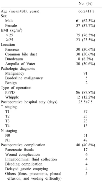
The demographic characteristics of the study population were summarized in Table 1. The study included 61 male and 37 female patients, with a mean age of 66.2 years (range: 18-78 years). Twenty-three patients (23.5%) had BMI of 25 or more. The tumor locations included the pan- creatic head (n=30, 30.6%), the common bile duct (n=30, 30.6%), ampulla of Vater (n=30, 30.6%), and the duode- num (n=8, 8.2%). PPPD was performed in 86 patients (87.8%) and the Whipple procedure in 12 patients (12.2%). Postoperative pathology confirmed: malignancy in 91 patients (adenocarcinoma, ductal adenocarcinoma, signet ring cell carcinoma, acinar cell carcinoma, meta- static renal cell carcinoma, and undifferentiated carcinoma with osteoclast-like giant cells); borderline malignancy in five patients (intraductal papillary mucinous adenoma [IPMN] with low/high grade dysplasia); and benign dis- ease (serous microcystic adenoma, and well differentiated endocrine tumor) in two patients. Postoperative complica-
Table 1. Demographic characteristics of the patients No. (%) Age (mean±SD, years)
Sex Male Female BMI (kg/m2)
<25
≥25 Location
Pancreas
Common bile duct Duodenum Ampulla of Vater Pathologic diagnosis
Malignancy
Borderline malignancy Benign
Type of operation PPPD
Whipple
Postoperative hospital stay (days) T staging
T1 T2 T3 T4 N staging
N0 N1
Postoperative complication Pancreatic fistula Wound complication
Intraabdominal fluid collection Bleeding complication Delayed gastric emptying
Others (ileus, pneumonia, pleural effusion, and voiding difficulty)
66.2±11.8 61 (62.3%) 37 (37.7%) 75 (76.5%) 23 (23.5%) 30 (30.6%) 30 (30.6%) 8 (8.2%) 30 (30.6%)
91 5 2 86 (87.8%) 12 (12.2%)
25.5±7.5 37 25 23 13 51 47 40 (40.8%)
17 6 4 4 4 5
BMI, body mass index; PPPD, pylorus-preserving pan- creatoduodenectomy
tions developed in 40 patients (40.8%), including pancre- atic fistula (n=19), delayed gastric emptying (n=4), intra- abdominal fluid collection (n=5), bleeding (n=4), and wound complication (n=6) (Table 2). The mean length of the postoperative hospital stay was 25.5 days (range:
15-36 days).
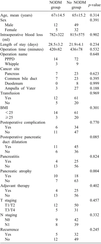
Risk factors for postoperative DM
In the follow-up of one year after the operation, post- operative new-onset DM (NODM) had developed in 17 (17.4%) of the 98 patients. Table 2 shows the comparative analysis of the risk factors for postoperative DM after PD two. Age, sex, BMI, type of operation, tumor site, patho-
logic diagnosis, T stage, N stage, amount of intraoperative blood loss, operation time, postoperative complication, postoperative hospital stay, postoperative pancreatic-duct size, underlying pancreatitis, history of adjuvant chemo- radiation therapy, and recurrence were not significantly different between the NODM group and no-NODM group.
Pancreatic atrophy was found to be the only significant risk factor for the development of postoperative DM after PD, with 10 patients (58.8%) in the NODM group vs. 18 patients (22.2%) in no-NODM group (p=0.004).
DISCUSSION
There have been contradictory reports on the develop- ment of pancreatogenic DM after PD.1,3,5,7-10 The degree of glucose metabolism impairment after pancreatectomy is related to the extent of pancreatic parenchyma resection, underlying pancreatic disease, and duration of fol- low-up.2,5,11 Assuming that PD involves the removal of similar volume (approximately 40-50%) of pancreatic pa- renchyma, the development of DM after PD can be main- ly determined by the latter two factors.1,4 However, most studies on this subject had variable follow-up durations after the operation or included patients with different en- docrine function of the pancreatic parenchyma prior to surgery. Some previous studies with a short-term fol- low-up suggested that DM was unlikely to develop after PD, while long-term follow-up studies reported the in- cidence of postoperative DM ranging from 20-50%.1 In addition, many studies included patients with underlying chronic pancreatitis or preoperative DM, making it diffi- cult to determine the effect of PD on the glucose metabo- lism of the remaining pancreatic parenchyma.6,12-18
To control the possible bias, this study was designed with patients who were non-diabetic preoperatively, with the evaluation of their glucose metabolism function at the same time-interval after operation. The results show that NODM occurred in 17% of the non-diabetic patients who underwent PD during the follow-up of one year after the operation. This is consistent with the results of the pre- vious studies that reported the incidence rate of 9-20% of pancreatogenic diabetes after PD.3,19 This finding indicates that the impairment of pancreatic endocrine function may develop after some degree of resection, unlike the exo- crine function that may not be hampered even after the
Table 2. Comparison of clinicopathologic factors between new-onset diabetes mellitus (NODM) group and no-NODM group
NODM group
No NODM group p-value Age, mean (years)
Sex Male Female
Intraoperative blood loss (ml)
Length of stay (days) Operation time (minutes) Operation name
PPPD Whipple Cancer site Pancreas
Common bile duct Duodenum Ampulla of Vater Transfusion
Yes No BMI
<25
≥25
Postoperative complication Yes
No
Postoperative pancreatic duct dilatation
Yes No Pancreatitis
Yes No
Pancreatic atrophy Yes
No
Adjuvant therapy Yes
No T staging
T1/T2 T3/T4 N staging
N0 N1 Recurrence
Yes No
67±14.5 12 5 782±322
28.5±3.2 420±82
14 3 7 7 0 3 12 5 14 3 6 11
11 6 4 13 10 7 4 13 12 5 9 8 5 12
65±15.2 49 32 815±575
21.9±4.1 436±78
72 9 23 23 8 27 61 20 61 20 34 47
45 36 25 56 18 63 25 56 50 31 42 39 32 49
0.314 0.391
0.902 0.234 0.532 0.648
0.623 0.395 0.999 0.188 0.969
0.301
0.770
0.085
0.824
0.004
0.402
0.457
0.332
0.245
BMI, body mass index; PPPD, pylorus-preserving pancreato- duodenectomy
resection of a considerable portion of the pancreas.
Moreover, this incidence is expected to increase through- out the remaining life of the patients, because glucose tol-
erance function naturally decreases with age, and the process of pancreatic atrophy may continue over time.9 Therefore, the endocrine function of patients with a long life expectancy after PD should be carefully monitored in order to early detect diabetes and to avoid the long-term complications caused by inadequate glycemic control.
Another important finding of this study is that among various clinicopathologic factors, postoperative pancreatic atrophy was the only risk factor for pancreatogenic DM after PD. The pancreatic atrophy after pancreatic head re- section resulted in the loss of endocrine function of pancreas.3,9 This finding is contradictory to the previous study of You et al.3 which showed that the volume of the remnant pancreas was not associated with pancreatogenic diabetes. These inconsistent results can be explained by the differences in the study design. The study of You et al.3 included 55 patients who had survived, after a median follow-up duration of no less than 55 months, among the 168 patients who had undergone PD; and the results of pancreatic endocrine function of the remaining patients were not presented. In comparison, this study evaluated the pancreatic endocrine function of all patients who had the follow-up period of more than one year. As such, the selection bias cannot be excluded in the previous study of You et al. Nonetheless, considering the small number of patients in this study, further prospective studies with a larger number of patients are needed to clarify the ef- fects of pancreatic atrophy on pancreatogenic DM after PD.
Atrophy of the remnant distal pancreas after PD com- monly occurs, and there have been several animal and hu- man studies in the attempts of preventing the pancreas atrophy.9 Although the mechanism is not well elucidated, a suggested mechanism involves the loss of gastro- intestinal hormones with the trophic effect on the pan- creas, such as cholecystokinin and gastrin, resulting from the resection of the duodenum and the distal stomach.
Jang et al. reported that induced hypergastrinemia can pre- vent pancreatic atrophy after PPPD, likely via a mecha- nism of the regenerative activity of the pancreas stimu- lated by gastrin.9 Additionally, a recent animal study showed that control of serum glucose levels play an im- portant role in preventing pancreatic atrophy.10 Based on the result of these studies, the efforts to prevent pancreatic atrophy should be continued and clinically applied to de-
crease the incidence of pancreatogenic DM after PD.
In conclusion, this study suggests that pancreatogenic DM is not an uncommon complication after PD, and that postoperative atrophy of the remaining pancreas sig- nificantly increases the risks of this complication. As such, patients who undergo PD need to be carefully fol- lowed up, in order to monitor the glucose metabolism function in avoiding the DM related long-term complica- tions and improving their quality of life. In addition, vari- ous efforts to prevent the atrophy of the remaining pan- creas are required to decrease this complication.
REFERENCES
1. Huang JJ, Yeo CJ, Sohn TA, et al. Quality of life and outcomes after pancreaticoduodenectomy. Ann Surg 2000;231:890-898.
2. Slezak LA, Andersen DK. Pancreatic resection: effects on glu- cose metabolism. World J Surg 2001;25:452-460.
3. You DD, Choi SH, Choi DW, et al. Long-term effects of pan- creaticoduodenectomy on glucose metabolism. ANZ J Surg 2012;
82:447-451.
4. Falconi M, Mantovani W, Crippa S, et al. Pancreatic in- sufficiency after different resections for benign tumours. Br J Surg 2008;95:85-91.
5. Ishikawa O, Ohigashi H, Eguchi H, et al. Long-term follow-up of glu- cose tolerance function after pancreaticoduodenectomy: Comparison between pancreaticogastrostomy and pancreaticojejunostomy. Surgery 2004;136:617-623.
6. Lemaire E, O’Toole D, Sauvanet A, et al. Functional and mor- phological changes in the pancreatic remnant following pan- creaticoduodenectomy with pancreaticogastric anastomosis. Br J Surg 2000;87:434-438.
7. Park YC, Kim SW, Jang JY, et al. Factor influencing delayed
gastric emptying after pylorus-preserving pancreatoduodenectomy.
J Am Coll Surg 2003;196:859-865.
8. Bock EA, Hurtuk MG, Shoup M, et al. Late complication after pan- creaticoduodenectomy with pancreaticogastrostomy. J Gastrointest Surg 2012;16:914-919.
9. Jang JY, Kim SW, Han JK, et al. Randomized prospective trial of the effect of induced hypergastrinemia on the Prevention of pancreatic atrophy after pancreatoduodenectomy in humans. Ann Surg 2003;237:522-529.
10. Yamamoto M, Jia DM, Fukumitsu K, Otsuki M. Treatment for hyperglycemia promotes pancreatic regeneration in rats without CCK-1 receptor gene expression. Pancreas 2003;26:368-374.
11. Jang JY, Kim SW, Park SJ, et al. Comparison of the functional outcome after pylorus-preserving pancreatoduodenectomy: pan- creatogastrostomy and pancreatojejunostomy. World J Surg 2002;
26:366-371.
12. Maeda H, Hanazaki K. Pancreatogenic diabetes after pancreatic resection. Pancreatology 2011;11:268-276.
13. Kim SW, Kim KW, Han JK, et al. Pylorus-preservation decreases the extent of atrophy of the remnant pancreas after pancreat- oduodenectomy. HPB 1999;1:65-70.
14. McPhee JT, Hill JS, Giles FW, et al. Perioperative mortality for pancreatectomy: a national perspective. Ann Surg 2007;246:
246-253.
15. Permert K, Ihse I, Jorfeldt L, et al. Improved glucose metabolism after subtotal pancreatectomy for pancreatic cancer. Br J Surg 1993;80:1047-1050.
16. Izbicki JR, Bloechle C, Knoefel WT, et al. Duodenum-preserving resection of the head of the pancreas in chronic pancreatitis: a prospective randomized trial. Ann Surg 1995;221:350-358.
17. Büchler M, Friess H, Muller M, et al. Randomized trial of duode- num-preserving pancreatic head resection versus pylorus-preserv- ing Whipple in chronic pancreatitis. Am J Surg 1995;169:65-69.
18. Grigore C, Sarbu V, Simion S, et al. Pancreatogenic diabetes pa- cient selection for pancreatic islet transplantation. J Med Life 2010;3:84-89.
19. King J, Kazanjian,K, Matusumoto J, et al. Distal Pancreatectomy:
Incidence of postoperative diabetes. J Gastrointest Surg 2008;12:
1548-1553.