Safe laparoscopic clearance of the common bile duct in emergently admitted patients with choledocholithiasis and cholangitis
Kristaps Atstupens, Haralds Plaudis, Vladimirs Fokins, Maksims Mukans, and Guntars Pupelis
Department of General and Emergency Surgery, Riga East University Hospital, Riga, Latvia
Backgrounds/Aims: Laparoscopic treatment of patients with choledocholithiasis and cholangitis is challenging due to mandatory recovery of the biliary drainage and clearance of the common bile duct (CBD). The aim of our study was to assess postoperative course of cholangitis and biliary sepsis after laparoscopic clearance of the CBD in emergently admitted patients with choledocholithiasis and cholangitis. Methods: Emergently admitted patients who underwent lapa- roscopic clearance of the CBD were included prospectively and stratified in 2 groups i.e., cholangitis positive (CH+) or negative (CH-) group. Patient demographics, comorbidities, preoperative imaging data, inflammatory response, surgi- cal intervention, complication rate and outcomes were compared between groups. Results: Ninety-nine of a total 320 patients underwent laparoscopic clearance of the CBD, of which, 60 belonged to the acute cholangitis group (CH+) and 39 to the cholangitis negative group (CH-). Interventions were done on average 4 days after admission, operation duration was 95-105 min, and the conversion rate was 3-7% without differences in the groups. Preoperative in- flammatory response was markedly higher in the CH+ group. Inflammation signs on intraoperative choledochoscopy were more evident in patients with cholangitis. Postoperative inflammatory response did not differ between the groups.
The overall complication rate was 8.3% and 5.1%, respectively. Laparoscopic clearance of the CBD resulted in 1 lethal case (CH+ group), resulting in 1% mortality rate and a similar 12-month readmission rate. Conclusions: Single-stage laparoscopic intraoperative US and choledochoscopy-assisted clearance of the CBD is feasible in emergently admitted patients with choledocholithiasis and cholangitis. (Korean J Hepatobiliary Pancreat Surg 2016;20:53-60)
Key Words: Choledocholithiasis; Laparoscopic surgery; Cholangitis; Ultrasound; Intraoperative; Choledochoscopy
Received: October 26, 2015; Revised: December 11, 2015; Accepted: December 13, 2015 Corresponding author: Kristaps Atstupens
Department of General and Emergency Surgery, Riga East University Hospital, Riga East University Hospital “Gailezers”, 2 Hipokrata St., LV 1038, Riga, Latvia
Tel: +371-29981662, Fax: +371-67042763, E-mail: [email protected]
Copyright Ⓒ 2016 by The Korean Association of Hepato-Biliary-Pancreatic Surgery
This is an Open Access article distributed under the terms of the Creative Commons Attribution Non-Commercial License (http://creativecommons.org/
licenses/by-nc/4.0) which permits unrestricted non-commercial use, distribution, and reproduction in any medium, provided the original work is properly cited.
Korean Journal of Hepato-Biliary-Pancreatic Surgery ∙ pISSN: 1738-6349ㆍeISSN: 2288-9213
INTRODUCTION
Cholelithiasis is one of the most common conditions that requires surgical intervention in Europe and the United States. According to the literature, roughly 10-15%
of the population may suffer from cholelithiasis, and 3-10% of patients who undergo cholecystectomy may have stones in the common bile duct (CBD).1
Laparoscopic cholecystectomy is one of the most fre- quently performed surgical interventions; however, intra- operative control of the patency of the CBD is not per- formed routinely in patients who are in the low risk cat- egory for probable choledocholithiasis. Simple laparo- scopic cholecystectomy is recommended if magnetic reso- nance cholangiopancreatography (MRCP) confirms the
absence of biliary stones in the CBD, or the patient has undergone endoscopic preoperative clearance of the CBD.
Endoscopic retrograde cholangiopancreatography (ERCP) typically combined with sphincterotomy, is a routine ther- apeutic procedure for cases with confirmed stones in the CBD. However, it is associated with a relatively high complication rate, including post-ERCP pancreatitis, and is therefore less useful in patients with biliary pancreatitis.2,3 Technical skill and proper clinical indications for the pro- cedure is a major determinant for success, rather than the age or the general medical condition of the patient.2 Laparoscopic treatment of emergent patients with a com- plicated gallstone disease is more challenging due to the limited time for preoperative MRCP or ERCP. Surgical intervention is often more complicated due to edema and
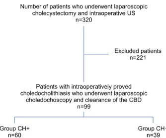
Fig. 1. Study population (US, ultrasonoscopy; CBD, common bile duct; CH, acute cholangitis).
inflammation. Although intraoperative cholangiography may improve the diagnostic accuracy, it is associated with a 5.1% unsuccessful procedure rate, increased operative time and a 15-20% rate of overlooked biliary stones.4-6 Laparoscopic intraoperative ultrasonoscopy (US) is an es- sential and reasonable alternative to intraoperative chol- angiography and, in combination with laparoscopic chol- edochoscopy, increases the options of exploring the CBD during the surgical intervention.
The aim of our study was to assess postoperative course of cholangitis and biliary sepsis after laparoscopic clearance of the CBD in emergently admitted patients with choledocholithiasis and cholangitis.
MATERIALS AND METHODS
Patients with symptomatic gallstone disease and sus- pected choledocholithiasis who were urgently admitted to the hospital and eligible for laparoscopic clearance of the common bile duct, were prospectively included in the study during the period from August 2012 to December 2014 (Fig. 1). Patients were stratified in 2 groups accord- ing to the preoperative presence or absence of cholangitis for assessment of the postoperative inflammatory response. Suspicion of choledocholithiasis was based on evidence of cholangitis, biliary pancreatitis and/or charac- teristic symptoms complex of Charcot’s triad frequently accompanied by pale stools and dark urine.
Preoperative diagnosis of cholangitis was based on cri-
teria recommended in the Tokyo Guidelines 2013, i.e., evidence of inflammatory response (increased leukocytes
>10×1000/l) or C-reactive protein more than 10 mg/L, cholestatic pattern presented by abnormal liver function tests (alkaline phosphatase, gamma-glutamyl transferase, aspartate aminotransferase, and alanine aminotransferase
≥1.5 of upper normal limit) or total bilirubin ≥34.2
mol/L and gallstones in the gallbladder and/or dilatation of the common bile duct >6 mm confirmed by trans- abdominal US.6,7 The diagnosis of biliary pancreatitis was made when 2 of the following revised Atlanta 2012 cri- teria were presented - abdominal pain consistent with acute pancreatitis; serum lipase activity >3 over the up- per limit of normal; and characteristic findings of acute pancreatitis on radiological investigations.8 Preoperative anesthesiological status was assessed in all patients using the American Society of Anaesthesiologists Physical Status classification system (ASA score).9,10
Patients were stratified in 2 groups according to the presence of acute cholangitis (CH+ group) or the absence of cholangitis (CH- group). Laparoscopic intervention was performed using the standard 4-troacar technique and start- ed with the dissection of Callot’s triangle. The cystic duct and artery were clipped and the cystic artery was divided.
The gallbladder was left in situ and retracted during the intraoperative US investigation, performed with BK Medical flex Focus 800 US machines and special flexible laparoscopic transducer 8666-RF. All examinations were performed by 2 specially trained surgeons. The US probe was inserted through the epigastric trocar and placed on the superior edge of the hepatoduodenal ligament and slid inferiorly to the distal end for the examination of the CBD.
The proximal part of CBD, left and right hepatic ducts and their junction were investigated through the right hepatic lobe. Diameter of the CBD and cystic duct, as well as size and number of stones, were measured for determining the choledochoscopy approach. The transcholedochal approach was chosen in cases where stones in CBD were larger than the obtainable diameter of cystic duct.
If transcholedochal approach was considered feasible, an incision was made on CBD longitudinally and the duct was flushed extensively with normal saline. Subsequently, a 2.5 mm flexible choledochoscope was inserted and bile ducts were examined distally and proximally under visual control. Stones were removed using baskets inserted
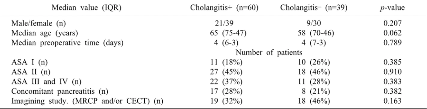
Table 1. Preoperative characteristic of patients
Median value (IQR) Cholangitis+ (n=60) Cholangitis– (n=39) p-value
Male/female (n) 21/39 9/30 0.207
Median age (years) 65 (75-47) 58 (70-46) 0.062
Median preoperative time (days) 4 (6-3) 4 (7-3) 0.789
Number of patients
ASA I (n) 11 (18%) 10 (26%) 0.385
ASA II (n) 27 (45%) 18 (46%) 0.910
ASA III and IV (n) 22 (37%) 11 (28%) 0.383
Concomitant pancreatitis (n) 17 (28%) 8 (21%) 0.382
Imagining study. (MRCP and/or CECT) (n) 19 (32%) 18 (46%) 0.163
ASA, American Society of Anaesthesiologists Physical Status classification system; MRCP, magnetic resonance chol- angiopancreatography; CECT, contrast-enhanced computed tomography
through the instrument channel of the choledochoscope or using Fogarty (No. 3 or 4) catheters. After removal of all stones, clearance of the common bile duct was confirmed with choledochoscopic visualisation and repeated in- tra-operative ultrasound. CBD was closed primarily with interrupted 3-0 absorbable sutures, except for cases of se- vere cholangitis when T-tubes were inserted or chol- edocho-duodenostomy in cases of dilatation of common bile duct >15 mm.
The age and gender of the patients, preoperative co- morbid conditions according to the ASA score, results of the preoperative imaging, and the status of inflammatory response including WBC count, CRP, liver enzyme levels and lipase activity, and presence of cholangitis were com- pared between groups. Diagnostic accuracy of the intra- operative US, success rate of the clearance of the CBD, magnitude of the inflammatory response, complication rate and main outcomes were the variables to compare treatment success.
Statistical analysis
Interval data was presented in median (Me) with inter- quartile range (IQR) and was confirmed by the Kolmogorov-Smirnov test for the asymmetrical dis- tribution of data. Comparison of the interval data in CH+
and CH- groups was performed with the Mann-Whitney U test, the nominal data comparison performed using the Pearson 2 test and Fisher’s exact test. Significant dynam- ics of laboratory analysis were revealed by the Wilcoxon test. The correlation between hospital stay and clinical da- ta was evaluated using the Spearman rho method. Also, the correlation coefficient was compared between groups.
A logistic regression analysis was performed to identify factors associated with a longer hospital stay. A p-value
<0.05 was considered as statistically significant. The stat- istical analysis was performed with SPSS version 20 and MedCalc version 15.
Ethics
The assessment and usage of all clinical data was ap- proved and permitted before the study by the ethics com- mittee of the Riga Stradins University. The study protocol conformed to the ethical guidelines of the “World Medical Association (WMA) Declaration of Helsinki - Ethical Principles for Medical Research Involving Human Subjects”
adopted by the 18th WMA General Assembly, Helsinki, Finland, June 1964 and amended by the 59th WMA General Assembly, Seoul, South Korea, October 2008.11
RESULTS
Out of a total of 320 patients who underwent laparo- scopic cholecystectomy and intraoperative US for suspected choledocholithiasis, 99 patients with intraoperatively con- firmed choledocholithiasis underwent laparoscopic chol- edochoscopy and clearance of the CBD (Fig. 1). In this co- hort, 60 patients had signs of cholangitis (CH+) and 39 had no signs of cholangitis (CH-) (Table 1, Fig. 1).
Preoperative findings
The incidence of comorbid conditions did not differ be- tween the groups. Concomitant acute pancreatitis was ob- served in 28% of patients in the CH+ group without sig- nificant difference, as compared to the CH- group.
Table 3. Inflammatory response
Cholangitis+ (n=60) Cholangitis– (n=39) p-value Median value at the time of admission (IQR)
WBC (×1000/l) 10.6 (12.7-8.1) 7.4 (8.8-5.7) 0.001
CRP (mg/L) 19 (57.0-6.7) 2.6 (5.5-1.7) 0.001
Total bilirubin (mol/L) 55.2 (88-30) 27 (58-11) 0.004
Direct bilirubin (mol/L) 46.1 (75-20) 19.1 (47-5) 0.013
ALT (IU/L) 234 (384-169) 201 (463-34) 0.067
AST (IU/L) 213.5 (371-124) 94.1 (298-33) 0.007
ALP (IU/L) 256 (368-161) 231 (387-111) 0.672
Lipase (U/L) 52 (188-35) 42 (78-31) 0.222
Median value before surgery (IQR)
WBC (×1000/l) 8 (11-6) 7.4 (10-6) 0.422
CRP (mg/L) 37 (99.0-13.3) 7.8 (42.0-3.3) 0.003
Total bilirubin (mol/L) 27 (66-16) 22 (84-10) 0.400
Direct bilirubin (mol/L) 19 (53-10) 12 (75-4) 0.279
ALT (IU/L) 155 (265-71) 182 (370-75) 0.572
AST (IU/L) 109 (175-58) 81 (180-45) 0.499
ALP (IU/L) 257 (330-184) 218 (339-120) 0.617
Lipase (U/L) 54 (92-34) 52 (79-26) 0.515
At the time of operation
Signs of infection during surgery, no. of patients* 36 (60%) 11 (28%) 0.002 Median value before discharge (IQR)
WBC (×1000/l) 7.1 (9.1-5.7) 7.7 (9.9-6.6) 0.187
CRP (mg/L) 33 (66-20) 27 (59-13) 0.396
Total bilirubin (mol/L) 11 (32-8.7) 17.5 (22-9) 0.341
Direct bilirubin (mol/L) 6.4 (21-3) 10.9 (19-5) 0.126
ALT (IU/L) 95 (184-44) 77 (166-39) 0.575
AST (IU/L) 58 (114-32) 46 (74-33) 0.289
ALP (IU/L) 129 173 (270-132) 0.718
Lipase (U/L) 37 (70-28) 69 (117-29) 0.367
WBC, white blood cell count; CRP, C-reactive protein; ALP, alkaline phosphatase; AST, aspartate aminotransferase; ALT, alanine aminotransferase. *Evidence of visible signs of acute cholecystitis during laparoscopy (histologically confirmed acute phlegmo- nous or gangrenous cholecystitis and/or empyema of gallbladder) and cholangitis during choledochoscopy (purulent or fibrinous bile)
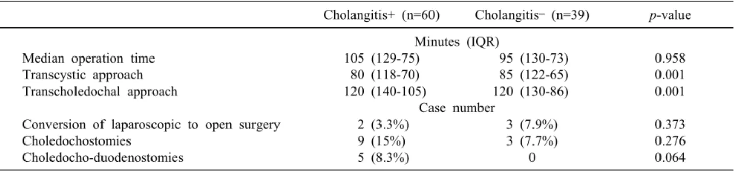
Table 2. Laparoscopic clearance of the common bile duct
Cholangitis+ (n=60) Cholangitis– (n=39) p-value Minutes (IQR)
Median operation time 105 (129-75) 95 (130-73) 0.958
Transcystic approach 80 (118-70) 85 (122-65) 0.001
Transcholedochal approach 120 (140-105) 120 (130-86) 0.001
Case number
Conversion of laparoscopic to open surgery 2 (3.3%) 3 (7.9%) 0.373
Choledochostomies 9 (15%) 3 (7.7%) 0.276
Choledocho-duodenostomies 5 (8.3%) 0 0.064
Preoperative MRCP and CECT were conducted in 32-46%
of patients without group wise difference. Preoperative preparation time was equal in both groups (Table 1).
Laparoscopy
Both groups showed a total laparoscopic intervention time of 95-105 minutes and 3-7% conversion rate. Choledocho-
duodenostomies for cholangitis and dilatation of the com- mon bile duct >15 mm, as well as most of the choledochos- tomies were performed the CH+ group. However, transcystic choledochoscopy was significantly shorter than the trans- choledochal approach in both groups. Moreover, transcystic approach and transcholedochal approach had equal median procedure time between groups (Table 2).
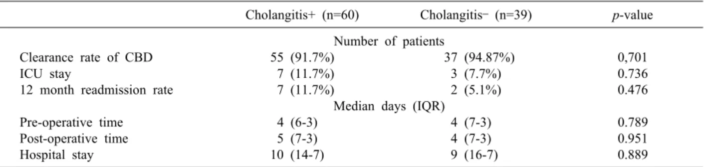
Table 4. Main outcomes
Cholangitis+ (n=60) Cholangitis– (n=39) p-value Number of patients
Clearance rate of CBD 55 (91.7%) 37 (94.87%) 0,701
ICU stay 7 (11.7%) 3 (7.7%) 0.736
12 month readmission rate 7 (11.7%) 2 (5.1%) 0.476
Median days (IQR)
Pre-operative time 4 (6-3) 4 (7-3) 0.789
Post-operative time 5 (7-3) 4 (7-3) 0.951
Hospital stay 10 (14-7) 9 (16-7) 0.889
CBD, common bile duct; ICU, intensive care unit
Fig. 2. Dynamics of C-reactive protein (CH, acute cholangitis).
Fig. 3. Dynamics of white blood cell count (CH, acute chol- angitis).
Dynamics of inflammation
Preoperative inflammatory response was markedly higher in the CH+ group; however, liver enzyme levels, lipase activity and alkaline phosphatase were not different.
Macroscopic signs of infection, inflammatory changes of the gallbladder and signs of the bile duct inflammation during the choledochoscopy were more evident in patients with cholangitis. Postoperative inflammatory response was not statistically different between groups (Table 3). The dynamics of C-reactive protein and white blood cell count were depicted in Figs. 2 and 3.
Complication rate and outcomes
The overall complication rate reached 8.3%, including bleeding from the liver bed in 3 patients from the CH+
group, requiring surgical intervention in 1 patient, 1 bile leak and 1 case of pulmonary thromboembolism, treated conservatively. One patient from the CH- group required open surgery due to injury of the diaphragm and 1 was treated conservatively due to postoperative bile leak, com-
prising 5.1% of the group, p=0.701. Postoperative in- tensive care unit stay and overall hospital stay was not statistically different between groups. Laparoscopic clear- ance of the CBD was associated with 1 lethal case (CH+
group) due to decompensated liver cirrhosis and progress- ing hepatocellular insufficiency, resulting in 1% mortality rate in the cohort of 99 patients. A similar 12-month read- mission rate was observed in both groups. Totally, com- plete laparoscopic clearance of the common bile duct was possible in 92.9% of patients (92 of 99); furthermore, the clearance rate was similar in both groups (Table 4).
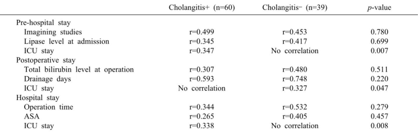
Correlation analysis
Correlation analysis revealed that patients with an ele- vated lipase level at the admission had a longer pre- operative stay; and patients with an elevated total bilirubin level preoperatively had a longer postoperative stay. The duration of the operation and the severity of the comorbid conditions according to ASA score correlated with the
Table 5. Correlation analysis
Cholangitis+ (n=60) Cholangitis– (n=39) p-value Pre-hospital stay
Imagining studies r=0.499 r=0.453 0.780
Lipase level at admission r=0.345 r=0.417 0.699
ICU stay r=0.347 No correlation 0.007
Postoperative stay
Total bilirubin level at operation r=0.307 r=0.480 0.511
Drainage days r=0.593 r=0.748 0.220
ICU stay No correlation r=0.327 0.047
Hospital stay
Operation time r=0.344 r=0.532 0.279
ASA r=0.265 r=0.405 0.457
ICU stay r=0.338 No correlation 0.008
ICU, intensive care unit; ASA, American Society of Anaesthesiologists Physical Status classification system
overall hospital stay. Prolongation of the hospital stay was mostly associated with the presence of the comorbid con- ditions in the CH- group, p=0.001 (CI 95% 1.79-5.66) (Table 5).
DISCUSSION
Preoperative recognition of the possible choledocholithiasis is crucial for planning of the treatment strategy in patients with a complicated course of gallstone disease. Existing recommendations for the management of patients with a complicated gallstone disease are controversial. Contrast- enhanced computed tomography scan or MRCP is recom- mended as a routine preoperative radiologic examination with high sensitivity for patients with suspected bile duct involvement, especially when malignant lesions cannot be excluded.12-17
However, MRCP may fail to visualise the sludge and stones <5 mm in diameter and bile duct stones in the cases of pancreatitis.1,18-21 The 2 recommended approaches include laparoscopic cholecystectomy with intraoperative evaluation of the CBD in the time of admission18,22,23 or early ERCP, followed by delayed cholecystectomy 6-8 weeks later, especially for patients with acute pancreatitis.24 Evidence suggests that the results on ERCP treatment are more dependent on the selection of appro- priate indications for ERCP and technical skills of the en- doscopy specialist than on the patients’ medical condition.2 Also, the reported mortality rate after chol- ecystectomy performed during the acute episode of biliary pancreatitis is higher.25
Contrary to a more routine concept of cholecystectomy in the interval after conservative treatment,24 several stud- ies have demonstrated good results from laparoscopic cholecystectomy in index hospital admission during the acute phase of the disease.18,22 The current study corrobo- rated these results, with similar general patient character- istics (mean age, gender and comorbidities) and complica- tion rate to those previously reported. This single-stage approach has become possible since the implementation of the intraoperative US visualisation of the CBD and choledochoscopy for the final clearing of the CBD when indicated.
The diagnostic value of the intraoperative US and the simplicity of performance has been demonstrated in sev- eral studies. It is currently a safe alternative to intra- operative cholangiography, excluding exposure to radia- tion and allowing a better anatomical visualisation.4,5,19,26-29
According to the results of the current and similar studies, laparoscopic CBD clearance does not significantly in- crease the duration of the surgery, and is not associated with an increased complication rate. Moreover, intra- operative US control is especially effective in patients with infiltrated tissue and difficult visualisation of the bili- ary tree.16,30 The conversion rate and reasons for con- version were not significantly different, but median oper- ation time and post-operative stay were even shorter in both groups of our cohort, as compared with the experi- ence from other studies.31
Thus, early laparoscopic clearance of the CBD follow- ing a single-stage strategy, is safe in patients with chol- angitis, as recently reviewed.32 This strategy may reduce
the risk of a recurrent attack and complications associated with delayed surgery. US visualisation and clearance of the CBD via transcystic or transcholedochal access re- quires better laparoscopic skills and more time spent in the operation theatre.33 This approach ensures post- operative control of CBD patency in patients with severe cholangitis or with incomplete clearance of the CBD, by adding T-tube drainage. It provides postoperative imaging of the biliary tree and for the removal of residual CBD stones and early postoperative ERCP when indicated.
Alternatively to t-tube drainage, laparoscopic chol- edocho-duodenostomies might be created according to se- lective indications when dilatation of the CBD is >15 mm in elderly patients. Low 12-month readmission rate indicates that laparoscopic clearance of the CBD in emer- gently admitted patients is feasible, as previously reported.34 The lack of randomisation and a relatively long learning curve are the weak aspects of our study. Another weak point is the lack of a uniform protocol considering the pre- operative standard of radiologic investigation, which was based on clinical, rather than scientific decision. However, single-stage laparoscopic interventions with provision of intraoperative US based choledochoscopic clearance of the CBD in emergently admitted patients resulted in a low complication rate and 1% mortality. The overall results and the low 12-month readmission rate indicated the safety and efficacy of this method.
We concluded that single-stage laparoscopic intra- operative US and choledochoscopy-assisted clearance of the CBD is feasible in emergently admitted patients with cholangitis. International prospective randomised study provided by specialists in the hepato-pancreatic-biliary surgery is justified to augment clinically based evidence.
REFERENCES
1. Freitas ML, Bell RL, Duffy AJ. Choledocholithiasis: evolving standards for diagnosis and management. World J Gastroenterol 2006;12:3162-3167.
2. Freeman ML, Nelson DB, Sherman S, Haber GB, Herman ME, Dorsher PJ, et al. Complications of endoscopic biliary sphincterotomy. N Engl J Med 1996;335:909-918.
3. Adler DG, Baron TH, Davila RE, Egan J, Hirota WK, Leighton JA, et al; Standards of Practice Committee of American Society for Gastrointestinal Endoscopy. ASGE guideline: the role of ERCP in diseases of the biliary tract and the pancreas.
Gastrointest Endosc 2005;62:1-8.
4. Hublet A, Dili A, Lemaire J, Mansvelt B, Molle G, Bertrand
C. Laparoscopic ultrasonography as a good alternative to intra- operative cholangiography (IOC) during laparoscopic chol- ecystectomy: results of prospective study. Acta Chir Belg 2009;
109:312-316.
5. Aziz O, Ashrafian H, Jones C, Harling L, Kumar S, Garas G, et al. Laparoscopic ultrasonography versus intra-operative chol- angiogram for the detection of common bile duct stones during laparoscopic cholecystectomy: a meta-analysis of diagnostic accuracy. Int J Surg 2014;12:712-719.
6. Takada T, Strasberg SM, Solomkin JS, Pitt HA, Gomi H, Yoshida M, et al; Tokyo Guidelines Revision Committee. TG13:
Updated Tokyo Guidelines for the management of acute chol- angitis and cholecystitis. J Hepatobiliary Pancreat Sci 2013;20:1-7.
7. Kiriyama S, Takada T, Strasberg SM, Solomkin JS, Mayumi T, Pitt HA, et al; Tokyo Guidelines Revision Committee. TG13 guidelines for diagnosis and severity grading of acute cholangitis (with videos). J Hepatobiliary Pancreat Sci 2013;20:24-34.
8. Banks PA, Bollen TL, Dervenis C, Gooszen HG, Johnson CD, Sarr MG, et al; Acute Pancreatitis Classification Working Group.
Classification of acute pancreatitis--2012: revision of the Atlanta classification and definitions by international consensus. Gut 2013;62:102-111.
9. Sankar A, Johnson SR, Beattie WS, Tait G, Wijeysundera DN.
Reliability of the American Society of Anesthesiologists physical status scale in clinical practice. Br J Anaesth 2014;113:424-432.
10. Aronson WL, McAuliffe MS, Miller K. Variability in the American society of anesthesiologists physical status classification scale.
AANA J 2003;71:265-274.
11. World Medical Association. World Medical Association Declaration of Helsinki: ethical principles for medical research involving hu- man subjects. JAMA 2013;310:2191-2914.
12. Hallal AH, Amortegui JD, Jeroukhimov IM, Casillas J, Schulman CI, Manning RJ, et al. Magnetic resonance chol- angiopancreatography accurately detects common bile duct stones in resolving gallstone pancreatitis. J Am Coll Surg 2005;
200:869-875.
13. Peng WK, Sheikh Z, Paterson-Brown S, Nixon SJ. Role of liver function tests in predicting common bile duct stones in acute cal- culous cholecystitis. Br J Surg 2005;92:1241-1247.
14. Taylor AC, Little AF, Hennessy OF, Banting SW, Smith PJ, Desmond PV. Prospective assessment of magnetic resonance cholangiopancreatography for noninvasive imaging of the biliary tree. Gastrointest Endosc 2002;55:17-22.
15. Topal B, Van de Moortel M, Fieuws S, Vanbeckevoort D, Van Steenbergen W, Aerts R, et al. The value of magnetic resonance cholangiopancreatography in predicting common bile duct stones in patients with gallstone disease. Br J Surg 2003;90:42-47.
16. Akisik MF, Jennings SG, Aisen AM, Sherman S, Cote GA, Sandrasegaran K, et al. MRCP in patient care: a prospective sur- vey of gastroenterologists. AJR Am J Roentgenol 2013;201:
573-577.
17. Chen W, Mo JJ, Lin L, Li CQ, Zhang JF. Diagnostic value of magnetic resonance cholangiopancreatography in choledocholithiasis.
World J Gastroenterol 2015;21:3351-3360.
18. Sinha R. Early laparoscopic cholecystectomy in acute biliary pancreatitis: the optimal choice? HPB (Oxford) 2008;10:332-335.
19. Costi R, Gnocchi A, Di Mario F, Sarli L. Diagnosis and manage- ment of choledocholithiasis in the golden age of imaging, endos- copy and laparoscopy. World J Gastroenterol 2014;20:13382-13401.
20. Srinivasa S, Sammour T, McEntee B, Davis N, Hill AG.
Selective use of magnetic resonance cholangiopancreatography in clinical practice may miss choledocholithiasis in gallstone pancreatitis. Can J Surg 2010;53:403-407.
21. Li P, Zhang Z, Li J, Jin L, Han W, Zhang J. Diagnostic value of magnetic resonance cholangiopancreatography for secondary common bile duct stones compared with laparoscopic trans-cystic common bile duct exploration. Med Sci Monit 2014;20:920-926.
22. Sangrasi AK, Syed B, Memon AI, Laghari AA, Talpur KA, Qureshi JN. Laparoscopic cholecystectomy in acute gallstone pancreatitis in index hospital admission: feasibility and safety.
Pak J Med Sci 2014;30:601-605.
23. Cuschieri A, Lezoche E, Morino M, Croce E, Lacy A, Toouli J, et al. E.A.E.S. multicenter prospective randomized trial com- paring two-stage vs single-stage management of patients with gallstone disease and ductal calculi. Surg Endosc 1999;13:952-957.
24. Nealon WH, Bawduniak J, Walser EM. Appropriate timing of cholecystectomy in patients who present with moderate to severe gallstone-associated acute pancreatitis with peripancreatic fluid collections. Ann Surg 2004;239:741-749.
25. Stone HH, Fabian TC, Dunlop WE. Gallstone pancreatitis: bili- ary tract pathology in relation to time of operation. Ann Surg 1981;194:305-312.
26. Tranter SE, Thompson MH. A prospective single-blinded con- trolled study comparing laparoscopic ultrasound of the common bile duct with operative cholangiography. Surg Endosc 2003;17:
216-219.
27. Metcalfe MS, Ong T, Bruening MH, Iswariah H, Wemyss- Holden SA, Maddern GJ. Is laparoscopic intraoperative chol-
angiogram a matter of routine? Am J Surg 2004;187:475-481.
28. Santambrogio R, Bianchi P, Opocher E, Mantovani A, Schubert L, Ghelma F, et al. Intraoperative ultrasonography (IOUS) during laparoscopic cholecystectomy. Surg Endosc 1996;10:622-627.
29. Overby DW, Apelgren KN, Richardson W, Fanelli R; Society of American Gastrointestinal and Endoscopic Surgeons. SAGES guidelines for the clinical application of laparoscopic biliary tract surgery. Surg Endosc 2010;24:2368-2386.
30. Rhodes M, Sussman L, Cohen L, Lewis MP. Randomised trial of laparoscopic exploration of common bile duct versus post- operative endoscopic retrograde cholangiography for common bile duct stones. Lancet 1998;351:159-161.
31. Naumowicz E, Białecki J, Kołomecki K. Results of treatment of patients with gallstone disease and ductal calculi by single-stage laparoscopic cholecystectomy and bile duct exploration.
Wideochir Inne Tech Maloinwazyjne 2014;9:179-189.
32. Bencini L, Tommasi C, Manetti R, Farsi M. Modern approach to cholecysto-choledocholithiasis. World J Gastrointest Endosc 2014;6:32-40.
33. Koc B, Adas G, Karahan S. Use of laparoscopic common bile duct exploration for failed endoscopic bile duct stone extractions.
Minerva Chir 2014;69:209-215.
34. Noble H, Norton S, Thompson M. Assuring complete laparo- scopic clearance of the bile duct. J Laparoendosc Adv Surg Tech A 2011;21:319-322.