• 검색 결과가 없습니다.

중증

N/A
N/A
Protected

Academic year: 2021

Share "중증"

Copied!
7
0
0

로드 중.... (전체 텍스트 보기)

전체 글

(1)

大 韓 不 妊 學 會 誌 : 第 32 卷 第 3 號 2005 Kor. J. Fertil. Steril., Vol. 32, No. 3, 2005, 9

중증 자궁내막증 환자의 내막에서 thrombospondin-1과 -2의 mRNA의 발현에 관한 연구

이화여자대학교 의과대학 산부인과학교실, 건국대학교 의과대학 산부인과학교실

1

허성은·이지영

1

·문혜성·정혜원

mRNA Expression of Thrombospondin-1 and -2 in Severe Endometriosis Patients in Korean Women

Sung Eun Hur, Lee Ji Young

1

, Hye-Sung Moon, Hye Won Chung

Department of Obstetrics and Gynecology, College of Medicine, Ewha Womans University, Seoul, Korea,

1

Department of Obstetrics and Gynecology, College of Medicine,

KonKuk University, Seoul, Korea

Objective: We investigated the expression of TSP-1 and -2 in eutopic endometrium of advanced endometriosis and control patients.

Methods: The 33 endometriosis patients and 32 controls were enrolled. Endometrial samples were obtained from 65 premenopausal women aged 29-44 years, undergoing laparoscopic surgery or hysterectomy for non-malignant lesions. Sufficient samples were collected from 33 patients with endometriosis stage III and IV and 32 controls without endometriosis confirmed by laparoscopic surgery.

The mRNA expression from eutopic endometrium for TSP-1 and -2 were analyzed by RT-QC PCR.

Results: The mRNAs of TSP-1 and -2 were expressed in eutopic endometrium from endometriosis and normal controls throughout the menstrual cycle. There were no significant differences in expression of TSP-1 and TSP-2 in eutopic endometrium between controls and endometriosis patients.

Conclusion: Our results indicated that TSP-1 and -2 had no crucial role compared to other molecules in the regulation of angiogenesis. These findings also suggest that dysregulation of other angiogenic regulators would be concerned in pathophysiologic role in endometriosis development.

Key Words: Thrombospondin, Endometriosis, Angiogenesis

자궁내막증은 자궁강 밖에서 자궁내막의 선과 간 질이 존재하는 질환으로 가임기 여성의 7~10%의 유병율을 보이지만 아직 정확한 원인과 병태생리는 명확하게 밝혀지진 않은 질환으로서 환경적 요인, 면역학적 요인, 호르몬 요인과 유전적 요인이 함께 작용하는 것으로 알려져 있다.

1

자궁내막증의 여러

병인 중에서 자궁내막세포가 월경시 역류된 생리혈 에 의해 복강내로 파종되고 착상된다는 가설이 가 장 유력한 실정이다.

2

이런 과정에서 세포외 단백 분해 (extracellular proteolysis)와 맥관 형성 (angio- genesis)이 중요한 과정으로 인식되고 있다. 이러한 일차적인 요인 외에 이차적인 요인으로는 유전적인

주관책임자: 정혜원, Department of Obstetrics and Gynecology, Ewha Womans University Mokdong Hospital, 911-1 Yang Chun Ku Mock 6 Dong 158-710 Seoul, Korea. Tel: 822-650-5568, Fax: 822-2647-9860, e-mail: [email protected]

*이 논문은 2003년도 한국과학재단의 지원에 의하여 연구되었음 (R0420030001004702004).

(2)

성향과 환경적인 원인, 면역학적인 원인 등이 제시 되어 왔다.

1,3,4

맥관 형성은 조직의 성장 및 재생에 필수적인 과 정으로 이미 존재하는 혈관에서 새로운 혈관을 만 들어 내는 과정이다.

5

이 과정에는 여러 가지 양성 또는 음성 조절인자가 관여하게 되고 그 외에도 여 러 종류의 분자와 세포들과도 관련이 된다. 이에 관 여하는 혈관생성인자에는 혈관내피세포성장인자 (vascular endothelial growth factor, VEGF), 섬유아세 포성장인자 (fibroblast growth factor, FGF), 표피성장 인자 (epidermal growth factor, EGF), angiopoietin 등이 있으며 , 혈관생성억제인자로서는 thrombospondin-1 (TSP-1), TSP-2, angiostatin, endostatin, vasostatin 등 이 알려져 있다.

6~10

TSP-1은 190,000 Da의 다양한 기능을 가지는 당 단백으로 1970년대에 처음으로 기술되었고,

11

그 후 에 420,000 Da의 삼합체로 발견되었다. 지혈전 (he- mostatic plug)을 만드는 중에 혈소판에서 분비되며, 그 외에도 대식구, 단핵구, 섬유아세포, 혈관근세 포, 종양세포 그리고 혈관내피세포 등에서 분비된 다 .

12

TSP-2는 섬유아세포에서 주로 분비되며, TSP-1 과 비슷하게 420,000 Da의 삼합체로 되어 있다.

13

TSP-2도 TSP-1과 유사하게 혈관생성억제인자의 기 능을 갖고 있으며 그 외의 부가적인 기능도 연구 중인데, TSP-2 결핍 생쥐에서 결합조직 이상과 골 밀도의 상승, 혈소판 기능의 이상 등으로 인한 출 혈성 경향을 보인다는 연구 결과가 있다.

14

맥관 형성이 일어나기 위해서는 혈관내피세포가 혈관 내강 구조 (capillary cord)를 형성하여야 하는 데 이 capillay cord를 형성하는데 TSP-1이 관여하며 cord 형성이 많은 경우는 TSP-1 뿐 아니라 TSP-1 mRNA가 적게 표현되고, 항TSP항체가 투여된 경 우는 capillary cord 형성이 30~50% 증가하였다는 실험 결과가 있으며

15

Iruela-Arispe 등도 유사한 결 과를 발표하였다.

16

본 연구에서는 자궁내막증에서 병태생리를 연구 함에 있어서 자궁내막증으로 확진된 환자와 자궁내 막증이 없음을 확인한 정상 대조군에서 혈관생성억 제인자라고 알려진 TSP-1와 TSP-2의 mRNA의 발 현에 차이가 있는지를 경쟁적 역전사 중합효소 연 쇄반응 (reverse transcription - quantitative competitive

polymerase chain reaction, RT-QC PCR)을 이용하여 분석해 보고자 하였다.

연구 대상 및 방법

1. 연구 대상

1996년 9월부터 2004년 12월까지 이화여대 목동 병원 산부인과를 방문한 한국인 여성 중 수술을 통 해 병리조직학적으로 자궁내막증을 확인한 환자에 서 revised American Society for Reproductive Medicine classification

17

분류에 따라 III기와 IV기인 33명을 대상으로 하였다. 대조군은 난소 낭종 등의 양성 질 환으로 개복술이나 골반경 수술을 시행한 환자 중 자궁내막증이 없음을 확인한 여성 32명을 대상으로 하였다 . 환자들은 최근 6주간 비스테로이드성 항염 증제재나 성선자극호르몬유리호르몬 작용제 (GnRH agonist), 그리고 스테로이드성 약물 등을 투여 받 은 적이 없었다. 본 연구는 임상시험 심사원 (Insti- tutional Review Board)에서 심의를 받았으며, 각각 의 환자에 대해 사전 동의를 받았다. 자궁내막조직 은 골반경을 시행한 경우는 수술 시행 전에, 개복 술을 시행한 경우는 자궁 적출 후 즉시 조직을 얻 었으며, 자궁내막조직은 Noyes 등의 분류에 따라 난포기와 황체기로 나누어 분류하였고,

18

인산염완 충식염수 (phosphate-buffered saline; PBS)에 세척한 후 , 실험할 때까지 -70℃에서 동결해 두었다.

2. 연구 방법 1) RNA 추출

동결해두었던 내막조직을 PBS로 세 번 세척한 후 , 100 mg의 조직을 RNA-STAT-60 시약 (Tel-Test

"B" Inc., Friendswood, TX, USA) 1 ml과 함께 균질화 시킨 후, chloroform을 첨가하고, isopropanol을 이용 하여 침전시킨 후, 75% ethanol로 두 번 세척한 후, diethylpycocarbonate (DEPC)-treated dH

2

O로 재희석 시켰다 . 추출된 RNA는 GenQuant RNA/DNA calcu- lator (Phamacia Biotech LTd., Cambridge, UK)를 사용 하여 spectrophotometry로 측정하였다. 평균 10~100 µg의 RNA가 추출되었다.

2) Reverse Transcription (RT) PCR

TSP-1, -2의 염기 서열을 National Institute of He-

(3)

alth의 National Center of Biotechnology Information의 Gene Bank Database에서 얻은 후 OLIGO 5.0 primer analysis software (National Bioscience, Plymouth, MN, USA)를 이용하여 TSP-1, -2에 대한 올리고 뉴클레 오타이드 시발체 (oligonucleotide primer)를 고안하 였고 (Table 1). RT PCR를 위해서는 Gen Amp RNA PCR kit (Perkin-Elmer, Foster City, CA, USA)를 사용 하였다 . 5 mmol/L MgCl

2

, 1X PCR buffer II, 1 mmol/L of each deoxy-NTP, 2.5 µl/L oligo (deoxythimidine)16, 20 IU ribonuclease inhibitor (이상은 모두, Perkin- Elmer Corp.), 100 IU Moloney murine leukemia virus reverse transcriptase (Gibco BRL)를 포함한 역전사용 혼합물 19 µl에 1 µl 당 1 µg이 되게 희석한 RNA 1 µl를 넣은 후 RT PCR 하였다. RT PCR은 Perkin- Elmer사의 DNA thermal cycler 9600을 이용하여 42℃에서 15분, 99℃에서 5분 반응시켜서 4℃로 냉 각시킨 후 PCR 전까지 -20℃에서 보관하였다. 음성 대조로는 RNA 1 µl 대신 DEPC로 처리한 물을 넣 고 같은 반응을 시켰다.

3) TSP-1 & -2의 competitive와 target cDNA 의 합성

자궁내막조직에서 추출한 RNA로부터 역전사 후 정상의 3'-, 5'-시발체를 넣고 PCR를 통하여 470 bp 의 TSP-1, 484 bp의 TSP-2의 target DNA를 얻은 후 agarose gel에 전기영동 시키고 Promega사의 DNA purification kit로 cDNA를 추출하였다. 정상 3'-, 5'-시발체 접합부위 사이의 염기 서열 중에서 3'- floating primer를 고안한 후 정상 5'-시발체와 함께 반응시켜서 target cDNA를 얻는 방법과 같은 방법

으로 cDNA를 추출하였다. 이렇게 만들어진 com- petitive cDNA는 TSP-1 307 bp, TSP-2 272 bp의 크기 였다.

4) TSP-1 & -2의 Standard curve 의 작성과 competitive PCR

TSP-1 & -2의 standard curve는 일정한 양의 com- petitive cDNA와 점차 감소시킨 양의 target cDNA 를 동시에 증폭시켜 만든다. 표준곡선은 1.9 mmol MgCl

2

, 10X PCR buffer II, 0.2 mmol/l of deoxy-NT, 2.5 U Taq-polymerase를 포함한 100 µl PCR 혼합 용액과 0.2 µmol/L의 시발체를 반응시킨 후 Perkin Elmer사의 DNA thermal cycler 9600으로 TSP-1의 경우에는 94℃ 2분간 모든 단백질을 변성시킨 후, 94℃ 45초, 59℃ 45초, 72℃ 1분으로 35주기, TSP-2 의 경우에는 95℃ 5분간 변성시킨 후, 94℃ 45초, 57℃ 45초, 72℃ 1분으로 35주기 반응시키고, 각각 7분간 elongation 시킨 후 4℃로 냉각시켰다. 1%

agarose gel에서 전기영동을 시행하였고, gel은 Ethi- dium Bromide로 염색하였다. 표준곡선 및 표본 PCR 산물 25 µl 씩을 100 bp의 DNA ladder와 함께 전기 영동 시킨 후 UV densitometry에서 gel blot을 분석 하였다. (Bio-Rad 사의 el-Doc 1000 system) 각각의 target cDNA의 양과 target cDNA/competitive cDNA 의 gel blot 농도의 비를 log로 전환하여 표준곡선 을 얻었으며, 이로부터, 농도를 알 수 없는 표본의 cDNA양을 계산하였다.

3. 통계 분석

각각의 결과는 mean ± SEM (standard error of mean)

Table 1. Oligonucleotide primers for TSP-1 and -2 mRNA amplification in eutopic endometrium

mRNA

Primer 5'-3' Size

(bp) Position on mRNA

Upstream 5'-TTC-TAC-GAG-CTG-TGG-CAA-TG-3' 470 1284~1303

Downstream 5'-TTG-GAC-AGT-CCT-GCT-TGT-TG-3' 1753~1772

TSP-1

Competitor 5'-TTG-GAC-AGT-CCT-GCT-TGT-TGC-TTT-CTT-GCA-GGC-TTT-GGT-C-3' 307 1753~1772 +1570~1589

Upstream 5'-CGT-GGA-CAA-TGA-CCT-TGT-TG-3' 484 2783~2802

Downstream 5'-GTC-CAC-AGA-CCC-AAA-CTC-GT-3' 3266~3285

TSP-2

Competitor 5'-GTC-CAC-AGA-CCC-AAA-CTC-GTC-AAA-TAT-CAC-CCC-GTC-CAT-3' 272 3266~3285 +3034~3053

(4)

으로 제시하였으며, 각 그룹간의 비교는 Kruskal- Wallis와 Mann-Whitney U test를 이용하였다. 각각 의 통계적 방법은 Statistical Package for Social Sci- ence version 11.5 (SPSS Inc., Chicago, IL, USA)를 사 용하였고 , p값이 <0,05에서 통계적으로 유의하다고 보았다.

결 과

1. 생리주기에 따른 자궁내막의 TSP-1 & -2의 mRNA의 발현

정상 대조군과 환자군 모두에서 생리주기 전반에 걸쳐 RT-PCR을 한 결과, 470 bp의 TSP-1과 484 bp 의 TSP-2 mRNA가 발현되었다. 대조군으로 실험한

β-actin mRNA도 정상 대조군과 자궁내막증 환자군 모두에서 생리주기 전반에 걸쳐 모두 838 bp로 발 현되었다.

2. TSP-1 & -2의 mRNA 정량 분석

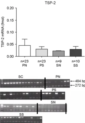
TSP-1 & -2의 QC-PCR 결과 정상 대조군과 자궁 내막증 환자군의 자궁내막조직에서 각각의 환자당, target과 competitive의 두 개의 band가 보였고, 이 를 표준곡선을 이용하여 정량하였다. 자궁내막증 환자에서의 TSP-1 & -2 mRNA는 생리주기 전반에 걸쳐 비교했을 때 정상 대조군에 비해서 통계적으 로 유의한 차이를 보이지 않았다 (Figure 1, 2).

고 찰

자궁내막증은 여성 양성 질환 중에서 가장 많은

Figure 1. The TSP-1 mRNA expression in eutopic

endometrium throughout the menstrual phase. The upper panel shows abundance of the TSP-1 mRNA in eutopic endometrium throughout the menstrual phase. PN: pro- liferative phase endometrium from normal patients. PS:

proliferative phase endometrium from endometriosis pa- tients. SN: secretory phase endometrium from normal patients. SS: secretory phase endometrium from endo- metriosis patients. SC: standard curve. Values are given as mean ± SEM.

Figure 2. The TSP-2 mRNA expression in eutopic

endometrium throughout the menstrual phase. The upper

panel shows abundance of the TSP-2 mRNA in eutopic

endometrium throughout the menstrual phase. Values are

mean ± SEM. All abbreviations and legends are same

as Figure 1.

(5)

빈도를 차지하는 질환 중의 하나로 그 원인과 발생 과정에 맥관 형성이 필수적임이 밝혀지고 있다. 맥 관 형성은 많은 조절인자들에 의해서 철저히 조절 되고 있다. 맥관 형성에 관여하는 많은 펩타이드와 단백질이 맥관 형성을 자극하거나 억제하는 기능을 가지고 있기에 angiogenic balance라는 개념이 대두 하게 되었다. 관여되는 많은 물질 중에서 본 연구에 서 다루었던 TSP-1의 경우, 다양한 종류의 종양세 포에서 유의하게 낮게 발현되었다는 연구도 있었

고,

12,19

종양세포의 맥관 형성과 TSP의 발현이 연관

성이 있지만 그 예후와는 연관성이 없었다는 연구 외에 ,

20

TSP-1의 경우에 그 다기능적인 특징에 의하 여 맥관 형성에 대해서는 자극과 억제의 양 기능이 공존할 수도 있다는 연구와 함께

12

최근에는 종양 세포에서의 antiangiogenic therapy에 대한 연구가 많이 발표되고 있다.

21,22

대부분의 경우에서, 종양 세포에서는 oncogene의 activation이나 tumor supp- ressor gene의 activation에 의해서 TSP-1의 발현을 down-regulation 시키는 것을 관찰할 수 있다. 그리 고 이런 TSP-1의 down-regulation이 pro-angiogenic phenotype을 획득하는 중요 단계가 된다고 할 수 있다 .

23

자궁내막증은 양성 종양이나 악성 종양과는 다른 질환이지만 자궁내막조직의 이소성으로 존재 하고 , 전이되며 성장한다는 점에서 유사하다고 할 수 있다. 이런 점에서 볼 때 자궁내막증을 가지는 환자군에서 자궁내막조직 내에서의 TSP-1 & -2의 발현이 정상 대조군보다 감소되어 있고 다른 맥관 형성인자의 경우는 과잉 발현되어 있어야 higher proliferative activity를 보일 것이며 전이의 가능성이 높아지게 되는 것이다. 자궁내막증의 이런 병태생리 와 관련하여 최근에는 종양세포 뿐 아니라 자궁내 막증에 대한 antiangiogenic therapy가 시험관내 (in vitro) 실험과 동물실험을 통하여 활발히 진행되고 있다 .

24~26

본 논문에서 저자는 중증 자궁내막증 환자와 정 상 대조군의 자궁내막에서 TSP-1 & -2의 mRNA 의 발현을 생리주기 전반에 걸쳐서 비교해 보았다.

TSP-1 & -2의 mRNA 발현은 생리주기 전반에 걸쳐 서 통계적으로 유의한 차이는 없었다. 자궁내막증에 대한 기본적인 병태생리로 볼 때, eutopic endome- trium 자체가 가장 중요한 역할을 하리라고 보며,

이를 맥관 형성과 연관지어 생각해 볼 때, TSP 발 현의 dysregulation이 자궁내막의 이소성 맥관 형성 에 가장 중요한 병태생리가 되지 않을 수 없다는 것이다 . 자궁내막증의 형성을 위한 맥관 형성의 과 정 , 조절, 기전 등이 복잡하고, 맥관 형성 과정에서 의 역할을 한가지로 규정하기가 어렵기 때문에 여 기에 첨가해서 신호전달체계에 여러 가지 다른 분 자들 - angiopoietins, Ties, Ephrins 등 - 에 대한 연 구가 필요할 것이다.

27~30

맥관 형성과 관련한 최근 연구에서 human glioma cell에서 p53 유전자의 변이 가 VEGF와 TSP-1의 발현에도 관여한다는 결과에 서도 볼 수 있듯이

31

여러 맥관 형성 인자들을 조 절하는 다양한 기전들이 존재하기 때문에, 마찬가 지로 자궁내막증의 병태생리를 연구하는 데에도 포 괄적인 접근이 필요할 것이라 사료된다.

자궁내막증의 병태생리와 관련하여 맥관 형성에 관련된 TSP-1, -2의 mRNA 발현에 대해서 조사한 결과, 자궁내막증 환자에서 그 발현이 월경주기에 따라 정상 대조군 보다 감소되어 있거나, 차이가 있으리라 예상하였으나, 두 군 사이의 차이가 통계 학적으로 유의하지 않았다. 이는 맥관 형성에 관련 된 TSP-1, -2의 mRNA 발현이 환자군과 정상 대조 군 사이에 차이가 있으리라는 가정과 반대된 결과 를 보이는 것이다. 그러나 맥관 형성에 관련된 인자 가 앞서 말한 바와 같이 여러 종류가 있으며, 그 인 자들 사이에도 상호 보완되거나, 상충되는 관계가 있으리라 예상되는 바, 앞으로 자궁내막증의 병태 생리와 밀접한 관련이 있다고 생각되는 맥관 형성 인자들의 상호 작용과 관련하여 연구가 진행되어야 할 것이다.

참 고 문 헌

1. Kennedy S. Is there a genetic basis to endometriosis?

Semin Reprod Endocrinol 1997; 15: 309-18.

2. Sampson JA. Peritoneal endometriosis due to men- strual dissemination of endometrial tissue into the peritoneal cavity. Am J Obstet Gynecol 1927; 14:

422-69.

3. Simpson JL, Elias S, Malinak LR, Buttram VC Jr.

Heritable aspects of endometriosis. I. Genetic studies.

(6)

Am J Obstet Gynecol 1980; 137: 327-31.

4. Berkkanoglu M, Arici A. Immunology and endome- triosis. Am J Reprod Immunol 2003; 50: 48-59.

5. Iruela-Arispe ML, Dvorak HF. Angiogenesis: a dy- namic balance of stimulator and inhibitors. Thrombo Haemost 1997; 78: 672-7.

6. Li J, Zhou J, Tota B, Scalia G, Gattuso A. Regulating effects of vascular endothelial growth factor and ANG II on frog's pericardial stomata, mesothelium and angiogenesis. Chin Med Sci J 2001; 16: 23-8.

7. Burns PA, Wilson DJ. Angiogenesis mediated by metabolites is dependent on vascular endothelial growth factor (VEGF). Angiogenesis 2003; 6: 73-7.

8. Sandberg T, Ehinger A, Casslen B. Paracrine sti- mulation of capillary endothelial cell migration by endometrial tissue involves epidermal growth factor and is mediated via up-regulation of the urokinase plasminogen activator receptor. J Clin Endocrinol Metab 2001; 86: 1724-30.

9. Jung SP, Siegrist B, Wang YZ, Wade MR, Anthony CT, Hornick C, et al. Effect of human Angiostatin protein on human angiogenesis in vitro. Angioge- nesis 2003; 6: 233-40.

10. Folkman J. Endogenous angiogenesis inhibitors.

APMIS 2004; 112: 496-507.

11. Baenziger NL, Brodie GN, Majerus PW. A thrombin- sensitive protein of human platelet membranes. Proc Natl Acad Sci USA 1971; 68: 240-3.

12. Esemuede N, Lee T, Pierre-Paul D, Sumpio BE, Gahtan V. The role of thrombospondin-1 in human disease. J Surg Res 2004; 122: 135-42.

13. Kyriakides TR, Zhu YH, Smith LT, Bain SD, Yang Z, Lin MT, et al. Mice that lack thrombospondin 2 display connective tissue abnormalities that are asso- ciated with disordered collagen fibrillogenesis, an increased vascular density, and a bleeding diathesis.

J Cell Biol 1998; 140: 419-30.

14. Kyriakides TR, Zhu YH, Yang Z, Bornstein P. The distribution of the matricellular protein thrombo- spondin 2 in tissues of embryonic and adult mice. J Histochem Cytochem 1998; 46: 1007-15.

15. Kanda S, Shono T, Tomasini-Johansson B, Klint P, Saito Y. Role of thrombospondin-1-derived peptide, 4N1K, in FGF-2-induced angiogenesis. Exp. Cell Res 1999; 252: 262-72.

16. Iruela-Arispe ML, Bornstein P, Sage H. Thrombo- spondin exerts an antiangiogenic effect on cord for- mation by endothelial cells in vitro. Proc Natl Acad Sci USA 1991; 88: 5026-30.

17. American Society for Reproductive Medicine. Re- vised American Society for Reproductive Medicine classification of endometriosis. Fertil Steril 1997;

67: 817-21.

18. Noyes RW, Hertig AT, Rock J. Dating the endo- metrial biopsy. Am J Obstet Gynecol 1975; 122:

262-3.

19. Grossfeld GD, Ginsberg DA, Stein JP, Bochner BH, Esrig D, Groshen S, et al. Thrombospondin-1 expre- ssion in bladder cancer: association with p53 altera- tions, tumor angiogenesis, and tumor progression. J Natl Cancer Inst 1997; 89: 219-27.

20. Fontana A, Filleur S, Guglielmi J, Frappart L, Bruno- Bossio G, Boissier S, et al. Human breast tumors override the antiangiogenic effect of stromal throm- bospondin-1 in vivo. Int J Cancer 2005; 116: 686 -91.

21. Rege TA, Fears CY, Gladson CL. Endogenous inhi- bitors of angiogenesis in malignant gliomas: nature's antiangiogenic therapy. Neuro-oncol 2005; 7: 106- 21.

22. Zhang X, Galardi E, Duquette M, Delic M, Lawler J, Parangi S. Antiangiogenic treatent with the three thrombospondin-1 type 1 repeats recombinant pro- tein in an orthotopic human pancreatic cancer model.

Clin Cancer Res 2005; 11: 2337-44.

23. Lawler J, Detmar M. Tumor progression: the effects of thrombospondin-1 and -2 Int J Biochem Cell Biol 2004; 36: 1038-45.

24. Nap AW, Griffioen AW, Dunselman GA, Bouma-Ter

Steege JC, Thijssen VL, Evers JL, et al. Antiangio-

genesis therapy for endometriosis. J Clin Endocrinol

Metab 2004 Mar; 89: 1089-95.

(7)

25. Taylor RN, Mueller MD. Anti-angiogenic treatment of endometriosis: biochemical aspects. Gynecol Obstet Invest 2004; 57: 54-6.

26. Nap AW, Dunselman GA, Griffioen AW, Mayo KH, Evers JL, Groothuis PG. Angiostatic agents prevent the development of endometriosis-like lesions in the chicken chorioallantoic membrane. Fertil Steril 2005;

83: 793-5.

27. Adams RH, Wilkinson GA, Weiss C, Diella F, Gale NW, Deutsch U, et al. Roles of ephrinB ligands and EphB receptors in cardiovascular development: de- marcation of arterial/venous domains, vascular mor- phogenesis, and sprouting angiogenesis. Genes Dev 1999; 13: 295-306.

28. Yancopoulos GD, Davis S, Gale NW, Rudge JS,

Wiegand SJ, Holash J. Vascular-specific growth fac- tors and blood vessel formation. Nature 2000; 407:

242-8.

29. Lobov IB, Brooks PC, Lang RA. Angiopoietin-2 displays VEGF-dependent modulation of capillary structure and endothelial cell survival in vivo. Proc Natl Acad Sci USA 2002; 99: 11205-10.

30. Thurston G. Role of Angiopoietins and Tie receptor tyrosine kinases in angiogenesis and lymphangio- genesis. Cell Tissue Res 2003; 314: 61-8.

31. Harada H, Nakagawa K, Saito M, Kohno S, Nagato

S, Furukawa K, et al. Introduction of wild-type p53

enhances thrombospondin-1 expression in human

glioma cells. Cancer Lett 2003; 191: 109-19.

수치

Figure 2. The TSP-2 mRNA expression in eutopic  endometrium throughout the menstrual phase

참조

관련 문서

Vertical ridge augmentation by means of deproteinized bovine bone block and recombinant human platelet-derived growth factor-BB: a histologic study in a

Since it is found that the self-regulation counseling program has an positive effect on the reduction of addiction in psychological factor, behavior․social

Bemiparin과 합성 물질의 FXa결과 (a) Bemiparin과 O-desulfation 시킨 Bemiparin의 Factor Xa assay 결과 비교, (b) Bemiparin과 EtDOCA와 결합시킨 Bemiparin의

Background : The E133K genetic variation of AGGF1 gene that is a potent angiogenetic factor have been assumed genetic susceptible factor for Klippel-Trenaunay

Objective :::: The neovascularization is an essential a factor for the growth of solid organ cancer, and especially vascular endothelial growth factor (VEGF)

It might also act in conjunction with a growth factor, such as bone morphogenic protein (BMP), insulin-like growth factor, or platelet-derived growth factor.

First, as for the relationship between interest factor and self-esteem formation, human relation factor as an interest factor had a significant effect at

Use Factor Pumping Storage Net Generation T &amp; D Losses Loss Factor(T &amp; D) Power Sold Growth Rate Peak Load Average Load Load Factor Plant Factor