Kor. J. Fertil. Steril., Vol. 30, No. 2, 2003, 6
Global Analysis of Estrogen-Regulated Genes in Mouse Uterus using cDNA Microarray and Laser Capture Microdissection
Seok Ho Hong
1,2, Hee Young Nah
2, Ji Yoon Lee
2, Chung Hoon Kim
2, Moon Kyoo Kim
11
Department of Life Science, College of Natural Sciences, Hanyang University, Seoul,
2
Department of Obstetrics and Gynecology, College of Medicine, Ulsan University, Asan Medical Center, Seoul, Korea
cDNA Microarray와 Laser Capture Microdissection을 이용한 생쥐 자궁에서 Estrogen에 의해 조절되는 유전자 발현에 관한 분석
한양대학교 자연과학대학 생명과학과1, 울산대학교 의과대학 서울아산병원 산부인과2
홍석호
1,2・나희영
2・이지윤
2・김정훈
2・김문규
1연구목적: Estrogen은 포유류의 생리주기와 착상과정에서 중요한 조절인자로 작용한다. 본 연구에서 는 난소 절제된 생쥐의 자궁에서 estrogen에 의해 직접 또는 간접적으로 조절되어 발현하는 유전자를 분석하고자 하였다.
연구재료 및 방법: 생후 8주된 생쥐의 양쪽 난소를 절제하고 14일 동안 회복기간이 지난 후, estrogen (300 ng/mouse)을 피하로 주사하였다. Estrogen 주사 후 6, 12시간째 자궁을 적출하여 cDNA microarray와 laser capture microdissection (LCM) 기술을 이용하여 estrogen에 의해 조절되는 유전자의 시공간적인 발현 양상을 조사하였다.
결 과: Estrogen 주사 후 6시간째에는 조사된 전체 유전자 가운데 0.9% (증가 22, 감소 49), 12시간째 에는 8.4% (증가 351, 감소 287)에 해당되는 유전자가 두 배 이상 증가 혹은 감소하는 결과를 보였다.
또한 일부 증감된 유전자를 선택한 후 LCM 기술을 이용하여 시공간적인 발현양상을 확인한 결과 자 궁내막상피세포에서만 estrogen에 의해 유전자의 발현이 증가되는 일부 유전자를 선별하였다.
결 론: 이상의 결과들을 종합해보면 1) estrogen에 의해 조절되는 유전자의 수나 증감의 정도는 12시 간 이후에 더 많고, 크게 조절되며, 2) 유전자의 조절부위가 자궁의 특이적인 세포층에서 시공간적으로 조절됨을 의미한다. 이러한 유전자의 정보는 생리주기 또는 착상과정의 분자생물학적 기작을 이해하는 데 도움이 될 것이다.
Key Words: Estrogen, Uterus, cDNA microarray, Laser capture microdissection
The steroid hormones, estrogen and progeste- rone, play a pivotal role in the development of
reproductive tract and regulation of implantation process. The cellular actions of these steroid hor-
주관책임자: 김문규, 우) 133-791 서울특별시 성동구 행당동 17, 한양대학교 자연과학대학 생명과학과 Tel: (02) 2290-0954, Fax: (02) 2290-1960, e-mail: [email protected]
연락저자: 홍석호, 우) 138-736 서울특별시 송파구 풍납동 388-1, 서울아산병원 산부인과 체외수정실
Tel: (02) 3010-5053, Fax: (02) 3010-5059, e-mail: [email protected]
mones are mediated through their ability to bind nuclear receptors, which are basically ligand-ac- tivated transcription factors. Estrogen receptors (ERs) are known to exist at least two types, ERα and ERβ, in mammals.
1,2Estrogen-ER complex directly binds to estrogen response elements on target genes and modulates gene transcription in a ligand-dependent manner, so-called genomic ac- tions of estrogen.
3However, some estrogen actions are mediated by intracellular second messengers or various signal-transduction cascades through the mechanism of nongenomic actions.
4,5Together with the brain, the uterus is a major estrogen target organ, which is composed of heterogeneous cell types that respond differentially to estrogen. To date, although relatively few genes have been identified in the uterus that are regulated by estro- gen, the exact molecular pathways of estrogen and the expression patterns of specific cell types are largely unknown.
6~10The aim of this study was to identify early- and late-responsive genes regulated by estrogen in the mouse uterus and determine whether estrogen-res- ponsive genes are differentially expressed accord- ing to the uterine specific cell types. The technique of cDNA microarray has been used to profile the genome-wide analysis of gene expression in vari- ous uterine tissues such as implantation-interim- plantaion sites, progesterone-induced and delayed- implanted uterus.
11,12We also employed a com- bination of laser capture microdissection (LCM) and RT-PCR to quantify estrogen-responsive genes in specific cell types of the ovariectomized mouse uterus. In recent, LCM was also used to isolate of specific cell types from tissue sections and examine the differentially expressed genes.
13Using these technique, we profiled both early- and late-respon- sive genes regulated by estrogen and demonstrated that several responsive genes was differentially expressed in specific cell types of the estrogen- induced uterus.
MATERIALS AND METHODS 1. Animals and Estrogen treatments
ICR mice were housed within temperature- and light-controlled conditions under the supervision of a licenced veterinarian. Mice were maintained on a 12L : 12D photoperiod and provided with food and water ad libitum. Female mice (6~7 weeks of age) were ovariectomized (OVX) and rested for 14 days before receiving estrogen treatment. They were injected with oil (0.1 ml) and 17β-estradiol (Sigma, St. Louis, MO; 300 ng/mouse) which was dissolved in sesame oil and injected (0.1 ml/mouse) subcutaneously. Mice were killed and uterine horns (n=9) were collected at 6 h or 12 h after injection.
All animal experiments were performed in accor- dance with the Guide of Ulsan University for Care and Use of Laboratory Animals.
2. RNA isolation for cDNA microarray
Uterine tissues from nine female mice were pooled, snap frozen and homogenized by mortar in liquid nitrogen. For cDNA microarray analysis, total RNA was extracted using TRIZOL reagent (InVitrogen, Carlsbad, CA) and purified using RNeasy total RNA isolation kit (Qiagen, Valencia, CA) according to the manufacturers instructions.
Total RNA was quantified by spectrophotometer and its integrity was assessed by running on a de- naturing 0.8% agarose gel. Prior to use in cDNA microarray analysis, each RNA sample via OVX/
estrogen treatment/6 h/12 h protocol was validated by assaying for up-regulation (Asparagine synthe- tase and Lactoferrin) or down-regulation genes (Glutathione S-transferase and Secreted frizzled- related sequence protein 2) as markers of estrogen efficacy.
3. cDNA microarray and Data analysis
Profiling of estrogen-regulated gene expression
was analyzed with a TwinChip Mouse-7.4 K (Di- gital Genomics, Seoul, Korea) consisting of 7616 mouse cDNA clones. Twenty µg RNA was re- verse-transcribed with Cy3- or Cy5-conjugated dUTP (Amersham Pharmacia Biotech, Piscataway, NJ) respectively, using SuperScript II (Gibco BRL, Rockville, MD) and oligo(dT)
18primer (Ambion, Austin, TX) in a reaction volume of 20 µl accor- ding to the method suggested by the manufacturer.
After the labeling reaction for 1 h at 42℃, unin- corporated fluorescent nucleotide was cleaned up using Microcon YM-30 column (Millipore, Bed- ford, MA). The Cy3- and Cy5-labeled cDNA pro- bes were mixed together and hybridized to a mi- croarray slide. After overnight at 65℃, the slide was washed twice with 2× SSC containing 0.1%
SDS for 5 min at 42℃, once with 0.1× SSC con- taining 0.1% SDS for 10 min at room temperature, and finally with 0.1× SSC for 1 min at room temperature. Slide was dried by centrifugation at 650 rpm for 5 min. Hybridization images on the slide were scanned by Scanarray lite (Packard Bio- science, Boston, MA) and analyzed by GenePix Pro3.0 software (Axon Instrument, Union City, CA). Three independent experiments were per- formed, and the ratio of Cy3 and Cy5 signal in- tensity was calculated for each spot. These ratio was log
2-transformed and normalized by subtrac- ting the average of log
2(Cy3/Cy5) values for internal control genes using Excel (Office 2000, Microsoft Corp.). For each gene, the mean values were then calculated and a difference of two-fold was applied to select up- or down-regulated genes by estrogen.
4. Uterine sections and Laser capture micro- dissection
Uterine horns were embedded in Tissue-Tek (Sakura Finetek, Torrance, CA) and frozen in li- quid nitrogen. Cryosections (thickness, 6 µm) were cut and mounted onto clean glass slides. After the
sections were conterstained with Mayers hema- toxylin, each population of uterine cells (luminal epithelial, muscle and stromal cells) was isolated from these sections using P.A.L.M. Robot-Micro- beam version 4.0 (P.A.L.M. Microlaser Techno- logies AG, Bernried, Germany). For each cell, an average of 150 laser shots were transferred onto 0.5 ml tube cap and stored at -70℃ until utilized for total RNA extraction.
5. Confirmation of microarray data with semiquantitative RT-PCR analysis
Total RNA was extracted from whole uterine
tissues or each population of uterine cells using
TRIZOL reagent and purified using RNeasy total
RNA isolation kit following the manufacturers
instructions. One µg of RNA was reverse-transcri-
bed at 42℃ for 60 min in 20 µl reaction mixture
consisting of oligo(dT)-adapter primer (Takara,
Shiga, Japan) and AMV reverse transcriptase XL
(Takara). The following PCR was performed in a
total volume of 40 µl with 2 µl of the RT reaction
mixture, 2 µl 25 mM MgCl
2, 4 µl 10× PCR bu-
ffer (100 mM Tris-HCl, 500 mM KCl, pH 8.3), 4
µl 2.5 mM dNTPs, 10 pmole forward and reverse
primer and 1.25 U Taq polymerase (Takara). The
sequences of the primers used were described in
Table 3. An increasing number of cycles was te-
sted to assess the best conditions of achieve linear
amplification. The thermal cycling parameters con-
sisted of 22~26 cycles of denaturing (94℃, 30
sec); annealing (60℃, 30 sec); and extension (72℃,
30 sec). The PCR products were separated by
electrophoresis on a 1.2% TBE agarose-ethidium
bromide gels and visualized under UV light. The
images were quantified by densitometric scanning
followed by BioID image analysis software (Vil-
ber-Lourmat, Mama La Vallee, Cedex, France) and
gene expression was nomalized against the den-
sity of the corresponding ribosomal protein L-7
(rpL7) PCR product as internal control.
Table 1. Genes up-regulated by estrogen in ovariectomized mouse uterus
Fold change Gene name6 H 12 H
Genbank No.
Cell cycle-related
Nuclear factor I/C -1.15 5.48 U57635
Protamine 2 1.21 3.11 X14004
Cyclin-dependent kinase inhibior 1A (P21) 1.28 2.51 BC002043 Ubiquitin-activating enzyme E1C 1.31 2.22 AY029181 FBJ ostosarcoma oncogene -1.05 2.22 BC029814 RAN, member RAS oncogene family 1.76 2.19 BC014829 Peroxisome prolifeator activated receptor binding protein 1.04 2.07 AF000294
Serum-inducible kinase -1.12 2.01 M96163
Immune-related Protein tyrosine phosphatase, receptor type, C 1.59 2.89 NM011210
Small chemokine (C-C motif) ligand 11 1.79 2.55 BC0027521 Proteasome subunit, beta type 5 (Psmb5) 2.29 2.43 AF060091 Tumor necrosis factor (ligand) super family, member 13b 1.01 2.01 AF119383 Signal transduction
Receptor (calcitonin) activity modifying protein 3 (Ramp3) 2.11 6.12 AF209907 Serine/arginine-ich proein specific kinase 2 1.26 5.96 BC020178 Chemokine orphan receptor 1 1.67 2.99 BC015254 Fibroblast growth factor 1 -1.19 2.94 U67610 Guanine nucleotide binding protein, alpha 12 1.49 2.68 M63659 Inositol polyphosphate-5-phosphatase, 145 kDa -1.17 2.57 AF228679 Purinergic receptor P2Y, G-protein coupled 2 1.06 2.49 BC006613 Guanine nucleotide binding protein, alpha 11 1.93 2.23 M55411
Debrin-like 1.18 2.22 BC046430
Mitogen activated protein kinase 13 1.04 2.21 U81823
Endoglin -1.04 2.14 X77952
MAD homolog 2 (Drosophila) 1.06 2.09 BC021342
Phosphatidylinositol 3-kinase, C2 domain containing, gamma 1.08 2.07 AB008792 Cell division cycle 42 homolog (S. cerevisiae) 1.19 5.79 L78075
Enzyme Branched chain keratoacid dehydrogenase E1 1.13 2.01 L16992
Branched chain aminotransferase 1, cytosolic 1.21 4.74 AK013888
Mevalonate (diphospho) decarboxylase 1.13 3.43 AJ309922
Mannosidase 1, beta 1.99 3.08 XM203179
Table 1. Continued
Fold change Gene name
6 H 12 H
Genbank No.
Apoptosis-related
Cytochome c, somatic 1.87 2.79 BC034363
Sphingosine-1-phosphate phosphatase 1 -1.15 2.14 AF247177 Programmed cell death 6 interacting protein 1.33 2.13 C026823
Transcription
Homeo box A5 1.73 9.63 X16840
Activating transcription factor 5 (AFT5) 1.44 4.03 BC010195
Zinc finger protein 1 -1.31 3.11 XM134378
Kruppel-like factor 4 (gut) 1.19 3.02 U20344 Paired-like homeodomain transcription factor 3 -1.19 2.86 AF005772 Williams-Beuren syndrome chromosome region 14 1.02 2.54 AF245479 General transcription factor II H, polypeptide 2 1.29 2.31 BC016231
CpG binding protein -1.05 2.29 AY099096
Activating transcription factor 4 (ATF4) -1.04 2.28 M94087 Paired related homeobox 1 -1.05 2.22 AK084387
Fibrillarin 1.16 2.21 Z22593
Ioquois related homeobox 3 (Drosophila) 1.14 2.17 Y15001 Tripartite motif protein 24 1.53 2.06 M64429
Activating transcription factor 1 (ATF1) 1.28 2.04 BC006871 Inhibitor of DNA binding 1 (Id-1) 1.23 2.01 M31885
Structure-related Small proline-rich protein 2A (Sprp2A) 1.95 75.86 BC010818
Small praline-rich protein 2H (Sprp2H) 1.27 12.07 AY158992 Procollagen, type I, alpha 1 -1.64 4.11 U03419 Small proline-rich protein A (SprpA) -1.15 2.72 X91824 Procollagen, type III, alpha 1 -1.58 2.39 X52046
Others Pleckstrin homology, Sec7 and coiled/coil domain 3 1.25 7.04 NM011182
B-ell CLL/lymphoma 7B 1.52 4.77 AJ011145
Heat shock 70 kDa protein 4 -1.19 3.38 D85904 Immediately early response 2 1.77 3.11 BC002067 SEC61, gamma subunit (S. cerevisiae) 2.85 2.52 U11027
Cystatin B 3.47 2.38 U59807
Eukaryotic translation initiation factor 2, subunit 2 (Eif2s2) 2.47 2.29 NM026030
Table 2. Genes down-regulated by estrogen in ovariectomized mouse uterus
Fold change Gene name6 H 12 H
Genbank No.
Cell cycle-related
Nuclear factor I/A -1.04 -3.79 Y07690
Epidermal growth factor receptor (EGFR) -1.33 -2.44 BC023729 Checkpoint kinase 1 homolog (S. pombe) -1.44 -2.28 AF16583
Topoisomerase (DNA) I -2.11 -1.23 D10061
Immune-related
Interleukin 15 ND -7.74 U14332
Chemokine (C-X-C motif) ligand 12 (CXC12) -3.19 -6.49 BC046827 Serine (or cysteine) proteinase inhibitor, Glade G, member 1 -1.54 -3.63 BC002026 Histocompatibility 2, class II antigen E beta -1.23 -3.09 M36940 Fc receptor, IgG, alpha chain transporter -1.19 -2.84 D37874 Immunoglobulin (CD79A) binding protein 1 -1.11 -2.75 XM196586 Complement component factor h -2.19 -2.65 NM009888 Proteasome (prosome, macropain) subunit, beta type 9 -1.19 -2.16 BC032210 Chemokine (C-X-C motif) ligand 14 (CXC14) -1.54 -2.13 AF192557 Chemokine (C-C motif) receptor 7 1.21 -2.04 L31580 Signal transduction
Lymphoid blast crisis-like 1 -1.46 -9.73 U28495 Secreted frizzled-related sequence protein 2 (SFR2) -1.41 -6.03 AF337040 Taste receptor, type 1, member 1 -1.29 -4.63 U36757 Coagulation factor II (thrombin) receptor -1.04 -3.37 BC047086 Protein tyrosine phosphatase, receptor type, B -1.30 -2.91 M84607 Platelet derived growth factor receptor, alpha polypeptide 1.23 -2.81 NM080428 F-box and WD-40 domain protein 7, archipelago homolog -1.89 -2.63 D14340 Tight junction protein 1 -1.34 -2.57 AB028143 Endothelial differentiation, sphingolipid G-protein -1.27 -2.41 U06924 Signal transducer and activator of transcription 1 -1.28 -2.29 AF194871
Interferon-inducible GTPase -1.48 -2.25 U07617
Growth factor receptor bound protein 2 -1.29 -2.24 BC037696 Platelet-deried actor, C polypeptide -1.25 -2.04 AF062484
Sorting nenxin 12 -1.37 -2.03 BC014722
Enzyme UDP-Gal:betaGlcNAc beta 1,3-galactosyltransferase -2.49 -11.97 BC046322
Putative phosphatase 1.24 -8.49 U96724
Thioether S-methyltransferase -1.19 -3.52 M88694 Ubiquitin specific protease 21 -1.23 -2.99 BC021903
Cysteine dioxygenase 1, cytosolic -1.72 -2.59 BC013638
RESULTS
1. Global analysis of estrogen- regulated genes in ovariectomized mouse uterus
To analyze early and late estrogen-responsive
genes in the uterus, total uterine RNA was ex- tracted at 6 h and 12 h after injection of estrogen to ovariectomized mice (OVX/estrogen treatment/
6 h/12 h protocol) and the differential expression patterns were examined using TwinChip Mouse- 7.4 K microarray. To confirm reproducibility, expe- Table 2. Continued
Fold change Gene name
6 H 12 H
Genbank No.
Apoptosis-related Programmed cell death 5 (Pcd5) -3.25 ND AF161074
Tumor necrosis factor (ligand) superfamily, member 12 -1.55 -2.31 AF030100
Transcription Neurogenic differentiation 1 (NeuroD) -2.28 ND U28068
Transcription factor Dp1 -2.05 -3.15 X72310 Snail homolog 3 (Drosophila) -1.62 -3.27 AF133714 Trichorhinophalangeal syndrome I (human) -1.11 -2.89 AF346836
Y box protein 2 1.01 -2.63 AF073954
Histone deacetylase 5 -1.05 -2.41 AF207748
Ring inger protein 12 1.02 -2.33 AF069992
Eleven-nineteen lysine-rich leukemia gene -1.14 -2.31 U80227 Lymphoid enhancer binding factor 1 -1.06 -2.29 X58636 Inhibitor of DNA binding 2 (Id-2) -1.26 -2.26 M69293
Homeo box D9 -1.21 -2.13 BC019150
BTB and CNC homology 2 (Bach2) -1.19 -2.08 D86604
WD40 protein Ciao 1 -1.01 -2.07 BC004089
Musculin (Msc) -1.38 -2.04 AF087035
Structure-related
Decorin -4.75 -7.03 X53929
Fibromodulin -1.37 -4.31 X94998
Tetranectin (plasminogen binding protein) -1.51 -3.13 U08595
Syndecan 2 1.03 -2.13 U00674
Others Selenoprotein P, plasma, 1 -1.91 -7.99 X99807
Integrin alpha 9 -1.23 -4.18 AJ344342
RNA binding motif protein 5 -1.01 -3.09 AY309168 Integral membrane protein 2B (Imp2B) -2.71 -2.91 U76253
Plexin B1 -1.02 -2.35 AB072381
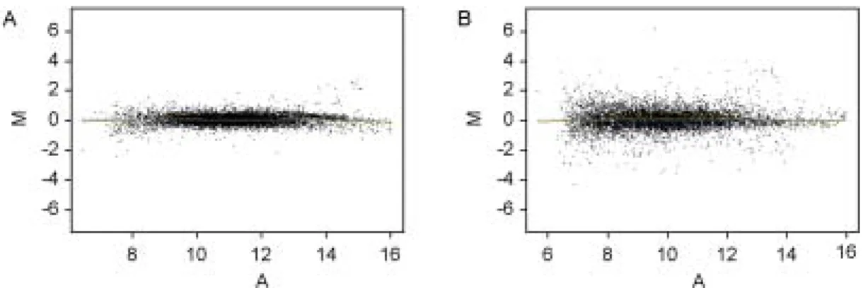
riments were repeated independently three times and fold changes were calculated. The mean values of each spot intensities in the three experiments were calculated and plotted (Figure 1). Of the 7616 genes examined in OVX/estrogen treatment/
6 h protocol, changes in mRNA expression were detected in 71 genes; 22 genes of these were activated and 49 genes were repressed (Table 1, 2).
Only 0.9% of all genes were activated or repre-
ssed more than two-fold and the remaining 99.1%
of the genes revealed no significant differences in their expression by estrogen. However, 638 genes showed differential expression ratio more than or less than two-fold in OVX/estrogen treatment/12 h protocol; 351 genes of these were activated and 287 genes were repressed. About 8.4% of all genes were activated or repressed (Table 1, 2).
Out of 638 activated or repressed genes, 433 Table 3. Primer sequences for RT-PCR
Gene Forward (F) and Reverse (S) primer sequences Product size (bp) F GTGTGGCCTGGGCCTTGTCG
Sprp2A
R GAGTCGGTGAGCTGGTGAG 240
F CGCGCCATCTGCACAAT Cystatin B
R GGCTTGTTTTCATGGGGGAG 225
F GGTGGTGTGGCGCAGCAAGC Ramp3
R GCAGGGGTCAGGGTCAGGAC 427
F GGCTGGGGTGCAGCGGAT Psmb5
R GGTAGATGGCTCGGCGGG 350
F CGTGATGGGGGTAAGGAGG Eif2s2
R CTAGAGCACAGGTTGGAG 489
F GATCATGAAGGTCGCCAGTG Id-1
R TCCATCTGGTCCTCAGTGC 476
F CCCCTACCGATGCCAGTGTC Decorin
R GCTCCGTTTTCAATCCCAGA 423
F GTGGCGGTGGATTGCAAGGA Imp2B
R GGGCGGCATACGATGGAAG 432
F TTGGCTTATACGTGCACT SFR-2
R TATTTGAGGGCATCATGCAA 295
F CCCATGTCACAAACCCTATC Bach2
R TGCTCACCTGACACCGTTCG 471
F CGTGGGAGATGCAAGGGCAG CXC12
R GAGGAGAATGGGGATGAAGC 247
F GTGACCAAGATGGAAATCCT Id-2
R TTTATTTAGCCACAGATAC 236
F TCAATGGAGTAAGCCCAAAG rpL7
R CAAGAGACCGAGCAATCAAG 246
genes were listed as known gene in GenBank, and 205 genes were unknown. These genes were cla- ssified into eight functional categories as based on biological functions; cell cycle-related, immune- related, signal transduction, transcription-related, enzyme, structure-related, apoptosis-related, and others (including expressed sequence tag and un-
known genes).
2. Confirmation of estrogen-regulated genes
The expression patterns of several genes up- or down-regulated by estrogen were confirmated to verify the results of microarray analysis using se- miquantitative RT-PCR. Several genes were ran- Figure 1. MA-plots represent genes activated and repressed by estrogen in ovariectomized mouse uterus. MA-plot is
used to represent the (R, G) data, where M = log2 R/G and A = log2 (R×G). (A) OVX/estrogen treatment/6 h protocol(B) OVX/estrogen treatment/12 h protocol. M, expression ratio; A, signal intensity; R, Red for Cy5; G, Green for Cy3.
Figure 2. Semiquantitative RT-PCR analysis confirming estrogen-regulated genes. Upper pannel, Expression patterns
of estrogen-activated genes; Lower pannel, Expression patterns of estrogen-repressed genes. Values of each band were normalized to rpL7 for the same sample. Data were mean±SEM from three replicate experiments.domly selected in different categories of genes;
Sprp2A, Cystatin B, Ramp3, Psmb5, Eif2s2 and Id-1 (up-regulated), and Decorin, Imp2B, SFR2, Bach2, Id-2 and CXC12 (down-regulated). The results of semiquantitative RT-PCR analysis were shown in Figure 2.
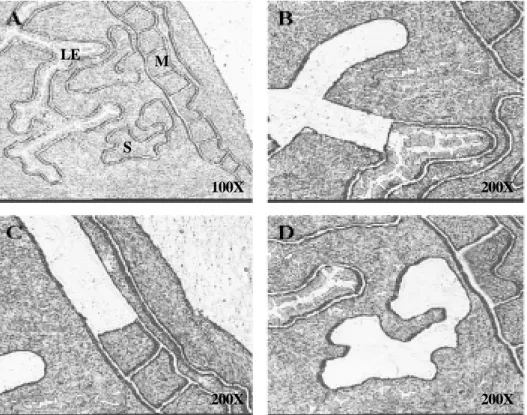
3. Analysis of selected gene expression pa- tterns in specific uterine cell types using LCM
To further analyze our findings we investigated gene expression of the selected up- or down re- gulated genes in uterine specific cell types using laser capture microdissection technique (LCM).
LCM allows the isolation of specific uterine cell types without contamination from other cell types.
We isolated the population of specific uterine cell types; luminal epithelial, stromal and muscle cells (Figure 3). While Sprp2A, Eif2s2, and Psmb5
mRNAs were strongly activated in luminal epithe- lial cells, Ramp3 and Cystatin B were activated in both luminal epithelial and stromal cells. On the other hand, decorin mRNA was mainly repressed in muscle cells and the rest of down-regulated genes were repressed in stromal cells (Figure 4).
DISCUSSIONS
Although estrogen plays an important role in various physiological events, the molecular me- chanisms that are regulated by estrogen in the uterus remain largely unknown. Therefore, the identification of novel estrogen-regulated gene pa- thway is essential to understanding how estrogen regulates various uterine physiology, such as es- trous cycle and implantation process. In the pre- sent study, we identified genes that are regulated LE
S M
100X 200X
200X 200X
Figure 3. Laser capture microdissection (LCM) procedure (A) Uterine sections stained with hematoxylin before
LCM. LE, luminal epithelial cells; S, stromal cells; M, muscle cells. (B) Capture of luminal epithelial cells, (C) muscle cells and (D) stromal cells.by estrogen in the ovariectomized mouse uterus exposed to 17β-estradiol (E2) for 6 h and 12 h using cDNA microarray. Prior to use in cDNA microarray analysis, each uterine RNA sample via OVX/estrogen treatment/6 h/12 h protocol was validated by assaying for up-regulated (Aspara- gine synthetase and Lactoferrin) or down-regula- ted genes (Glutathione S-transferase and Secreted frizzled-related sequence protein 2) which were previously known to regulate by estrogen in mo- use uterus.
14,15Our results showing that only 0.9% (71 genes) of all genes spotted in microarray slide were acti- vated or repressed more than two-fold in OVX/
estrogen treatment/6 h protocol suggest that direct or fast-responses by estrogen is not many in mRNA levels in uterus. However, 638 genes sh- owed differential expression ratio more than or less than two-fold in OVX/estrogen treatment/12 h protocol. About 8.4% of all genes were activated
or repressed. It is possible that many indirect or nongenomic pathways affected the patterns of mRNA expression in latter protocol in addition to direct or genomic pathways. Recently, Watanabe et al. also perfomed the analysis of estrogen- responsive genes in ovariectomized mouse uterus.
14They reported that generally 5% of all genes were activated or repressed more than three-fold by estrogen in OVX/estrogen treatment/6 h protocol;
616 genes were selected as estrogen-affected genes, of which 299 genes were activated and 317 genes were repressed. It is likely that this difference between their and our results is due to the species difference (they used C57/BL6/J strain) used in microarray analysis and the gene difference spo- tted in microarray.
Although our results to identify estrogen-regu- lated genes is quite different from that employed by Watanabe et al., two genes were shown the same expression pattern, chemokine orphan recep- tor 1 and receptor activity modifying protein 3 (Ramp3), which were slightly increased in OVX/
estrogen treatment/6 h protocol and highly incre- ased in OVX/estrogen treatment/12 h protocol.
Calcitonin (CT), CT-gene-related peptide (CGRP), adrenomedullin (AM) and amylin are related hor- mones and neuropeptides. Ramp 1, 2 and 3, th- rough interaction with calcitonin receptor (CTR), are required for the recognition of CGRP, AM and amylin by these receptor.
16,17These relationship is elevated by estrogen and suggested a role for these genes in uterine estrogen response.
Activating transcription factor (ATF) 1, 4 and 5 were activated by estrogen in OVX/estrogen treat- ment/12 h protocol. It is known that ATF activate gene expression by binding as homo- or heterodi- mers to the cAMP response element in regulatory region of target genes. Although ATF family was not well characterized in uterine physiological events, such as estrous cycle and implantation process, its functions in other tissues suggest an Figure 4. Expression patterns of estrogen-regulated
genes in specific uterine cell types obtained with LCM.
(A) Expression patterns of estrogen-activated genes. (B)
Expression patterns of estrogen-repressed genes. C, oil- treated group; E 6H, post-estrogen 6 hr; E 12H, post- estrogen 12 hr; LE, luminal epithelial cells; S, stromal cells; M, muscle cells.important role in cell proliferation and various signaling mechanisms.
18,19Small proline-rich protein 2A (Sprp2A) showed the highest level of up-regulation by estradiol in OVX/estrogen treatment/12 h protocol. This gene has been reported to express mainly in stratified squamous epithelial cells and control the diffe- rentiation of several cell types.
20,21In our study, Sprp2A mRNA was dominantly activated in lu- minal epithelial cells (LE) compared to stromal and muscle cells using LCM techniques. This re- gulation suggested that the expression of Sprp2A in LE has a significance relationship between the epithelial cell function and developing embryos.
Although the clarification of the function of Sprp 2A and the significance of estrogen-responsive- ness in relation to uterine physiological events are not identified, recent studies suggested that Sprp 2A gene family may play a pivotal role in estrous cycle and implantation process.
12,14To the best of our knowledge this is the first study to profile the early- and lately-reponsive ge- nes by estrogen involved in estrous and implan- tation process in mouse using cDNA microarray and LCM techniques. Our results indicate diffe- rential expression in spatiotemporal manner of genes regulated by estrogen and implicate various genes not previously known to express in ova- riectomized mouse uterus. In conclusion, further analysis including to progesterone regulation, spa- tiotemporal expression, protein expression and function of these genes may be necessary to un- derstand the exact mechanisms of estrogen effects in estrous and implantation process.
Acknowledgements
We thank Prof. Kang Hee-Gyoo for contributing to several experiments and helpful suggestions in Department of Biomedical Laboratory Science, Seoul Health College. We are also grateful to Lim Hee-Joung for excellent technical assistance of
laser capture microdissection.
REFERENCES
1. Green S, Walter P, Kumar V. Human oestrogen re- ceptor cDNA: sequence, expression and homology to v-erb-A. Nature 1986; 320: 134-9.
2. Kuiper GG, Enmark E, Pelto-Huikko M, Nilsson S, Gustafsson JA. Cloning of a novel receptor expre- ssed in rat prostate and ovary. Proc Natl Acad Sci 1996; 93: 5925-30.
3. Kousteni S, Bellido T, Plotkin LI. Nongenotropic, sex-nonspecific signaling through the estrogen or androgen receptors: dissociation from transcriptional activity. Cell 2001; 104: 719-30.
4. Angel N, Ana BR, Ouahiba L, Marjorie M, Esther F, Bernat S. Nongenomic actions of estrogens and xenoestrogens by binding at a plasma membrane re- ceptor unrelated to estrogen receptor α and estrogen receptor β. Proc Natl Acad Sci 2000; 97: 11603-8.
5. Ralf L, Martin W. Nongenomic actions of steroid hormones. Mol cell Biol 2003; 4: 46-56.
6. McMaster MT, Teng CT, Dey SK, Andre GK. Lac- toferrin in the mouse uterus: Analysis of the preim- plantation period and regulation by ovarian steroids.
Mol Endo 1992; 5: 101-11.
7. Wang X, Das SK, Damm D, Klagsbrun M, Abraham JA, Dey SK. Differential regulation of heparin-bin- ding epidermal growth factor-like growth factor in the adult ovariectomized mouse uterus by progeste- rone and estrogen. Endocrinol 1994; 135: 1264-71.
8. Das SK, Tsukamura H, Paria BC, Andrews GK, Dey SK. Differential regulation of epidermal growth fac- tor receptor (EGF-R) gene and regulation of EGF-R bioactivity by progesterone and estrogen in the adult mouse uterus. Endocrinol 1994; 134: 971-81.
9. Diane MK, Sylvia CH, Kenneth SK, Richard PD.
Activation of a uterine insulin-like growth factor I signaling pathway by clinical and environmental es- trogens: requirement of estrogen receptor-α. Endo- crinol 2000; 141: 3430-9.
10. Brannvall K, Korhonen L, Lindholm D. Estrogen- receptor-dependent regulation of neural stem cell proliferation and differentiation. Mol Cell Neurosci 2002; 21: 512-20.
11. Reese J, Das SK, Paria BC, Lim H, Song H, Ma- tsumoto H, et al. Global gene expression analysis to identify molecular markers of uterine receptivity and embryo implantation. J Biol Chem 2001; 276:
44137-45.
12. Cheon YP, Li Q, Xu X, Demayo FJ, Bagchi IC, Bagchi MK. A genomic approach to identify novel progesterone receptor regulated pathways in the uterus during implantation. Mol Endo 2002; 16:
2853-71.
13. Green AR, Edwards RE, Greaves P, White INH.
Comparison of the effect of oestradiol, tamoxifen and raloxifene on nerve growth factor-α expression in specific neonatal mouse uterine cell types using laser capture microdissection. J Mol Endo 2003; 30:
1-11.
14. Watanabe H, Suzuki A, Mizutani T, Khono S, Lu- bahn DB, Iguchi T. Genome-wide analysis of cha- nges in early gene expression induced by estrogen.
Genes to Cells 2002; 7: 497-507.
15. Das SK, Tan J, Johnson DC, Dey SK. Differential spatiotemporal regulation of lactoferrin and proge- sterone receptor genes in the mouse uterus by pri- mary estrogen, catechol estrogen, and xenoestrogen.
Endocrionol 1998; 139: 2905-15.
16. Fischer JA, Muff R, Born W. Functional relevance of G-protein-coupled- receptor-associated proteins, exemplified by receptor-activity-modifying proteins (RAMPs). Biochem Soc Trans 2002; 30: 455-60.
17. Gangula PR, Wimalawansa SJ, Yallampalli C. Pre- gnancy and sex steroid hormones enhance circu- lating calcitonin gene-related peptide concentrations in rats. Hum Reprod 2000; 5: 949-53.
18. Sabbah M, Courilleau D, Mester J, Redeuilh G.
Estrogen induction of the cyclin D1 promoter: invol- vement of a cAMP response-like element. Proc Natl Acad Sci 1999; 96: 11217-22.
19. Bleckmann SC, Blendy JA, Rudolph D, Monahan AP, Schmid SG. Activating transcription factor 1 and CREB are important for cell survival during early mouse development. Mol Cell Biol 2002; 22:
1919-25.
20. Song HJ, Poly G, Darwiche N, Lichti U, Kuroki T, Kartasova T. Mouse Sprr2 genes: A clustered family of genes showing differential expression in epithelial tissues. Genomics 1999; 55: 28-42.
21. Fischer DF, Gibbs S, Putte P, Backendorf C. In- terdependent transcription control elements regulate the expression of the SPRP2A gene during kerati- nocyte terminal differentiation. Mol Cell Biol 1996;
16: 5365-74.