Korean J Thorac Cardiovasc Surg 2012;45:251-253 □ Case Report □ http://dx.doi.org/10.5090/kjtcs.2012.45.4.251 ISSN: 2233-601X (Print) ISSN: 2093-6516 (Online)
− 251 −
Department of Thoracic and Cardiovascular Surgery, Seoul National University Hospital, Seoul National University College of Medicine Received: December 5, 2011, Revised: December 20, 2011, Accepted: January 25, 2012
Corresponding author: Ki-Bong Kim, Department of Thoracic and Cardiovascular Surgery, Seoul National University Hospital, Seoul National University College of Medicine, 101 Daehak-ro, Jongno-gu, Seoul 110-744, Korea
(Tel) 82-2-2072-3482 (Fax) 82-2-747-5245 (E-mail) [email protected]
C The Korean Society for Thoracic and Cardiovascular Surgery. 2012. All right reserved.
CC This is an open access article distributed under the terms of the Creative Commons Attribution Non-Commercial License (http://creative- commons.org/licenses/by-nc/3.0) which permits unrestricted non-commercial use, distribution, and reproduction in any medium, provided the original work is properly cited.
Redo-Coronary Artery Bypass due to Progression of the Celiac Axis Stenosis
Sang Yoon Yeom, M.D., Ho Young Hwang, M.D., Ph.D., Ki-Bong Kim, M.D., Ph.D.
We report a redo coronary artery bypass grafting (CABG) in a 55-year-old man. Angina recurred 7 years after the initial surgery. Coronary angiography showed all patent grafts except a faint visualization of the in situ right gastro- epiploic artery (RGEA) graft, which was anastomosed to the posterior descending coronary artery, associated with celiac axis stenosis. Redo-CABG was performed at postoperative 10 years because of aggravated angina and de- creased perfusion of the inferior wall in the myocardial single photon emission computed tomography. The saphe- nous vein graft was interposed between the 2 in situ grafts used previously; the right internal thoracic artery and RGEA grafts. Angina was relieved and myocardial perfusion was improved.
Key words: 1. Coronary artery bypass surgery 2. Reoperation
3. Ischemic heart disease
CASE REPORT
A 55 year-old man visited our emergency department be- cause of increasing frequency of chest pain. He had under- gone off-pump coronary artery bypass grafting (CABG) 10 years ago because of unstable angina associated with three vessel coronary artery disease. At the initial operation, the in situ right internal thoracic artery (ITA), in situ left ITA and in situ right gastroepiploic artery (RGEA) grafts were used to revascularize the left anterior descending coronary artery, two obtuse marginal coronary branches, and posterior descending coronary artery, respectively. An excess segment of the distal right ITA was connected to the side of left ITA as a Y-com- posite graft and anastomosed to the first diagonal coronary artery. Coronary angiography and myocardial single photon emission computed tomography (SPECT) were performed at
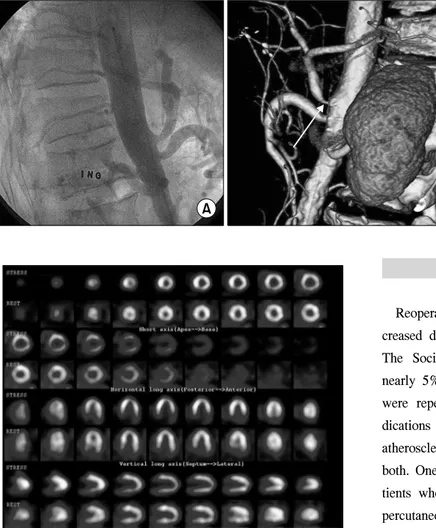
5 years after surgery as a follow-up study. The 5-year an- giography showed all patent grafts and the myocardial SPECT demonstrated no perfusion decrease. Exertional chest pain recurred at 7 years after surgery, and a repeated coro- nary angiography showed patent previous grafts including faint visualization of the in situ RGEA graft associated with significant stenosis at the os of the celiac axis. The computed tomographic angiogram also demonstrated a 90% stenosis at the celiac os, which had been without stenosis on abdominal angiography taken before the surgery (Fig. 1). The my- ocardial SPECT, however, demonstrated no perfusion decrease. Redo off-pump CABG was performed 10 years af- ter the initial surgery because of an increasing frequency of angina and an aggravated finding of the follow-up myocardial SPECT, which was a newly developed reversible perfusion decrease in the inferior wall (Fig. 2).
Sang Yoon Yeom, et al
− 252 −
Fig. 1. (A) Abdominal aortography demonstrating no celiac axis stenosis before the initial surgery. (B) Com- puted tomography angiogram showed a 90% stenosis of the celiac axis os at postoperative 7 years.
Fig. 2. Postoperative 10-year myocardial single photon emission computed tomography demonstrating a newly developed reversible perfusion decrease in the inferior wall.
At reoperation, the great saphenous vein was harvested from the lower leg and interposed between the middle part of in situ right ITA and distal part of in situ RGEA grafts used previously, to supply blood flow from the right ITA graft to the posterior descending coronary artery. The patient was dis- charged without any complication on the 9th postoperative day. One year after redo surgery, the patient had no symp- toms of angina and coronary angiogram was performed and revealed patent grafts, including an interposed saphenous vein graft (Fig. 3A). The myocardial SPECT test was also per- formed and demonstrated that there was no perfusion de- crease including the inferior wall (Fig. 3B).
DISCUSSION
Reoperations for coronary artery disease have been in- creased due to the increased number of isolated CABG [1].
The Society of Thoracic Surgeons statistics indicated that nearly 5% of the current CABG procedures done in the US were repeat surgical revascularization [2]. Angiographic in- dications for reoperation are progression of native coronary atherosclerosis, previous graft failure or a combination of both. One previous study demonstrated that 4 out of 400 pa- tients who underwent CABG using the RGEA graft needed percutaneous interventions due to the RGEA graft failure dur- ing postoperative follow-up of 22±11 months [3]. One of those 4 patients required an angioplasty for a newly devel- oped stenosis of the celiac trunk. In the present case, an in- dication for reoperation was recurred angina symptom even in the presence of all patent grafts. The patient had been free of angina, and the angiographic and myocardial SPECT fol- low-up studies revealed no abnormal findings at postoperative 5 years. When the patient suffered from recurred angina at postoperative 7 years, coronary angiography showed a faint visualization of the in situ RGEA graft associated with sig- nificant stenosis at the os of the celiac axis. The 10-year fol- low-up myocardial SPECT test demonstrated a newly devel- oped reversible perfusion decrease in the inferior wall.
The prevalence of celiac axis stenosis was 7.3% in a Korean population although it was lower than the previously reported incidence of celiac axis stenosis in Western pop- ulations ranged from 12.5% to 24% [4]. In the present case, significant celiac ostial stenosis was newly detected in a pa-
Redo CABG due to Celiac Axis Stenosis
− 253 −
Fig. 3. (A) Postoperative 1-year cor- onary angiogram showing patent grafts including an interposed saphe- nous vein graft. (B) Postoperative 1-year single photon emission com- puted tomography demonstrating an improved perfusion in the inferior wall. RITA, right internal thoracic ar- tery; SVG, saphenous vein graft;
LAD, left anterior descending coro- nary artery; PDA, posterior descend- ing coronary artery.
tient with recurred angina. Celiac artery stenting could be an alternative option in such a case. However, we performed a redo operation because celiac axis stenting was associated with a high incidence of late restenosis [5]. There are several strategies in redo CABG. The aorta or another in situ arterial graft could be chosen as a blood source. Alternatively, patent in situ grafts used previously may be re-used as an inflow conduit [6]. With regards to our patient, the 3 in situ arterial grafts had already been used. We used the patent in situ right ITA graft as an inflow conduit. The saphenous vein graft was interposed between the middle part of right ITA and distal part of in situ RGEA grafts used previously.
REFERENCES
1. Ferguson TB Jr, Hammill BG, Peterson ED, DeLong ER, Grover FL; STS National Database Committee. A decade of change--risk profiles and outcomes for isolated coronary ar-
tery bypass grafting procedures, 1990-1999: a report from the STS National Database Committee and the Duke Clini- cal Research Institute. Society of Thoracic Surgeons. Ann Thorac Surg 2002;73:480-9.
2. Zacharias A, Schwann TA, Riordan CJ, et al. Late outcomes after radial artery versus saphenous vein grafting during re- operative coronary artery bypass surgery. J Thorac Cardio- vasc Surg 2010;139:1511-8.e4.
3. Jegaden O, Eker A, Montagna P, et al. Technical aspects and late functional results of gastroepiploic bypass grafting (400 cases). Eur J Cardiothorac Surg 1995;9:575-80.
4. Park CM, Chung JW, Kim HB, Shin SJ, Park JH. Celiac axis stenosis: incidence and etiologies in asymptomatic individuals. Korean J Radiol 2001;2:8-13.
5. AbuRahma AF, Stone PA, Bates MC, Welch CA. Angio- plasty/stenting of the superior mesenteric artery and celiac trunk: early and late outcomes. J Endovasc Ther 2003;10:
1046-53.
6. Min HK, Lee YT, Kim WS, et al. Complete revasculariza- tion using a patent left internal thoracic artery and variable arterial grafts in multivessel coronary reoperation. Heart Surg Forum 2009;12:E244-9.