ABSTRACT
Objective: We investigated the clinical and radiological outcomes of a cement augmented anterior reconstruction and decompression without pedicle screw fixation in patients with osteoporotic thoracolumbar vertebral fracture with myelopathy.
Methods: There were 2 male and 6 female patients with thoracolumbar fracture and myelopathy included in the study. The mean follow-up period was more than 1 years.
The anterolateral decompression and cement augmented anterior reconstruction with poly(methyl methacrylate) (PMMA) was performed. Demographic data, clinical outcomes, perioperative parameters and radiologic parameter were retrospectively evaluated.
Results: The symptoms due to myelopathy were improved in all patients. The preoperative median visual analog scale score for lower back and leg were 8.5 that improved 4.25 and 3 at last follow up. The preoperative function state showed a median Oswestry Disability Index score 61.5 that improved 33. After surgery, preoperative encroachment of the spinal canal (5.12 mm, 37%) was disappeared. The median height of fractured vertebral body significantly increased from 7.83 to 12.63 mm. At the last follow-up point, the median height was 9.91 mm. The median kyphotic deformity was improved from 22.12° to 14.31°. At the final follow- up, the improvement was preserved (median value: 15.03). The acute complication according to PMMA such as leakage and embolization was none, but adjacent compression fracture as late complication according to cement augmentation was. One patient developed surgical site infection.
Conclusion: On the basis of the preliminary results, we considered that anterolateral
decompression and PMMA augmentation might be an optimal method for treating osteoporotic fracture with myelopathy in elderly patients or those with multiple medical comorbidities.
Keywords: Fracture; Spine; Osteoporosis; Vertebroplasty; Polymethyl methacrylate
Clinical Article
Received: Feb 10, 2020 Revised: Aug 13, 2020 Accepted: Sep 23, 2020 Address for correspondence:
Byeong-Wook Hwang
Department of Neurosurgery, Dongrae Wooridul Spine Hospital, 286, Chungnyeol- daero, Dongnae-gu, Busan 47879, Korea.
E-mail: hwangbre@hanmail.com
Copyright © 2020 Korean Neurotraumatology Society
This is an Open Access article distributed under the terms of the Creative Commons Attribution Non-Commercial License (https://
creativecommons.org/licenses/by-nc/4.0/) which permits unrestricted non-commercial use, distribution, and reproduction in any medium, provided the original work is properly cited.
ORCID iDs Sang-Min Lee
https://orcid.org/0000-0002-2021-9128 Hyeong Seok Oh
https://orcid.org/0000-0001-7608-3920 Sang-Ho Lee
https://orcid.org/0000-0002-8526-0260 Hyung-Chang Lee
https://orcid.org/0000-0001-5641-2475 Byeong-Wook Hwang
https://orcid.org/0000-0001-6041-9063 Conflict of Interest
The authors have no financial conflicts of interest.
Sang-Min Lee 1, Hyeong Seok Oh 1, Sang-Ho Lee 2, Hyung-Chang Lee 3, and Byeong-Wook Hwang 4
1Department of Neurosurgery, Busan Wooridul Spine Hospital, Busan, Korea
2Department of Neurosurgery, Spine Health Wooridul Hospital (SHWH) Gangnam, Seoul, Korea
3Department of Cardiovascular Surgery, Busan Wooridul Spine Hospital, Busan, Korea
4Department of Neurosurgery, Dongrae Wooridul Spine Hospital, Busan, Korea
Cement Augmented Anterior
Reconstruction and Decompression without Posterior Instrumentation:
A Less Invasive Surgical Option for
Osteoporotic Thoracolumbar Fracture
with Cord Compression
INTRODUCTION
Fracture of the thoracolumbar vertebra is more common, and severe lesions would lead to disastrous changes in the quality of life of patients with osteoporosis.2) The gold standard for the treatment of thoracolumbar fractures still remains controversial.2)
An important concept in thoracolumbar fractures is anterior column reconstruction.5) With advancement of the devices used in verterbroplasty and kyphoplasty, anterior column reconstruction can be performed by a single approach, offering adequate axial support immediately after operation and preventing collapse of the vertebral body into a kyphotic deformity, which could cause new neurologic problems.5,17)
Some article reported that posterior stabilization with pedicle screws and cement
augmentation were effective in thoracolumbar fracture, but the fixation with pedicle screws decreased the motion of spine because of facet joint involvement.5)
This study was the first to investigate the clinical and radiological outcomes of a cement augmented anterior reconstruction and decompression without pedicle screw fixation, thereby preserving the movement of segments involved in osteoporotic thoracolumbar vertebral fractures with myelopathy.
MATERIALS AND METHODS
We retrospectively collected data on patients with osteoporotic single thoracolumbar fracture who visited our hospital between August 2007 and January 2010. The Institutional Review Board of Wooridul Hospital Research Institutes approved the study protocol (WRD- IRB-2017-05-006). The inclusion criteria were as follows: (1) one thoracolumbar fracture, (2) the lesion compressed the spinal cord or thecal sac, as detected on the patient's magnetic resonance imaging photograph, (3) age >65 years, (4) osteoporosis (T score) <−2.5, and (5) anterior decompression and reconstruction with poly(methyl methacrylate) (PMMA) cement but without posterior fixation. After surgery, all patients were followed up clinically and radiologically for more than 1 year. At 5 years postoperatively, data on clinical manifestations were collected via phone interviews.
All operations were performed by one thoracic surgeon (H.C. Lee) and one neurosurgeon (B.W. Hwang). After induction of general anesthesia, the patients were placed in a lateral decubitus position. In patients with fractures at T10–12, surgeons performed the procedure using the extrapleural approach, whereas those with fractures located at the L1–2 vertebrae, the lesion was approached using the retroperitoneal method. After approaching the site of fracture, PMMA cement was injected into the vertebral body, with median PMMA cement volumes of 8 mL (interquartile range [IQR]: 6–10 mL). After some minutes from vertebroplasty, the neurosurgeon performed ventral decompression using a microscope and microscopic instruments and preserved the posterior longitudinal ligament. (FIGURE 1).
Perioperative parameters such as operative time and blood loss were collected. We assessed the clinical manifestations using the visual analog scale (VAS), Oswestry Disability Index (ODI), and American Spinal Injury Association (ASIA) scale. To evaluate the VAS results, the degree of lower back pain and radiating pain in the lower extremities was quantified from
0 to 10. The ODI was used to assess the disability due to low back pain. The ASIA scale was used to evaluate neurologic status.
Radiologic parameters included the morphology of the fracture, the height of the fractured body, the kyphotic angle of fractured segment, spinal canal encroachment, and injury of the posterior ligamentous complex in preoperative images. After surgery, we collected information on the height of the vertebral body, encroachment of the spinal canal, and kyphotic change of the segment. Data on the kyphotic angle and height of the segments were collected at routine postoperative intervals of 1, 3, and 12 months.
Data collection was performed by 2 independent spine surgeons, and the mean values were used for the statistical analysis. An independent t test was used to analyze continuous variables, whereas a χ2 test or Fisher's exact test was used to analyze categorical data.
Wilcoxon's signed-rank test was used to assess changes between preoperative and
postoperative parameters. All statistical analyses were performed using SPSS (SPSS, Chicago, IL, USA) 12.0 version. All p-values <0.05 were considered statistically significant.
RESULTS
Clinical characteristics of the study population
TABLE 1 presents the clinical data of all study participants. There were 2 male and 6 female patients included in the study, with the median age of 71 years (IQR: 65.25–76.75 years). The
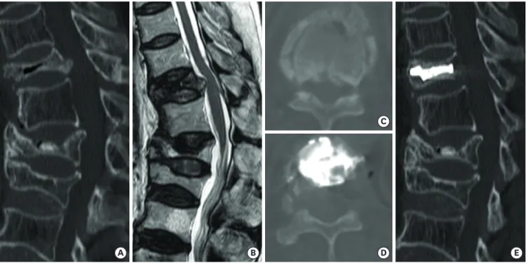
A B
C
D E
FIGURE 1. A 66-year-old female patient (case 7) had a T11 busting fracture after a slippage accident. (A) Preoperative CT scanning shows a T11 bursting fracture, with the loss of vertebral body height of 62% and a change in the kyphosis angle of 22.10°. (B) Preoperative magnetic resonance imaging shows incomplete injury of the posterior ligament complexes and compression of the spinal cord by the lesion. (C) The canal compromise was observed to be 32% on the axial CT scan. (D & E) The patient underwent a decompression procedure via the extra-pleural approach and vertebroplasty. The postoperative CT scan shows a well- decompressed state and recovery of the height and kyphotic angle of the segments.
CT: computed tomography.
most commonly injured vertebral body was T12. Slippage was the most common mechanism of vertebral fracture. The median point of thoracolumbar injury classification and severity score (TLICS) was 6 points (IQR: 5–7 points). The median operative time and estimated blood loss were 250 minutes (IQR: 212.5–300.0 minutes) and 195 mL (IQR: 167.5–200.0 mL), respectively. The median hospital day was 23 days (IQR: 13.75–33.50 days) and the median follow up months was 13.5 months (IQR: 12.0–17.75 months).
Clinical outcome
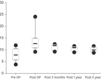
We collected the clinical outcomes preoperatively and at 3, 12, and 60 months postoperatively; the clinical outcome at the final follow-up was collected via telephone interview at least after 60 months. After operation, 6 patients had good outcomes but 2 patients had complications, with one patient developing surgical site infection and the other patient developing new compressive fracture above the operative site, because the lesion compressed the thecal sac. Thus, these patients underwent revision corpectomy and posterior pedicle fixation. The patient's neurologic problem caused by compression of the thecal sac improved after surgery. TABLE 2 shows the patients' preoperative and postoperative neurologic status. The median preoperative VAS scores for back pain and lower leg pain were 8.5 (IQR: 3–10) and 8.5 (IQR: 7.25–10), respectively, whereas the postoperative VAS scores were 1.75 (IQR: 1.75–3.25) and 3.5 (IQR: 3–4.25), respectively (p=0.027). At the last follow-up, the median VAS scores for back pain and lower leg pain were 4.25 (IQR: 2.75–5) and 3 (IQR:
2–4), respectively (p=0.027). FIGURE 2 presents the severity of back pain and lower extremity pain using VAS at each follow-up time point. The median ODI score was 61.5 (IQR: 54.75–
88.50) preoperatively and 34 (IQR: 27–39) postoperatively (p=0.047). At the final follow-up done via telephone interview, the median ODI score was 33 (IQR: 26.25–42.5). FIGURE 3 also shows the change of ODI score at each follow-up timepoint.
Radiologic outcome
Preoperative encroachment of the spinal canal was 5.12 mm (IQR: 4.49–7.18 mm) and 37%
(IQR: 31%–56%). After surgery, the MRI scan showed that the fragments compressing the thecal sac totally disappeared in all cases.
TABLE 1. The demographic characteristics of all patients
No. Sex Age Level PLC NEx TLIC OP time EBL Duration of hospitalization F/U
1 M 76 T12 Intact D 5 250 200 64 12
2 M 77 L1 Intact D 5 300 200 23 14
3 F 81 T12 Incomplete C 7 200 225 29 12
4 F 65 L2 Incomplete D 7 250 150 13 18
5 F 72 T12 Intact D 5 200 195 16 17
6 F 66 T12 Incomplete C 7 250 190 23 12
7 F 70 T11 Incomplete D 7 300 160 35 84
8 F 60 L11 Intact C 5 320 195 11 13
PLC: The injury of posterior ligament complex, NEx: American Spinal Injury Association grade, TLIC: thoracolumbar injury scale, OP: operation, EBL: estimated blood loss, F/U: follow-up.
TABLE 2. The preoperative and postoperative neurologic state using ASIA scale
Postoperative Preoperative
Grade A Grade B Grade C Grade D Grade E
Grade A 1
Grade B Grade C
Grade D 2 3
Grade E 2
ASIA: American Spinal Injury Association.
The median height of the fractured vertebral body was 7.83 mm (IQR: 5.74–10.75), and the height significantly increased after surgery (median value: 12.63, IQR: 10.87–14.93, p=0.012).
At 1 year postoperatively, the median height of the segment was 9.91 (IQR: 9.02–11.10, p=0.046). FIGURE 4 represents the change of the vertebral height at each follow-up time point.
The median kyphotic angle of the segments was 22.12° (IQR: 19.72°–28.00°) preoperatively and 14.31° (IQR: 8.31°–15.70°, p=0.012) postoperatively. At the final follow-up evaluation, the median kyphotic angle at the operative level was 15.03 (IQR: 11.02–18.02, p=0.028). The change in kyphotic angle is shown in FIGURE 5.
DISCUSSION
In this study, we investigated the clinical and radiological outcomes of a cement augmented anterior reconstruction and decompression without pedicle screw fixation in patients with 8
6
4
2
0 10
A
Pre OP Post OP Post 3 months Post 1 year Post 5 years
8
6
4
2
0 10
B
Pre OP Post OP Post 3 months Post 1 year Post 5 years FIGURE 2. Change in visual analog scale scores at each follow-up timepoint for lower back pain (A) and lower extremity pain (B).
OP: operation.
80
60
40
20
0 100
Pre OP Post OP Post 3 months Post 1 year Post 5 years FIGURE 3. Change in ODI at each follow-up timepoint.
ODI: Oswestry Disability Index, OP: operation.
osteoporotic thoracolumbar vertebral fracture with myelopathy. Our findings revealed that this technique may be a potential method for treating osteoporotic fracture with myelopathy in elderly patients or those having medical comorbidities.
Thoracolumbar junctional lesions are located between the rigid vertebral column of the associated rib cage and relatively movable lumbar vertebrae. As a result, these lesions were the most common site of vertebral fractures.6) In fact, our patients had bursting fractures at these lesions. Traumatic bursting fractures account for more than 10% of all spine injuries.21) The axial forces and flexion movements were caused by the failure of both anterior and middle columns.21) There are multiple classifications of thoracolumbar fractures, but there has been no absolute consensus on which fracture should be treated surgically and which surgical method is the best approach. Operation with posterior pedicle screw and rod constructs is commonly performed for thoracic and lumbar traumatic injuries.12) This was performed to prevent the fractured vertebrae from collapsing, which could result in kyphotic deformity. The long implantation for up and down 2 levels was the traditional operative method. However, this method required extensive exposure of the spine and prolonged operative time, involved profuse estimated blood loss, and could markedly damage the
25
20
15
10
5
0 30
Pre OP Post OP Post 3 months Post 1 year Post 5 years FIGURE 4. Change in the vertebral height at each follow-up timepoint.
OP: operation.
25 20 15 10 5 0 35 30
Pre OP Post OP Post 3 months Post 1 year Post 5 years
FIGURE 5. Change in the kyphotic angle at the operated segment at each follow-up timepoint.
OP: operation.
paraspinal muscles.17-19) Short segment pedicle screw fixation was an alternative method for fast stabilization of the vertebral body, which is inserted above and below the vertebrae.
This method had many benefits such as minimal blood loss, short operative time, and early fast mobilization, but some articles have reported a high incidence of instrumental failure in this procedure.17) Although pedicle screw instrumentation was developed a few decades ago, the pedicle screw instrumentation method, which lacked anterior column supports, resulted in delayed kyphosis, nonunion, and loosening of the pedicle screw.7,20) Given that anterior column reconstruction is the most important point of treatment for thoracolumbar fractures, mechanical instruments and cement augmentation using PMMA were used for anterior column support.13,21) There are three article on the standalone cement augmentation method for bursting fractures. Two articles present the method using PMMA cement and the other utilized calcium phosphate cement. Chen and Lee performed vertebroplasty using the standalone cement augmentation method in 6 patients with bursting fractures; the patients did not show effects of conservative care for more 3 months. In this article, pain was reduced immediately after the operation, and the effect was prolonged even beyond 3 months (p<0.005).6) They also reported 4 cases of cement leakage into the discal and/or paravertebral space, but all cases were asymptomatic. Kyphotic change of the fractured segments was evaluated, and the correction was preserved until the last follow-up. The second study on standalone PMMA cement augmentation was conducted by Huwart et al.,11) who investigated 62 patients with vertebral split fractures and without neurologic defects. The authors performed CT-guided vertebroplasty with PMMA cements. The article presented statistically significant improvements in the patients' VAS score and ODI at the final follow-up (p<0.001).
They also achieved significant height restoration and kyphosis correction immediately after the operation, but the kyphosis correction decreased to 6° at the last follow-up. Despite the loss of the correction, the clinical outcome was not affected. In our study, the correction of kyphosis was not also preserved but the change did not affect the clinical results. The third study on standalone cement augmentation was conducted by Maestretti et al.14) who included traumatic compression and bursting fractures in their cohort; they used calcium phosphate cements (CPC). Interestingly, 20% of these patients treated with CPC showed cement resorption at the last follow-up.
PMMA cement has been used in the treatment of osteoporotic compression fractures since a long time because it functions as a solid bone void filler and immediately provides stability by interdigitating within the vertebral trabecular bone through an exothermic reaction.1,21) This exothermic reaction ablates the intraosseous nerve ending; thus, this reaction has analgesic effects.1) However, this thermogenic reaction decreases bone healing and stimulates thermal necrosis of surrounding soft tissue and inflammation.9,16)
Cement augmentation is not absolutely a safe procedure, and complications may occur in this type of procedure. Cements injected under pressure can lead to leakage out of the vertebral body. In the majority of these problems, cements leak into the paravertebral soft tissue or intradiscal space, but rarely into the spinal canal and intervertebral foramina.21) After bone cement leakage into neural structures, these structures were damaged by direct compression and thermal and chemical reactions.8) In Benneker and Hoppe's study, the incidence of leakage into the intervertebral foramen and spinal canal were 0%–3.7% and 0%–0.5%, respectively.3) Another problem was leakage into the venous system. In cases with cement leakage into the azygous vein or inferior vena cava, the particles migrated to the lung parenchyma, which is a life-threatening complication.8) The report also represented that the risk of pulmonary embolism was 3.5%–23.0%.3) The major risk factor for these leakage cases was the viscosity
of PMMA.20) After the development of highly viscous PMMA, the incidence of cement leakage decreased significantly.10) Given that we used the highly viscous PMMA produced by DePuy Synthes, there was no leakage of PMMA cement in our cases. Another method to reduce the incidence of cement leakage was the use of continuous pulsed fluoroscopy.17)
The late complications of PMMA cement augmentation were incident fractures in adjacent vertebrae and recollapse of augmented vertebrae. On average, 20% of patients treated with cement augmentation developed new osteoporotic fractures within 1 year.15) Biomechanically, the pressure over the vertebral body adjacent to the site augmented with cement is increased;
thus, over 50% of incidental fractures occurred adjacent to the treated vertebral body.15) The most popular theory about the occurrence of these adjacent fractures was that the stiffness in the augmented vertebrae was caused by the collapse of osteoporotic vertebrae around the lesion. However, another opinion about this phenomenon was that adjacent fractures might be the natural manifestations of severe osteoporotic spine.15) Cao et al.4) showed that low bone mineral density, the presence of multiple treated vertebrae, and a history of steroid use were associated with the development of this new fracture around the augmented vertebra.
In our study, one case of new compression fracture adjacent to the augmented vertebrae occurred at 1 month postoperatively. This patient also had multiple augmented vertebral bodies and severe osteoporosis, which required corpectomy and posterior fixation with pedicle screw.
Our surgical method for thoracolumbar junctional bursting fracture has very limited
indications. First, the posterior longitudinal ligaments at fracture site should be intact, which can be confirmed by the preoperative MRI scan with 2-mm axial interval at bursting fracture site. Moreover, it also should be preserved during the decompressive surgery. Second, all patients underwent this surgery had sclerotic change, but other defect or fracture, at upper and lower endplate at surgical site. Third, the posterior compartments of the fractured vertebrae including interarticular facet, lamina and spinous process were fully preserved.
There are a few points to be aware during this procedure. The surgeons are recommended to use highly viscous PMMA to prevent the leakage toward spinal canal during the cement augmentation, which was performed before the decompressive operation. Because the operator could remove the PMMA particle during decompression when the leakage takes place. To maintain the spinal stability, it is important to preserve the two-thirds of vertebral body and the contralateral pedicle at the surgical level.
There are a few limitations in our study. We included a very small number of patients (n=6), and this study was retrospective in nature. Another limitation of this study was the length of radiological follow-up. We did not analyze the changes of patient's spine with severe osteoporosis beyond 1 year. Despite these limitations, our results suggest that anterolateral decompression with PMMA augmentation might be another treatment method for osteoporotic fracture with myelopathy in elderly patients with medical comorbidities.
CONCLUSION
On the basis of the preliminary results, we considered that anterolateral decompression and PMMA augmentation might be an optimal method for treating osteoporotic fracture with myelopathy in elderly patients or those with multiple medical comorbidities.
REFERENCES
1. Aebli N, Goss BG, Thorpe P, Williams R, Krebs J. In vivo temperature profile of intervertebral discs and vertebral endplates during vertebroplasty: an experimental study in sheep. Spine 31:1674-1678, 2006 PUBMED | CROSSREF
2. Alpantaki K, Bano A, Pasku D, Mavrogenis AF, Papagelopoulos PJ, Sapkas GS, et al. Thoracolumbar burst fractures: a systematic review of management. Orthopedics 33:422-429, 2010
PUBMED | CROSSREF
3. Benneker LM, Hoppe S. Percutaneous cement augmentation techniques for osteoporotic spinal fractures.
Eur J Trauma Emerg Surg 39:445-453, 2013 PUBMED | CROSSREF
4. Cao J, Kong L, Meng F, Zhang Y, Shen Y. Risk factors for new vertebral compression fractures after vertebroplasty: a meta-analysis. ANZ J Surg 86:549-554, 2016
PUBMED | CROSSREF
5. Chen C, Lv G, Xu B, Zhang X, Ma X. Posterior short-segment instrumentation and limited segmental decompression supplemented with vertebroplasty with calcium sulphate and intermediate screws for thoracolumbar burst fractures. Eur Spine J 23:1548-1557, 2014
PUBMED | CROSSREF
6. Chen JF, Lee ST. Percutaneous vertebroplasty for treatment of thoracolumbar spine bursting fracture.
Surg Neurol 62:494-500, 2004 PUBMED | CROSSREF
7. Cheng LM, Wang JJ, Zeng ZL, Zhu R, Yu Y, Li C, et al. Pedicle screw fixation for traumatic fractures of the thoracic and lumbar spine. Cochrane Database Syst Rev 5:CD009073, 2013
PUBMED | CROSSREF
8. Cotten A, Dewatre F, Cortet B, Assaker R, Leblond D, Duquesnoy B, et al. Percutaneous vertebroplasty for osteolytic metastases and myeloma: effects of the percentage of lesion filling and the leakage of methyl methacrylate at clinical follow-up. Radiology 200:525-530, 1996
PUBMED | CROSSREF
9. Dean JR, Ison KT, Gishen P. The strengthening effect of percutaneous vertebroplasty. Clin Radiol 55:471- 476, 2000
PUBMED | CROSSREF
10. Gonschorek O, Hauck S, Weiß T, Bühren V. Percutaneous vertebral augmentation in fragility fractures- indications and limitations. Eur J Trauma Emerg Surg 43:9-17, 2017
PUBMED | CROSSREF
11. Huwart L, Foti P, Andreani O, Hauger O, Cervantes E, Brunner P, et al. Vertebral split fractures: technical feasibility of percutaneous vertebroplasty. Eur J Radiol 83:173-178, 2014
PUBMED | CROSSREF
12. Kallemeier PM, Beaubien BP, Buttermann GR, Polga DJ, Wood KB. In vitro analysis of anterior and posterior fixation in an experimental unstable burst fracture model. J Spinal Disord Tech 21:216-224, 2008
PUBMED | CROSSREF
13. Liao JC, Fan KF, Chen WJ, Chen LH, Kao HK. Transpedicular bone grafting following short-segment posterior instrumentation for acute thoracolumbar burst fracture. Orthopedics 32:493-500, 2009 PUBMED | CROSSREF
14. Maestretti G, Cremer C, Otten P, Jakob RP. Prospective study of standalone balloon kyphoplasty with calcium phosphate cement augmentation in traumatic fractures. Eur Spine J 16:601-610, 2007 PUBMED | CROSSREF
15. Nagad P, Rawall S, Kundnani V, Mohan K, Patil SS, Nene A. Postvertebroplasty instability. J Neurosurg Spine 16:387-393, 2012
PUBMED | CROSSREF
16. Oner FC, Verlaan JJ, Verbout AJ, Dhert WJ. Cement augmentation techniques in traumatic thoracolumbar spine fractures. Spine (Phila Pa 1976) 31:S89-S95, 2006
PUBMED | CROSSREF
17. Rahamimov N, Mulla H, Shani A, Freiman S. Percutaneous augmented instrumentation of unstable thoracolumbar burst fractures. Eur Spine J 21:850-854, 2012
PUBMED | CROSSREF
18. Schinkel C, Anastasiadis AP. The timing of spinal stabilization in polytrauma and in patients with spinal cord injury. Curr Opin Crit Care 14:685-689, 2008
PUBMED | CROSSREF
19. Schinkel C, Frangen TM, Kmetic A, Andress HJ, Muhr G; German Trauma Registry. Timing of thoracic spine stabilization in trauma patients: impact on clinical course and outcome. J Trauma 61:156-160, 2006 PUBMED | CROSSREF
20. Shen YX, Zhang P, Zhao JG, Xu W, Fan ZH, Lu ZF, et al. Pedicle screw instrumentation plus augmentation vertebroplasty using calcium sulfate for thoracolumbar burst fractures without neurologic deficits.
Orthop Surg 3:1-6, 2011 PUBMED | CROSSREF
21. Zaryanov AV, Park DK, Khalil JG, Baker KC, Fischgrund JS. Cement augmentation in vertebral burst fractures. Neurosurg Focus 37:E5, 2014
PUBMED | CROSSREF