ABSTRACT
Purpose: Although several reports have described the relationship between periodontal disease and cardiovascular disease, information about the association between periodontal disease and the progression of degenerative aortic stenosis (AS) is lacking. Therefore, we performed a retrospective, single-center, pilot study to provide insight into this potential association.
Methods: Data from 45 consecutive patients (19 men; median age, 83 years) with mild or moderate degenerative aortic stenosis were analyzed for a mean observation period of 3.3±1.9 years. The total amount of Aggregatibacter actinomycetemcomitans and Porphyromonas gingivalis and titers of serum immunoglobulin G (IgG) against periodontal bacteria and high-sensitivity C-reactive protein (hs-CRP) were evaluated. Aortic valve area (AVA), maximal velocity (Vmax), mean pressure gradient (mean PG), and the Doppler velocity index (DVI) were evaluated. The change in each parameter per year ([ParameterLATEST–ParameterBASELINE]/Follow-up Years) was calculated from the retrospective follow-up echocardiographic data (baseline vs. the most recently collected data [latest]).
Results: No correlation was found between the concentration of periodontopathic bacteria in the saliva and AS status/progression. The anti-P. gingivalis antibody titer in the serum showed a significant positive correlation with AVA and DVI. Additionally, there was a negative correlation between the anti-P. gingivalis IgG antibody titer and mean PG. The hs-CRP concentration showed positive correlations with Vmax and mean PG. Meanwhile, a negative correlation was observed between the anti-P. gingivalis IgG antibody titer and ΔAVA/year and Δmean PG/year. The hs-CRP concentration showed positive correlations with Vmax and mean PG, and it was significantly higher in patients with rapid aortic stenosis progression (ΔAVA/year <−0.1) than in their counterparts.
Conclusions: Our results suggest that periodontopathic bacteria such as A. actinomycetemcomitans and P. gingivalis are not directly related to the status/progression of degenerative AS. However, inflammation and a lower immune response may be associated with disease progression.
Keywords: Aortic valve stenosis; C-reactive protein; Periodontal diseases; Porphyromonas gingivalis
Research Article
Received: Sep 6, 2020 Revised: Dec 15, 2020 Accepted: Mar 5, 2021
*Correspondence:
Akihisa Kataoka
Division of Cardiology, Department of Medicine, Teikyo University, 2-11-1 Kaga, Itabashiku, Tokyo 173-8606, Japan.
E-mail: kataoaki@sd5.so-net.ne.jp Tel: +81-3-3964-1211
Fax: +81-3-3964-6022 Sayaka Katagiri
Department of Periodontology, Tokyo Medical and Dental University (TMDU), 1-5-45 Yushima, Bunkyoku, Tokyo 113-8549, Japan.
E-mail: katagiri.peri@tmd.ac.jp Tel: +81-3-5803-5488 Fax: +81-3-5803-0196
Copyright © 2021. Korean Academy of Periodontology
This is an Open Access article distributed under the terms of the Creative Commons Attribution Non-Commercial License (https://
creativecommons.org/licenses/by-nc/4.0/).
ORCID iDs Akihisa Kataoka
https://orcid.org/0000-0002-1857-709X Sayaka Katagiri
https://orcid.org/0000-0002-5765-2742 Hideyuki Kawashima
https://orcid.org/0000-0002-2847-1248 Fukuko Nagura
https://orcid.org/0000-0001-9021-7681 Yugo Nara
https://orcid.org/0000-0002-9099-6336 Hirofumi Hioki
https://orcid.org/0000-0001-7189-8450
Akihisa Kataoka 1,*, Sayaka Katagiri 2,*, Hideyuki Kawashima 1,
Fukuko Nagura 1, Yugo Nara 1, Hirofumi Hioki 1, Makoto Nakashima 1, Naoki Sasaki 2, Masahiro Hatasa 2, Shogo Maekawa 2, Yujin Ohsugi 2, Takahiko Shiba 2, Yusuke Watanabe 1, Tomoki Shimokawa 3,
Takanori Iwata 2, Ken Kozuma 1
1Division of Cardiology, Department of Medicine, Teikyo University, Tokyo, Japan
2Department of Periodontology, Graduate School of Medical and Dental Sciences, Tokyo Medical and Dental University, Tokyo, Japan
3Department of Cardiovascular Surgery, Teikyo University, Tokyo, Japan
Association between periodontal bacteria and degenerative aortic stenosis: a pilot study
Periodontal Science
Makoto Nakashima
https://orcid.org/0000-0001-6835-6669 Naoki Sasaki
https://orcid.org/0000-0002-1831-4863 Masahiro Hatasa
https://orcid.org/0000-0001-9103-5898 Shogo Maekawa
https://orcid.org/0000-0002-6207-7826 Yujin Ohsugi
https://orcid.org/0000-0002-2843-173X Takahiko Shiba
https://orcid.org/0000-0002-8388-6868 Yusuke Watanabe
https://orcid.org/0000-0003-2297-5303 Tomoki Shimokawa
https://orcid.org/0000-0002-7692-9452 Takanori Iwata
https://orcid.org/0000-0002-5461-7298 Ken Kozuma
https://orcid.org/0000-0003-3977-5973 Funding
The author(s) disclose receipt of the following financial support for the research, authorship, and/or publication of this article: the work was supported by the Japan Society for the Promotion of Science (16K21382 to A.K.
and 26463128 to S.K.) and grants from the Cardiovascular Research Foundation, Tokyo, Japan, and the Epidemiological Research grant from St. Luke's International University, Tokyo, Japan, to A.K.
Author Contributions
Conceptualization: Akihisa Kataoka, Sayaka Katagiri; Formal Analysis: Akihisa Kataoka, Sayaka Katagiri, Masahiro Hatasa;
Investigation: Hideyuki Kawashima, Fukuko Nagura, Yugo Nara, Hirofumi Hioki, Makoto Nakashima, Yusuke Watanabe, Tomoki Shimokawa, Ken Kozuma; Methodology: Naoki Sasaki, Masahiro Hatasa, Shogo Maekawa, Yujin Ohsugi, Tomoki Shimokawa, Takanori Iwata ; Project Administration: Ken Kozuma, Takanori Iwata; Writing - original draft: Akihisa Kataoka, Sayaka Katagiri; Writing - review
& editing: Akihisa Kataoka, Sayaka Katagiri, Hideyuki Kawashima, Fukuko Nagura, Yugo Nara, Hirofumi Hioki, Makoto Nakashima, Naoki Sasaki, Masahiro Hatasa, Shogo Maekawa, Yujin Ohsugi, Takahiko Shiba, Yusuke Watanabe, Tomoki Shimokawa, Takanori Iwata, Ken Kozuma.
Conflict of Interest
No potential conflict of interest relevant to this article was reported.
INTRODUCTION
Periodontal disease is an inflammatory disorder caused by pathogenic Gram-negative oral microorganisms that can cause both local inflammation, which destroys the alveolar bone and soft tissues around the teeth, and systemic inflammation [1-3]. Periodontal bacteria present in the dental plaque possess various virulence factors, including lipopolysaccharide, fimbriae, and enzymes, that can trigger inflammation in the periodontal tissues [4]. A previous study reported elevated levels of systemic inflammatory mediators in patients with severe periodontal disease [5]. In addition, bacteria originated from the periodontal pocket can cause bacteremia [6]. Furthermore, considering that the bacterial flora of the oral cavity differs from that of the gut [7], swallowed bacteria may affect the composition of the gut microbiome. Therefore, periodontal infection has long been associated with an increased risk of various diseases, including cardiovascular diseases [2,3,8], type 2 diabetes [9], and non-alcoholic fatty liver disease [10].
Degenerative aortic stenosis (AS), which is a common and progressive disease that makes a large contribution to mortality in the current aging population, is most often caused by an active disease process characterized by inflammation, lipid accumulation, and calcification [11]. Leaflet calcification and fibrosis eventually occur once AS is initiated, resulting in reduced leaflet motion and leading to a progressive reduction of the aortic valve area (AVA) [12].
Given that degenerative AS is a systemic inflammatory disorder, we hypothesized that it could be associated with periodontal disease, similarly to other cardiovascular diseases [2,3,8,13,14]. However, in contrast to other cardiovascular diseases, information regarding the relationship between degenerative AS and periodontal disease as a chronic inflammatory disorder is lacking.
Therefore, the aim of the present study was to conduct a preliminary analysis using retrospective patient data to evaluate the potential association between periodontal disease and AS by focusing on Porphyromonas gingivalis and Aggregatibacter actinomycetemcomitans as representative periodontal bacteria.
MATERIALS AND METHODS
Study design and population of the An evaluation of the association between PeRIodontal baCteria and prOgression of degenerative aorTic stenosis (APRICOT) study
In this retrospective pilot study, we screened 57 consecutive patients who were diagnosed with degenerative AS at Teikyo University Hospital between November 2017 and April 2018 and were part of the larger APRICOT study, which is a collaboration network on AS involving the Department of Medicine, Division of Cardiology, Teikyo University, and the Department of Periodontology, Tokyo Medical and Dental University. Among the 57 eligible patients, 12 were excluded due to bicuspid AS (n=5) and severe AS at baseline (n=7). Therefore, a total of 45 patients (median age, 83 years; interquartile range, 77–86 years; 42% men) were included for the main analysis (Figure 1A). In addition, 2 patients with severe AS who were excluded from the main analysis and underwent surgical aortic valve replacement were included for analyses of the aortic valve specimen to evaluate the presence of periodontal bacteria in the valves.
The study protocol was developed in accordance with the 1975 Declaration of Helsinki (revised in 2013) and was approved by the Institutional Review Board committees of Teikyo University and the Tokyo Medical and Dental University, Department of Dentistry (approval numbers TEIRIN15-236, TEIRIN16-102, and D2016-066). All patients provided informed written consent for participating in the study. This trial was registered with the University Hospital Medical Information Network with the number UMIN000024251.
Data collection
The data collection protocol is outlined in Figure 1B. All patients were diagnosed with AS using standard 2-dimensional B-mode and Doppler transthoracic echocardiography at baseline and then followed up every 6 to 12 months according to the guidelines of the American College of Cardiology/American Heart Association [15]. Conventional and AS diagnostic parameters such as the AVA, maximal velocity (Vmax), mean pressure gradient (mean PG), and Doppler velocity index (DVI) were measured at each visit according to the guidelines of the American Society of Echocardiography [16]. The change in each parameter per year ([ParameterLATEST–ParameterBASELINE]/Follow-up Years) was calculated from the retrospective follow-up echocardiographic data using baseline and the most recently collected data during follow-up (latest). Saliva and blood samples were collected at the time of enrollment after signed consent forms were provided. Saliva and plasma components obtained from centrifuged blood samples were frozen at −80°C and subsequently were sent to the laboratory at Tokyo Medical and Dental University for analysis.
Cultivation of P. gingivalis and A. actinomycetemcomitans
The ATCC 33277 strain of P. gingivalis was cultured as described previously [17]. In brief, P.
gingivalis cells were maintained on trypticase soy agar (Difco Laboratories, Detroit, MI, USA) 57 consecutive aortic stenosis
5 excluded 7 excluded
45 less than severe degenerative AS
2 patients undergo SAVR Bicuspid
Severe at baseline
• Consent
• Saliva sample
→ measurement of Pg and Aa
• Blood sample
→ serum IgG antibody titer
→ hs-CRP
(baseline)Echo Echo
(latest) Echo
6 to 12 months
A
B Figure 1. Diagram showing patient details and data collection protocols.
(A) Subjects were selected according to the criteria shown in the flowchart and (B) data were collected according to the protocols.
SAVR: surgical aortic valve replacement, AS: aortic stenosis, Echo: echocardiography, Pg: Porphyromonas gingivalis, Aa: Aggregatibacter actinomycetemcomitans, IgG: immunoglobulin G, hs-CRP: high-sensitivity C-reactive protein.
supplemented with 10% defibrinated horse blood, hemin (5 μg/mL), and menadione (0.5 μg/mL) at 37°C under anaerobic conditions (10% CO2, 10% H2, and 80% N2). After 2 days of incubation, the cells were inoculated into trypticase soy broth supplemented with 0.5% yeast extract, menadione, and hemin under anaerobic conditions.
A. actinomycetemcomitans (the ATCC 43718 strain) was inoculated in ATCC medium 44 (brain heart infusion broth) and cultured anaerobically (AnaeroPackR-Anaero for Susceptibility, Mitsubishi Gas Chemical Company Inc., Tokyo, Japan) at 37°C for 24 hours [18].
Measurement of serum anti-P. gingivalis/A. actinomycetemcomitans immunoglobulin G (IgG) antibody titers
Specific serum IgG titers were measured using enzyme-linked immunosorbent assay (ELISA) as described previously [18,19]. In brief, 96-well microplates (EIA plates; Costar, Corning, NY, USA) were coated with 10 μg/mL sonicated P. gingivalis or A. actinomycetemcomitans extracts in carbonate buffer and incubated for 2 hours at 37°C. After blocking with 2% bovine serum albumin in carbonate buffer, the plates were washed with phosphate-buffered saline (PBS)- Tween® (1× PBS, 0.05% Tween 20®, pH 7.2). Serially diluted, reference-pooled, positive- control serum samples obtained from healthy subjects (25–215, 200 µL per well) and single diluted patient serum samples (210, 200 µL per well) were added to each well, and the plates were further incubated for 1 hour at 37°C and washed again. Subsequently, 200 µL of alkaline phosphatase-conjugated goat anti-human IgG (Sigma, St. Louis, MO, USA) was added to each well. After incubation, the plates were washed and developed with phosphatase substrate (Sigma), and the optical density at 450 nm was read using a microplate reader (SoftMAX; Molecular Devices, Sunnyvale, CA, USA). Antibody titers were calculated according to a previously described method [20].
DNA isolation and detection of P. gingivalis and A. actinomycetemcomitans in saliva samples
A NucleoSpin DNA tissue kit (Takara Bio Inc., Kusatsu, Japan) was used to extract DNA from the saliva according to the manufacturer's instructions. To detect P. gingivalis and A.
actinomycetemcomitans DNA, the extracted DNA was subjected to quantitative polymerase chain reaction (qPCR) using a TaqMan probe (5′-FAM-TGCGTAACGCGTATGCAACTTGCC- TAMRA-3′ for P. gingivalis and 5′-FAM-ACACGTGCTACAATGGCGTATACAGAGGGT-TAMRA-3′
for A. actinomycetemcomitans) and primers (forward: 5′-TAGCTTGCTAAGGTCGATGG-3′, reverse: 5′-CAAGTGTATGCGGTTTTAGT-3′, for P. gingivalis; and forward:
5′-GTCATCATGGCCCTTACGAGTAG-3′, reverse: 5′-CCCCATCGCTGGTTGGT-3′ for A.
actinomycetemcomitans) using the thermal cycler.Dice Real-time System II.
Evaluation of high-sensitivity C-reactive protein (hs-CRP) concentrations in serum samples
The concentration of hs-CRP in the serum was measured using a commercially available kit (C-reactive protein human ELISA kit; Helica Biosystems, Inc., Santa Ana, CA, USA) according to the manufacturer's protocols.
Detection of periodontal bacteria in the aortic valve
Detection of P. gingivalis and A. actinomycetemcomitans in the exenterate aortic valve was performed in specimens from the patients who underwent surgical aortic replacement due to symptomatic severe AS. DNA was extracted from the aortic valve using the NucleoSpin DNA tissue kit. qPCR was performed according to the same protocol described above.
Statistical analysis
Data distribution was assessed using the Shapiro-Wilk test. Correlations between the amount of periodontal bacteria, anti-periodontal bacteria IgG titers, and AVA, Vmax, mean PG, and DVI were evaluated using Spearman rank correlation coefficients. The Wilcoxon signed- rank test was used to compare the status between baseline and follow-up, while the Mann- Whitney U test was used for comparisons between the rapid and non-rapid AS progression groups. The statistical analysis was performed using SPSS version 22.0 (IBM Corp., Armonk, NY, USA). A P value <0.05 was considered to indicate statistical significance.
RESULTS
Subject characteristics
The mean observation period in this study was 3.3±1.9 years. Table 1 and Table 2 show the basic characteristics of the subjects along with their echocardiographic data at baseline and at the most recent follow-up. Both the AVA and DVI were significantly lower at follow-up than at baseline (P<0.001), whereas the Vmax and mean PG values were significantly higher at follow-up than at baseline (P<0.001).
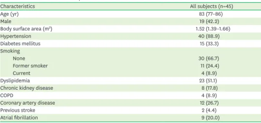
Table 1. Clinical characteristics of patients
Characteristics All subjects (n=45)
Age (yr) 83 (77–86)
Male 19 (42.2)
Body surface area (m2) 1.52 (1.39–1.66)
Hypertension 40 (88.9)
Diabetes mellitus 15 (33.3)
Smoking
None 30 (66.7)
Former smoker 11 (24.4)
Current 4 (8.9)
Dyslipidemia 23 (51.1)
Chronic kidney disease 8 (17.8)
COPD 4 (8.9)
Coronary artery disease 12 (26.7)
Previous stroke 2 (4.4)
Atrial fibrillation 9 (20.0)
Values are median (interquartile range) or number (%).
COPD: chronic obstructive pulmonary disease.
Table 2. Echocardiographic data of patients at baseline and follow-up
Echocardiographic data Baseline Latest follow-up P value
AVA (cm2) 1.34±0.36 1.03±0.28a) <0.001
Peak velocity (m/s) 2.8±0.6 3.4±0.7a) <0.001
Mean PG (mmHg) 18.7±10.4 25.5±11.4a) <0.001
DVI 0.43±0.13 0.32±0.10a) <0.001
LVEF (%) 60.4±8.8 60.4±7.5 0.856
Moderate or severe AR 4 (8.9) 3 (6.7) 0.695
Moderate or severe MR 4 (8.9) 1 (2.2) 0.169
Moderate or severe TR 3 (6.7) 3 (6.7) 1.000
Values are number (%) or mean±standard deviation.
AVA: aortic valve area, PG: pressure gradient, DVI: Doppler velocity index, LVEF: left ventricular ejection fraction, AR: aortic regurgitation, MR: mitral regurgitation, TR: tricuspid regurgitation.
a)P<0.05 compared with baseline.
Correlation between periodontal biochemical parameters and AS parameters
First, we evaluated the correlation between the amount of P. gingivalis or A. actinomycetemcomitans in the saliva and AS parameters at the latest follow-up (when saliva and serum samples were collected) and we found no significant correlation (Figure 2A-D, and Supplementary Figure 1A-D).Interestingly, the anti-P. gingivalis antibody titer in the serum showed a significant positive correlation with AVA (Figure 3A) and DVI (Figure 3D). In addition, there was a negative correlation between the anti-P. gingivalis IgG antibody titer and mean PG (Figure 3C). No significant correlation was observed between the anti-P. gingivalis IgG antibody titer and Vmax (Figure 3B). On the other hand, the anti-A. actinomycetemcomitans IgG antibody titer did not show any significant correlation with AS parameters (Supplementary Figure 2A-D).
Correlation between hs-CRP concentration and AS parameters
The hs-CRP concentration showed positive correlations with Vmax (Figure 4B) and mean PG (Figure 4C). Conversely, there were no significant correlations between the hs-CRP concentration and AVA (Figure 4A) or DVI (Figure 4D).
Correlation between the anti-P. gingivalis IgG antibody titer and AS progression
Since the anti-P. gingivalis IgG antibody titer showed a significant correlation with ASparameters, we evaluated the relationship between anti-P. gingivalis IgG antibody titer and AS progression. Although the anti-P. gingivalis IgG antibody titer showed no significant
AVA
1.25 1.50
1.00 0.75 0.50
103 105 107 109 1.75
Pg (cells/mL) A
Vmax
3 4
103 105 107 109 5
Pg (cells/mL) B
mean PG
20 40
103 105 107 109 60
Pg (cells/mL) C
DVI 0.4
0.5
0.3 0.2
103 105 107 109 0.6
Pg (cells/mL) D Figure 2. Correlations between the amount of Pg in the saliva and AS parameters.
Correlations between Pg in the saliva and (A) AVA, (B) Vmax, (C) mean PG, and (D) DVI.
Pg: Porphyromonas gingivalis, AS: aortic stenosis, AVA: aortic valve area, Vmax: maximal velocity, PG: pressure gradient, DVI: Doppler velocity index.
AVA
1.25 1.50
1.00 0.75 0.50 1.75
Anti-Pg IgG antibody titer
4 6 8 10 12
P=0.02 ρ=0.36
A
Vmax
3 4
4 6 8 10 12
5
Anti-Pg IgG antibody titer P=0.06
ρ=−0.28
B
4 6 8 10 12
mean PG
20 40 60
Anti-Pg IgG antibody titer P=0.03
ρ=−0.32
C
4 6 8 10 12
DVI 0.4
0.5
0.3 0.2 0.6
Anti-Pg IgG antibody titer P=0.03
ρ=0.32
D Figure 3. Correlations between anti-Pg IgG antibody titer in the serum and AS parameters.
Correlations between the anti-Pg IgG antibody titer and (A) AVA, (B) Vmax, (C) mean PG, and (D) DVI.
Pg: Porphyromonas gingivalis, IgG: immunoglobulin G, AS: aortic stenosis, AVA: aortic valve area, Vmax: maximal velocity, PG: pressure gradient, DVI: Doppler velocity index.
correlation with ΔAVA/year (Figure 5A) or ΔDVI/year (Figure 5D), a negative correlation was observed with ΔAVA/year (Figure 5B) and Δmean PG/year (Figure 5C).
Correlation between the hs-CRP concentration and AS progression
The hs-CRP concentration did not show a significant correlation with ΔAVA/year (Figure 6A), ΔVmax/year (Figure 6B), or ΔDVI/year (Figure 6D). However, Δmean PG showed a non- significant positive correlation (P=0.07) with the hs-CRP concentration (Figure 6C).
AVA
1.25 1.50
1.00 0.75 0.50 1.75
hs-CRP (ng/mL)
0 5,00010,00015,000
A hs-CRP (ng/mL)
0 5,000 10,00015,000
Vmax
3 4
5 P=0.046
ρ=0.30
B hs-CRP (ng/mL)
0 5,00010,00015,000
mean PG
20 40
60 P=0.04
ρ=0.31
C hs-CRP (ng/mL)
0 5,00010,00015,000
DVI 0.4
0.5
0.3 0.2 0.6
D Figure 4. Correlations between the hs-CRP concentration and AS parameters.
Correlations between the hs-CRP concentration and (A) AVA, (B) Vmax, (C) mean PG, and (D) DVI.
hs-CRP: high-sensitivity C-reactive protein, AS: aortic stenosis, AVA: aortic valve area, Vmax: maximal velocity, PG: pressure gradient, DVI: Doppler velocity index.
ΔAVA/year −0.25 0
−0.50
−0.75
−1.00 0.25
Anti-Pg IgG antibody titer
4 6 8 10 12
A Anti-Pg IgG antibody titer
4 6 8 10 12
ΔVmax/year
0 2
1
3 P=0.01
ρ=−0.38
B Anti-Pg IgG antibody titer
4 6 8 10 12
Δmean PG/year
20 40
0
−20
P=0.004 ρ=−0.42
C Anti-Pg IgG antibody titer
4 6 8 10 12
ΔDVI/year
−0.10
−0.05
−0.15
−0.20 0.05 0
D Figure 5. Correlations between the anti-Pg IgG antibody titer in the serum and AS progression.
Correlations between the anti-Pg IgG antibody titer and (A) ΔAVA, (B) ΔVmax, (C) Δmean PG, and (D) ΔDVI.
Pg: Porphyromonas gingivalis, IgG: immunoglobulin G, AS: aortic stenosis, AVA: aortic valve area, Vmax: maximal velocity, PG: pressure gradient, DVI: Doppler velocity index.
ΔAVA/year −0.25 0
−0.50
−0.75
−1.00 0.25
hs-CRP (ng/mL)
0 5,00010,00015,000
A
ΔVmax/year
0 2
1 3
hs-CRP (ng/mL)
0 5,00010,00015,000
B
Δmean PG/year
20 40
0
−20
P=0.07 ρ=0.28
hs-CRP (ng/mL)
0 5,00010,00015,000
C
ΔDVI/year
−0.10
−0.05
−0.15
−0.20 0.05 0
hs-CRP (ng/mL)
0 5,00010,00015,000
D Figure 6. hs-CRP levels according to the progression of degenerative AS.
Correlations between the hs-CRP concentration and (A) ΔAVA, (B) ΔVmax, (C) Δmean PG, and (D) ΔDVI.
hs-CRP: high-sensitivity C-reactive protein, AS: aortic stenosis, AVA: aortic valve area, Vmax: maximal velocity, PG: pressure gradient, DVI: Doppler velocity index.
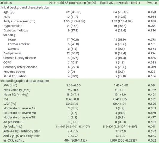
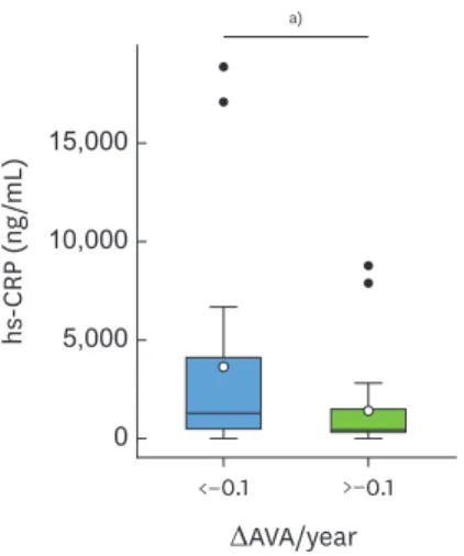
Twenty-one subjects with ΔAVA/year below −0.1 cm2/year were categorized into the rapid AS progression group, and 24 subjects with ΔAVA/year greater than or equal to −0.1 cm2/year were categorized into the non-rapid AS progression group (Table 3). Among all characteristics evaluated, only the hs-CRP level was significantly higher in the rapid AS progression group than in the non-rapid AS progression group (P=0.022) (Table 3 and Figure 7).
Evaluation of periodontal bacteria in the aortic valve
One of the severe degenerative AS cases was a 71-year old woman, and the other was a 74- year old woman. The pathology of both valve specimens showed collagenous fiber growth with vitrification and formation of nodular calcification nests that were consistent with degenerative AS. The exenterate aortic valve specimens from both subjects were negative for P. gingivalis and A. actinomycetemcomitans DNA.
DISCUSSION
P. gingivalis and A. actinomycetemcomitans are representative periodontal bacteria. P. gingivalis belongs to the “red complex,” a group of bacteria associated with periodontal disease [21],
Table 3. Differences between rapid and non-rapid AS progression groups
Variables Non-rapid AS progression (n=24) Rapid AS progression (n=21) P value Clinical background characteristics
Age (yr) 82 (76–86) 84 (78–86) 0.828
Male 10 (41.7) 9 (42.9) 0.936
Body surface area (m2) 1.50 (1.43–1.63) 1.57 (1.35–1.68) 0.963
Hypertension 21 (87.5) 19 (90.5) 0.754
Diabetes mellitus 9 (37.5) 6 (28.6) 0.530
Smoking
None 17 (70.8) 13 (61.9) 0.278
Former smoker 5 (20.8) 6 (28.6) 0.551
Current 2 (8.3) 2 (9.5) 0.889
Dyslipidemia 12 (50.0) 11 (52.4) 0.874
Chronic kidney disease 4 (16.7) 4 (19.0) 0.836
COPD 3 (12.5) 1 (4.8) 0.368
Coronary artery disease 6 (25.0) 6 (28.6) 0.789
Previous stroke 0 (0) 2 (9.5) 0.126
Atrial fibrillation 4 (16.7) 5 (23.8) 0.554
Echocardiographic data at baseline
AVA (cm2) 1.26±0.30 1.43±0.40 0.091
Peak velocity (m/s) 2.7±0.5 2.9±0.7 0.362
Mean PG (mmHg) 18.2±11.8 19.1±8.9 0.425
DVI 0.43±0.11 0.42±0.15 0.532
LVEF (%) 60.3±7.8 60.4±10.1 0.606
Moderate or severe AR 3 (12.5) 1 (4.8) 0.368
Moderate or severe MR 1 (4.2) 3 (14.3) 0.239
Moderate or severe TR 1 (4.2) 2 (9.5) 0.477
Aa (cells/mL) 0 (0–0) 0 (0–0) 0.598
Pg (cells/mL) 1.4×10⁶ (4.8×10⁴–6.1×10⁶) 5.5×10⁵ (5.3×10⁴–1.4×10⁷) 0.741
Anti-Aa IgG antibody titer 9.4±1.5 9.7±2.0 0.592
Anti-Pg IgG antibody titer 9.4±1.7 8.7±1.8 0.245
hs-CRP, ng/mL 464 (266–1,452) 1,760 (506–6,293)a) 0.022
Values are median (interquartile range), mean±standard deviation, or number (%).
AS: aortic stenosis, COPD: chronic obstructive pulmonary disease, AVA: aortic valve area, PG: pressure gradient, DVI: Doppler velocity index, LVEF: left ventricular ejection fraction, AR: aortic regurgitation, MR: mitral regurgitation, TR: tricuspid regurgitation, Aa: Aggregatibacter actinomycetemcomitans, Pg: Porphyromonas gingivalis, IgG: immunoglobulin G, hs-CRP: high-sensitivity C-reactive protein.
a)P<0.05 compared with non-rapid AS progression subjects.
and is the most common bacterium in periodontal infections. A previous study showed that periodontitis due to P. gingivalis increased the risk of developing peripheral arterial disease by 5-fold [13]. In contrast, A. actinomycetemcomitans, which harbors both endotoxins and exotoxins [22], was frequently detected in patients with severe periodontitis and was associated with aggressive periodontitis [23,24]. A. actinomycetemcomitans infection was suggested to play an important role in the development of acute coronary syndrome in the Japanese population [14]. In this study, saliva samples were collected and evaluated for the presence of P. gingivalis and A. actinomycetemcomitans by qPCR. Microbial composition depends on the collection site and sample type [25]. Therefore, subgingival plaque might be more suitable than saliva for evaluating periodontal status. However, the presence and relative abundance of P. gingivalis in the saliva have been found to be associated with periodontitis [26]. In addition, unstimulated saliva was found to be representative of pooled subgingival plaque samples, making it a useful tool for the detection of A. actinomycetemcomitans in the mouth [27].
Many reports have shown the relationship between periodontal disease and cardiovascular disease [3,5,10,11]. According to the American Heart Association, periodontal disease is associated with cardiovascular disease independently of other confounding factors, although the causative relationship is unclear [28]. Interestingly, the anti-P. gingivalis IgG antibody titer showed a negative correlation with the progression or clinical status of AS, unlike previous reports about the relationship between periodontal disease and cardiovascular disease. Patients with coronary heart disease showed higher anti-P. gingivalis serum IgG antibody titers than those without coronary heart disease [29]. In addition, the prevalence of heart failure was higher in subjects with high anti-P. gingivalis serum IgG antibody titers [30]. The discrepancy among these results may reflect differences in host immunity. In this study, there was no significant correlation between the anti-P. gingivalis IgG antibody titer and the amount of P. gingivalis in the saliva (data not shown). However, generally, P. gingivalis infection increases the anti-P. gingivalis IgG antibody titer, which decreases after periodontal treatment [31]. IgG antibody production is based on host immunity. Subjects with AS are generally quite old and their immune system may be disrupted [32]. Therefore, the titers of IgG antibodies against periodontopathic bacteria might not have reflected periodontopathic bacterial infections in this study. This might explain why our results are different from those of previous studies on the relationship between periodontitis and cardiovascular diseases.
<−0.1 >−0.1 10,000
hs-CRP (ng/mL)
5,000 15,000
0
ΔAVA/year
a)
Figure 7. hs-CRP concentrations between rapid AS progression and non-rapid AS progression groups.
hs-CRP: high-sensitivity C-reactive protein, AS: aortic stenosis, AVA: aortic valve area.
a)P=0.022
hs-CRP is a well-known biomarker of low-grade inflammation, and several studies have reported hs-CRP to be a prognostic marker of cardiovascular events, including for patients with AS [11,33-35]. Furthermore, a high hs-CRP level was associated with an increased risk of aortic valve replacement in patients with AS [36]. However, in the present study, only ΔVmax/
year and Δmean PG/year showed significant associations with hs-CRP, whereas ΔAVA/year and ΔDVI/year did not. AVA and DVI are calculated using a formula with echocardiographic parameters such as the time velocity index at the aortic valve and left ventricular outflow tract, and the left ventricular outflow tract diameter. Therefore, AVA and DVI are more vulnerable to measurement errors than Vmax and mean PG, and are not recommended as a first choice for the diagnosis of severe AS by the American College of Cardiology/American Heart Association guideline [15]. This may also be a reason for the inconsistent correlations with hs-CRP in this study. Our results also showed that the hs-CRP level in the rapid AS progression group was significantly higher than that in the non-rapid AS progression group, indicating that some inflammatory mechanisms might be associated with disease progression. Although we could not identify the cause of the inflammation, including the indirect involvement of periodontal bacteria, a previous report showed that degenerative AS had 2 distinct phases: early initiation and propagation [37,38]. Pathophysiologically, the early initiation phase is dominated by valvular lipid deposition, injury, and inflammation, with similarities to atherosclerosis, while the propagation phase is characterized by the emergence of pro-calcific and pro-osteogenic factors, which initiate the self-perpetuating processes of calcification and ultimately drive AS progression. Taken together, a prospective study in the future using calcium-detecting imaging modalities such as cardiac computed tomography (CT) and 18F positron emission CT will be required to clarify the mechanism of AS progression.
Several significant findings emerged from this pilot study. First, echocardiographic parameters during follow-up showed that AS had progressed significantly. Second, the anti-P. gingivalis IgG antibody titer showed a negative correlation with the progression of degenerative AS in this population. Third, despite the absence of any significant correlation between the hs-CRP level and ΔAVA/year, the hs-CRP level increased significantly in the rapid AS progression group compared with the non-rapid AS progression group. Finally, the DNA of periodontal bacteria was not detected in the 2 aortic valve specimens, which is in line with a previous pilot study reporting lack of periodontal pathogens in the aortic valve specimens and blood samples of patients with AS [39]. Therefore, irrespective of the progression of degenerative AS, these bacteria did not emerge as significant risk factors in our cohort.
This study has several limitations. First, this was a retrospective, single-center, pilot study. The sample size was thus small and the observation period was not long enough to comprehensively test the hypothesis. Second, we investigated the concentrations of periodontopathic bacteria and the serum IgG titers at a single time point. Oral bacteria in the saliva are considered to be stable without causing acute disease or immune deficiencies, which were not present in any of the patients included in this study cohort. Third, we did not perform standard dental examinations, as the study was performed at our institution’s cardiology department. However, a previous report showed that the anti-P. gingivalis IgG antibody levels of periodontitis patients were significantly higher than those of healthy controls, and the anti-P. gingivalis IgG titer was associated with the severity of periodontitis [40]. A multi-center study with a larger population, longer follow-up period, and dental examinations should address these limitations.
To the best of our knowledge, this is the first study to examine the relationship between periodontal bacteria and the progression of degenerative AS using periodontological and immunological approaches. Our results suggest that periodontopathic bacteria such as P. gingivalis and A. actinomycetemcomitans are not directly related to the progression of degenerative AS, in contrast to their well-known association with other atherosclerotic cardiovascular diseases. However, some inflammatory mechanisms may be associated with AS progression and warrant further investigation.
ACKNOWLEDGEMENTS
We thank Ms. Aki Takahashi, who is a technician at Teikyo University, for providing technical support.
SUPPLEMENTARY MATERIALS
Supplementary Figure 1
Correlations between amount of Aa in the saliva and AS parameters.
Click here to view
Supplementary Figure 2
Correlations between anti-Aa IgG antibody titer in the serum and AS parameters.
Click here to view
REFERENCES
1. Pihlstrom BL, Michalowicz BS, Johnson NW. Periodontal diseases. Lancet 2005;366:1809-20.
PUBMED | CROSSREF
2. Genco RJ, Van Dyke TE. Prevention: reducing the risk of CVD in patients with periodontitis. Nat Rev Cardiol 2010;7:479-80.
PUBMED | CROSSREF
3. Bahekar AA, Singh S, Saha S, Molnar J, Arora R. The prevalence and incidence of coronary heart disease is significantly increased in periodontitis: a meta-analysis. Am Heart J 2007;154:830-7.
PUBMED | CROSSREF
4. Kolenbrander PE, Andersen RN, Blehert DS, Egland PG, Foster JS, Palmer RJ Jr. Communication among oral bacteria. Microbiol Mol Biol Rev 2002;66:486-505.
PUBMED | CROSSREF
5. Katagiri S, Nitta H, Nagasawa T, Uchimura I, Izumiyama H, Inagaki K, et al. Multi-center intervention study on glycohemoglobin (HbA1c) and serum, high-sensitivity CRP (hs-CRP) after local anti-infectious periodontal treatment in type 2 diabetic patients with periodontal disease. Diabetes Res Clin Pract 2009;83:308-15.
PUBMED | CROSSREF
6. Page RC. The pathobiology of periodontal diseases may affect systemic diseases: inversion of a paradigm.
Ann Periodontol 1998;3:108-20.
PUBMED | CROSSREF
7. Koren O, Spor A, Felin J, Fåk F, Stombaugh J, Tremaroli V, et al. Human oral, gut, and plaque microbiota in patients with atherosclerosis. Proc Natl Acad Sci U S A 2011;108 Suppl 1:4592-8.
PUBMED | CROSSREF
8. Humphrey LL, Fu R, Buckley DI, Freeman M, Helfand M. Periodontal disease and coronary heart disease incidence: a systematic review and meta-analysis. J Gen Intern Med 2008;23:2079-86.
PUBMED | CROSSREF
9. Salvi GE, Carollo-Bittel B, Lang NP. Effects of diabetes mellitus on periodontal and peri-implant conditions: update on associations and risks. J Clin Periodontol 2008;35:398-409.
PUBMED | CROSSREF
10. Yoneda M, Naka S, Nakano K, Wada K, Endo H, Mawatari H, et al. Involvement of a periodontal pathogen, Porphyromonas gingivalis on the pathogenesis of non-alcoholic fatty liver disease. BMC Gastroenterol 2012;12:16.
PUBMED | CROSSREF
11. Freeman RV, Otto CM. Spectrum of calcific aortic valve disease: pathogenesis, disease progression, and treatment strategies. Circulation 2005;111:3316-26.
PUBMED | CROSSREF
12. Pellikka PA, Sarano ME, Nishimura RA, Malouf JF, Bailey KR, Scott CG, et al. Outcome of 622 adults with asymptomatic, hemodynamically significant aortic stenosis during prolonged follow-up. Circulation 2005;111:3290-5.
PUBMED | CROSSREF
13. Chen YW, Umeda M, Nagasawa T, Takeuchi Y, Huang Y, Inoue Y, et al. Periodontitis may increase the risk of peripheral arterial disease. Eur J Vasc Endovasc Surg 2008;35:153-8.
PUBMED | CROSSREF
14. Sakurai K, Wang D, Suzuki J, Umeda M, Nagasawa T, Izumi Y, et al. High incidence of Actinobacillus actinomycetemcomitans infection in acute coronary syndrome. Int Heart J 2007;48:663-75.
PUBMED | CROSSREF
15. Nishimura RA, Otto CM, Bonow RO, Carabello BA, Erwin JP 3rd, Guyton RA, et al. 2014 AHA/ACC guideline for the management of patients with valvular heart disease: a report of the American College of Cardiology/
American Heart Association Task Force on Practice Guidelines. J Am Coll Cardiol 2014;63:e57-185.
PUBMED | CROSSREF
16. Lang RM, Badano LP, Mor-Avi V, Afilalo J, Armstrong A, Ernande L, et al. Recommendations for cardiac chamber quantification by echocardiography in adults: an update from the American Society of Echocardiography and the European Association of Cardiovascular Imaging. J Am Soc Echocardiogr 2015;28:1-39.e14.
PUBMED | CROSSREF
17. Udagawa S, Katagiri S, Maekawa S, Takeuchi Y, Komazaki R, Ohtsu A, et al. Effect of Porphyromonas gingivalis infection in the placenta and umbilical cord in pregnant mice with low birth weight. Acta Odontol Scand 2018;76:433-41.
PUBMED | CROSSREF
18. Komazaki R, Katagiri S, Takahashi H, Maekawa S, Shiba T, Takeuchi Y, et al. Periodontal pathogenic bacteria, Aggregatibacter actinomycetemcomitans affect non-alcoholic fatty liver disease by altering gut microbiota and glucose metabolism. Sci Rep 2017;7:13950.
PUBMED | CROSSREF
19. Ohtsu A, Takeuchi Y, Katagiri S, Suda W, Maekawa S, Shiba T, et al. Influence of Porphyromonas gingivalis in gut microbiota of streptozotocin-induced diabetic mice. Oral Dis 2019;25:868-80.
PUBMED | CROSSREF
20. Wang D, Kawashima Y, Nagasawa T, Takeuchi Y, Kojima T, Umeda M, et al. Elevated serum IgG titer and avidity to Actinobacillus actinomycetemcomitans serotype c in Japanese periodontitis patients. Oral Microbiol Immunol 2005;20:172-9.
PUBMED | CROSSREF
21. Rôças IN, Siqueira JF Jr, Santos KR, Coelho AM, de Janeiro R. “Red complex” (Bacteroides forsythus, Porphyromonas gingivalis, and Treponema denticola) in endodontic infections: a molecular approach. Oral Surg Oral Med Oral Pathol Oral Radiol Endod 2001;91:468-71.
PUBMED | CROSSREF
22. Baehni P, Tsai CC, McArthur WP, Hammond BF, Taichman NS. Interaction of inflammatory cells and oral microorganisms. VIII. Detection of leukotoxic activity of a plaque-derived gram-negative microorganism.
Infect Immun 1979;24:233-43.
PUBMED | CROSSREF
23. Mandell RL, Socransky SS. A selective medium for Actinobacillus actinomycetemcomitans and the incidence of the organism in juvenile periodontitis. J Periodontol 1981;52:593-8.
PUBMED | CROSSREF
24. Fine DH, Markowitz K, Furgang D, Fairlie K, Ferrandiz J, Nasri C, et al. Aggregatibacter actinomycetemcomitans and its relationship to initiation of localized aggressive periodontitis: longitudinal cohort study of initially healthy adolescents. J Clin Microbiol 2007;45:3859-69.
PUBMED | CROSSREF
25. Human Microbiome Project Consortium. Structure, function and diversity of the healthy human microbiome. Nature 2012;486:207-14.
PUBMED | CROSSREF
26. Damgaard C, Danielsen AK, Enevold C, Massarenti L, Nielsen CH, Holmstrup P, et al. Porphyromonas gingivalis in saliva associates with chronic and aggressive periodontitis. J Oral Microbiol 2019;11:1653123.
PUBMED | CROSSREF
27. Cortelli SC, Feres M, Rodrigues AA, Aquino DR, Shibli JA, Cortelli JR. Detection of Actinobacillus actinomycetemcomitans in unstimulated saliva of patients with chronic periodontitis. J Periodontol 2005;76:204-9.
PUBMED | CROSSREF
28. Lockhart PB, Bolger AF, Papapanou PN, Osinbowale O, Trevisan M, Levison ME, et al. Periodontal disease and atherosclerotic vascular disease: does the evidence support an independent association?: a scientific statement from the American Heart Association. Circulation 2012;125:2520-44.
PUBMED | CROSSREF
29. Aoyama N, Kobayashi N, Hanatani T, Ashigaki N, Yoshida A, Shiheido Y, et al. Periodontal condition in Japanese coronary heart disease patients: a comparison between coronary and non-coronary heart diseases. J Periodontal Res 2019;54:259-65.
PUBMED | CROSSREF
30. Aoyama N, Kure K, Minabe M, Izumi Y. Increased heart failure prevalence in patients with a high antibody level against periodontal pathogen. Int Heart J 2019;60:1142-6.
PUBMED | CROSSREF
31. Horibe M, Watanabe H, Ishikawa I. Effect of periodontal treatments on serum IgG antibody titers against periodontopathic bacteria. J Clin Periodontol 1995;22:510-5.
PUBMED | CROSSREF
32. Frasca D, Blomberg BB. Aging affects human B cell responses. J Clin Immunol 2011;31:430-5.
PUBMED | CROSSREF
33. Emerging Risk Factors CollaborationKaptoge S, Di Angelantonio E, Pennells L, Wood AM, White IR, et al. C-reactive protein, fibrinogen, and cardiovascular disease prediction. N Engl J Med 2012;367:1310-20.
PUBMED | CROSSREF
34. Ridker PM. Clinical application of C-reactive protein for cardiovascular disease detection and prevention.
Circulation 2003;107:363-9.
PUBMED | CROSSREF
35. Blyme A, Asferg C, Nielsen OW, Sehestedt T, Kesäniemi YA, Gohlke-Bärwolf C, et al. High sensitivity C reactive protein as a prognostic marker in patients with mild to moderate aortic valve stenosis during lipid-lowering treatment: an SEAS substudy. Open Heart 2015;2:e000152.
PUBMED | CROSSREF
36. Blyme A, Asferg C, Nielsen OW, Boman K, Gohlke-Bärwolf C, Wachtell K, et al. Increased hsCRP is associated with higher risk of aortic valve replacement in patients with aortic stenosis. Scand Cardiovasc J 2016;50:138-45.
PUBMED | CROSSREF
37. Pawade TA, Newby DE, Dweck MR. Calcification in aortic stenosis: the skeleton key. J Am Coll Cardiol 2015;66:561-77.
PUBMED | CROSSREF
38. New SE, Aikawa E. Molecular imaging insights into early inflammatory stages of arterial and aortic valve calcification. Circ Res 2011;108:1381-91.
PUBMED | CROSSREF
39. Raffaelli L, Santangelo R, Falchetti P, Galluccio F, Luciani N, Anselmi A, et al. Examination of periodontal pathogens in stenotic valve specimens and in whole blood samples in patients affected by aortic valve stenosis and chronic periodontitis. Int J Immunopathol Pharmacol 2010;23:561-6.
PUBMED | CROSSREF
40. Kudo C, Naruishi K, Maeda H, Abiko Y, Hino T, Iwata M, et al. Assessment of the plasma/serum IgG test to screen for periodontitis. J Dent Res 2012;91:1190-5.
PUBMED | CROSSREF