Vo1. 9, No. 1, May, 2000
Vascularized Osteocutaneous Fibular free Flap for Reconstruction of Mid Foot
Yoon-Kyu Chung, M.D., Joon Pio Hong, M.D., Sug-Won Kim, M.D.
Department of Plastic and Reconstructive Surgery Yonsei University Wonju College of Medicine Wonju, Korea
─ Abstract ─
The foot plays a vital role in standing and gait. This function results from harmonious interac- tion of bones, joints, and soft tissue. An imbalance or a defect in such structures can lead to impaired function of the foot. The mid foot, composed of cunieforms, navicular and cuboid bone, plays a vital role in maintaining longitudinal and transverse arches and injury or defects to this region can cause instability of the foot. This paper reports a case of complex foot injury; soft tissue defect of dorsum of foot, and medial and intermediate cuneiform bone defect, reconstructed in a single stage using vascularized osteocutaneous fibular free flap. Segmented to fit the defects of medial and intermediate cuneiform bones and a skin paddle providing adequate coverage, restored the stability to the arches and function of the midfoot. The fibula osteocutaneous free flap has appealing characteristics for reconstruction of the foot and the complex mid foot injuries can be considered to the long list of indications.
Key Words : Complex foot injuries, Vascularized fibular free flap, Cunieform bone defect
I N T R O D U C T I O N
The foot plays a vital role to support the weight in standing and provide a smooth interface between body and the ground during gait. This function results from harmonious interactions of numerous small bones, joints, soft tissue envelope, and appropriate sensory feedback. The cuneiform bones of the foot consists of three bones;
medial, intermediate, and lateral. It plays
an important role in maintaining the skeletal arch and thus to withstand the stress of the body weight. The defect or dislocation can disrupt the distribution of weight bearing complex of the foot and lead to difficult gait. Despite the importance, reports regarding the reconstruction of the midfoot has been few most likely due to the rarity of its injury.
Since the first report of vascularized fibular free flap by Talor et al, it has been widely used in reconstruction of long bone defects,
congenital pseudoarthrosis of tibia, mandible, and after tumor resection3 , 5 , 1 0 - 1 2 ). Its anatomy including the vascular supply to the skin has been clarified through multiple studies making it reliable for reconstruction1 - 3 ) T h e vascularized graft tolerates infection, provides shorter healing period, rapid union, and reduction of absorption making it a good option for complex foot reconstruction. But reconstruction of the foot using vascularized fibula free flap has been seldom reported8 ) The reconstruction of cuneiform bones(medial and intermediate) using vascularized fibula bone has not been reported. This paper reports a case of complex foot injury with soft tissue defect of dorsum of foot, and medial and intermediate cuneiform bone defect.
CASE REPORT
A 16-year-old male patient sustained an
open lateral malleolar fracture and cuneiform fracture with dislocation of the left foot. He was treated primarily by orthopedic surgeon who performed open reduction with multiple percutaneous pinning. Due to the nature of the dirty wound, coverage had been delayed. After three weeks, aggravated by staphylococcus aureus infection, the regional skin was necrotic with active discharge and bone exposure. The patient was referred to our department for reconstruction.
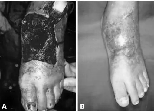
The initial surgery consisted of debridement of necrotic bone and soft tissue including the medial and intermediate cuneiform bone. The total size of the soft tissue defect was 15×1 0㎝ and the bone gap after removal of medial and intermediate cuneiform bone was maintained with silicone blocks to prevent collapse(Fig 1-A and Fig 2, Above). Multiple debridement and irrigation Fig. 1-A. After serial debridements and removal of silicone block prior to reconstruction.
Fig 1. B. 2 months after reconstruction.
A B
took place till infection was controlled and bacterial culture was negative. The second operation was staged after two weeks.
The osteocutaneous fibular free flap was elevated from the right leg with fibular bone length of 11 cm and skin paddle of 15
×1 0㎝. The fibular bone was divided into two segments, each being 3.5㎝ long with a gap of 4㎝ in between. The fibula bone was inset in a double barrel shape. Arthrodesis was performed. The first segment of the fibular bone was fixed between the first metatarsal bone distally and the navicular bone proximally. The second segment was fixed between the second metatarsal bone and the navicular bone. Microanastomosis was performed between the peroneal artery and anterior tibial artery. The skin paddle of the flap covered the defect. The remaining stump of fibula was stitched through the periosteum to the tibia to
prevent valgus deformity. Venous congestion of the flap was noted postoperatively resulting in minor skin loss requiring additional skin graft. The patient was discharged 22 days after surgery. Bone union was noted at 8 weeks after surgery and the patient was able to stand weight bearing with functional cast bracing(Fig 1-B and Fig 2, Below). During the follow-up period of 18 months, the contour of the reconstructed feet was satisfactory and the arches of the foot were reestablished.
D I S C U S S I O N
The bone and soft tissues of the foot are arranged to support the weight of the body and be flexible enough to absorb the shock at gait. Skeletally, it has an arch structure composed of joints and ligaments arranged in longitudinal and transverse arches. The Fig. 2-A. Note the defect of medial and intermediate cuneiform bone spaced with silicone block.
B. After 2 month of vascularized fibular free flap reestablishing the arch of the foot.
Union is noted at the arthrodesis site.
A B
longitudinal arch is composed of medial and lateral arches. The medial arch consists of navicular, cuneiforms and medial three metatarsals showing a much higher arch and considerable elasticity. The transverse arch results from the shapes of distal tarsals and the bases of metatarsals. It is the cuneiforms that are in the middle of these arches playing a vital role in maintaining the form and functions of these arches4 , 6 , 1 3 )
Without the bridging of the cuneiform bones, the arch would collapse and abnormal distribution of weight would ensue. Before reconstruction, a silicone block was used to prevent collapse. Placed in a open wound and irrigated daily, the silicone block was not considered a factor to aggravate infection. The cuneiform is not an independent moving bone but a relative fixed bone in maintaining the overall structure of the arches. The arthrodesis of these joints is sometimes the treatment of choice in severe dislocation or fractures. The conventional treatment for small bone defects or fractures with soft tissue defects would comprise of active antibiotic treatment(including antibiotic beads) covered by flaps under temporary external fixation followed by a second stage pelvic bone graft upon resolution of infection. This requires long period of hospital admission and time is consumed before weight bearing is tolerable.
The fibula, a well recognized vascularized bone for reconstruction of long bones and mandible, composed of primarily with cortical bone is able to withstand and remodel to the stress of weight bearing. The natural triangular shape that it poses also resembles the wedge shape of cuneiform bones and thus can resist angular and rotational stress. The vascularized bone and soft tissue, with its
abundant blood flow, can resist infection and augment bone union. The fibula is also easy to dissect with predictable anatomy and rather a low donor site morbidity. The fibula with such advantages can be a candidate for reconstruction of mutiple cuneiform defect to maintain proper arch forms and function of foot. If a small bone defect such as single or partial cuneiform defect was the case, it can be interfixed, grafted with non-vascularized bone grafts or isoplastic materials. But multiple cuneiform bone defects which can lead to disruption of arch stability, requires a bone graft with large dimension. Nonvas- cularized cancellous bone grafts are best utilized for nonunions or small bone gaps of less than few centimeters. The case presented utilized total of 7㎝ long, two 3.5㎝ s e g m e n t s , fibula bone to reconstruct the cunieform bone. If free bone grafts were to be performed, it would require a two stage operation in which the stabilization of the flap covering the foot is essential prior to second stage requiring addition 6 to 12 weeks.
The complex injury can benefit from a one stage reconstruction of fibula bone that can endure the stress of body weight, withstand infection, and achieve faster union with minimal bone resorption. During the follow- up period, the patient has not shown signs of stress fractures to the reconstructed region and foot scans revealed relatively even pressure distributions of both feet. The fibula bone can be segmented to fit the defects of medial and intermediate cuneiform bones, it also can be arranged to reconstruct single or multiple defects of other distal tarsal bones and metatarsal bones. There has been a report of reconstruction of metatarsal bone using fibular free flap and defects extending to the cuneiform could be reconstructed using a longer bone flap8 ). A case where osteoten
dofascicutaneous lateral arm free flap was used was reported to reconstruct a small partial bone defect of the first cunieform bone with soft tissue defect6 ). This case also demon strated the effectiveness of a single stage procedure. Although the importance of midfoot can not be overemphasized, the indication for vascularized fibula flap to reconstruct cunieform can be controversial.
But the success of reconstruction of foot can be judged from the restoration of function influenced by factors as soft tissue loss, soft tissue contracture, malunion, nonunion, and avascular changes9 ). The use of vascularized osteocutaneous fibula flap in cases of multiple cunieform bone with extensive soft tissue defect that requires a relatively large bone harvest and flap coverage, it can be considered for reconstruction based on the advantages it provides to minimize such risks.
C O N C L U S I O N
This paper reports a rare case of cuneiform bone and soft tissue recons- truction of the dorsum of the midfoot using vascularized osteocutaneous fibular free flap. With the advantages it provides, the fibular bone can be considered for recons- truction of complex foot injuries. The merit of one stage procedure reduces the cost in hospitalization and results in early weight bearing ambulation. The high cortical density and rapid union of the fibula bone makes it sufficient enough to withstand the body weight and maintain the form and function of the midfoot.
REFERENCES
11) Carr A, MacDonald D, Waterhouse A : The blood supply of the osteocutaneous free fibular graft. J Bone Joint Surg(Br) 70B:319-324, 1988.
12) Carriquiry C, Costa A, Vasconez L : An anatomic study of the septocutaneous vessels of the leg. Plast Reconstr Surg 76:354-363, 1985.
13) Chen ZW, Yan W : The study and clinical applica - tion of the osteocutaneous flap of fibula.
Microsurgery 4:11-16, 1983.
14) Clark DF, Quint HA : Dislocation of a single cuneiform bone. J Bone Joint Surg 15:237-239, 1993.
15) Hidalgo D : Fibular free flap: a new method of mandibular reconstruction. Plast Reconstr Surg 84:71-78, 1989.
16) Jahss MH : Disorders of the anterior tarsus, mid - tarsus, and Lisfrancs joint. In Jahss M.H.(ed), Disorders of Foot and Ankle, Vol. 2. Philadelphia:
W.B. Saunders, 1991, pp. 1284-1332.
17) Moshammer HE, Hellbom BA, Schwar키 FX, et al : Reconstruction of a complex defect on the foot with an osteotendofasciocutaneous lateral arm flap.
Scand J Plast Reconstr Hand Surg 31:271-273, 1997.
18) Rajacic N, Ebrahim MKH, Grgurinovic S, et al : Foot reconstruction using vascularized fibula. Br J Plast Surg 46:317-321, 1993.
19) Sangeorzan BJ, Hansen ST : Early and late post - traumatic foor reconstruction. Clin Orthop 243:86- 91, 1989.
10) Talor IG : The current status of free vascularized bone grafts. Clin Plast Surg 10:185-209, 1983.
11) Taylor IG, Miller GDH, Ham FJ : The free vascu - larized bone graft: a clinical extension of microvas - cular techniques. Plast Reconstr Surg 55:533-541, 1975.
12) Townsend PLG : Vascularized fibular graft using reverse peroneal flow in the treatment of congenital pseudoarthrosis of the tibia. Br J Plast Surg 43:261-265, 1990.
13) Vuori J, Aro HT : Lisfranc joint injuries: trauma mechanisms and associated injuries. J Trauma 35:40-45, 1993.