Targeted next-generation sequencing for
locally advanced prostate cancer in the Korean population
Jungyo Suh1,2,* , Chang Wook Jeong1,* , Seongmin Choi3 , Ja Hyeon Ku1 , Hyeon Hoe Kim1 , Kwang Soo Kim3 , Cheol Kwak1
1Department of Urology, Seoul National University College of Medicine, Seoul, 2Department of Urology, SMG-SNU Boramae Medical Center, Seoul, 3Department of Biomedical Research, Biomedical Research Institute, Seoul National University Hospital, Seoul, Korea
Purpose: This study aimed to evaluate the feasibility of pan-cancer panel analysis for locally advanced prostate cancer in the Ko- rean population.
Materials and Methods: We analyzed 20 patients with locally advanced prostate cancer who underwent radical prostatectomy.
A pan-cancer panel (1.9 Mbp) developed by Seoul National University Hospital (SNUH), composed of 183 target genes, 23 fusion genes, and 45 drug target regions was used for this analysis. We compared the SNUH pan-cancer panel results with The Cancer Ge- nome Atlas (TCGA) database to search for different mutations in the Korean population. Clinical data were analyzed with univariate and multivariate analysis, and p-values <0.05 were considered statistically significant. Kaplan–Meier curve and log-rank tests were performed to evaluate survival.
Results: The average age of the patients and initial prostate-specific antigen values were 69.3±7.8 years and 66.3±16.9 ng/dL, respectively. Average sequencing depth was 574.5±304.1×. Ninety-nine genetic mutations and 5 fusions were detected. SPOP (25%), KMT2D (20%), and BRAF (15%) were frequently detected. ERG fusions were recurrently detected in 20% of the patients, with SLMAP and SETD4 as novel fusion partners. BRAF mutation was frequently detected in this study, but not in the TCGA database.
Multivariate analysis showed BRAF mutation as an independent prognostic factor for biochemical recurrence (hazard ratio, 9.84;
p=0.03).
Conclusions: The pan-cancer panel comprising genes related to prostate cancer is a useful tool for evaluating genetic alterations in locally advanced prostate cancers. Our results suggest that the BRAF mutation is associated with biochemical recurrence in the Korean population.
Keywords: Genetics; Prostate-specific antigen; Prostatic neoplasms; Recurrence
This is an Open Access article distributed under the terms of the Creative Commons Attribution Non-Commercial License (http://creativecommons.org/licenses/by-nc/4.0) which permits unrestricted non-commercial use, distribution, and reproduction in any medium, provided the original work is properly cited.
Received: 11 August, 2019 • Accepted: 29 September, 2019
Corresponding Author: Cheol Kwak https://orcid.org/0000-0002-1987-2111
Department of Urology, Seoul National University Hospital, Seoul National University College of Medicine, 101 Daehak-ro, Jongno-gu, Seoul 03080, Korea TEL: +82-2-2072-2999, FAX: +82-2-742-4665, E-mail: [email protected]
*These authors contributed equally to this study and should be considered co-first authors.
ⓒ The Korean Urological Association
www.icurology.org
Investig Clin Urol 2020;61:127-135.
https://doi.org/10.4111/icu.2020.61.2.127 pISSN 2466-0493 • eISSN 2466-054X
INTRODUCTION
Precision medicine is a novel paradigm in clinical prac- tice that determines personalized treatment based on a patient’s genetic and environmental factors. Before it can be applied clinically; however, precision medicine still has many issues to overcome. A massive amount of genetic and clinical data is required for the implementation of precision medicine, and recent advances in next-generation sequencing (NGS) make implementation more likely [1].
Compared with other sequencing platforms, NGS tech- nology is beneficial in obtaining large-scale genetic infor- mation using single inputs [2]. Although whole-genome and whole-exome sequencing techniques yield more information, targeted NGS (tNGS) is more promising for clinical applica- tion because of its cost-effectiveness, rapid sequencing time, and deep sequencing read depth [3]. Because various solid cancers share major molecular pathways, the pan-cancer panel may provide additional useful information for specific cancers [4,5]. Routine application of pan-cancer panels is not a distant prospect. For example, MSK-IMPACTTM (Memorial Sloan Kettering, New York, NY, USA) and Foundation One are Food and Drug Administration (FDA)-approved multi- cancer NGS panels designed to obtain genetic information on various malignancies that can be applied for therapeutic purposes.
Prostate cancer is the most common malignancy in men and a major cause of cancer-related mortality in Western countries [6-8]. Geographic and racial differences in the incidence and disease characteristics of prostate cancer sug- gest that genetic and environmental factors affect disease development and progression [9]. With advances in genetic analysis, genetic factors in prostate cancer development and progression have been exponentially studied over the past decade [9-12]. However, most genetic studies have focused on metastatic or castration-resistant prostate cancer. Further- more, almost all genetic studies constituted a negligible pro- portion of the Asian population [13,14].
Therefore, we performed a pan-cancer panel assay that contained prostate cancer-related genes for locally advanced prostate cancer in the Korean population. By performing a comparison with The Cancer Genome Atlas (TCGA) data- base, we aimed to find racial differences in Korean patients that result in aggressive tumor behavior. Using combined clinical data and genetic alterations, we performed univari- ate and multivariable analysis to evaluate potential risk factors for biochemical recurrence.
MATERIALS AND METHODS
1. Ethics approval and informed consent
This study was approved by the Seoul National Uni- versity Hospital (SNUH) Institutional Review Board (IRB) (approval number: 1706-103-860). The prospectively collected registry was approved by the SNUH IRB (approval number:
1506-121-682) for use of clinical data for scientific purposes.
Informed consent for NGS analysis was obtained from each participant. Fresh frozen tissues were stored in the SNUH biobank. All experiments were performed in accordance with relevant guidelines and regulations.
2. Patient selection
All patients were selected from a prospective, multidis- ciplinary, and biobank lined cohort, the Seoul National Uni- versity Prospectively Enrolled Registry for Prostate Cancer- Radical Prostatectomy (SUPER-PC-RP) [15]. This prospective cohort collects comprehensive clinical information, including functional and oncological outcomes, patient-reported quality of life, and bio-specimens. We selected 20 patients with lo- cally advanced prostate cancer at stage T3a or T3b without metastasis. Patients underwent radical prostatectomy from March 2016 to June 2016, with a follow-up of 25.6 months (median). We used tumor and normal tissues collected from the operating room or frozen biopsy room and immediately stored the removed prostates at -195ºC in liquid nitrogen at the SNUH Cancer Tissue Bank.
3. Cancer panel information
The SNUH cancer panel, named the FIRST-panel ver- sions 3 and 3.1, was used for this analysis. This panel con- tains exons of 183 genes, specific introns of 23 fusion genes, the TERT promoter region, 8 MSI markers, and 45 drug tar- get regions, with a total length of ~1.949 Mbp. We assessed the FIRST-panel composition for major urological malignan- cies—prostate, bladder, and kidney cancers. We determined prostate cancer-related genes on the basis of a literature review [11,12,16,17] and finally selected 40 prostate cancer- related genes, including AR, FOXA1, TMPRSS2, and ERG, in the FIRST-panel v3.0. and 3.1. Among these targets, we reviewed potentially applicable targets for prostate cancer from the OncoKB database [18].
4. DNA extraction
Fresh frozen tumor tissues were homogenized and lysed with proteinase K. Next, total DNA was isolated from each target with the Maxwell 16 CSC DNA Blood kit (Promega Corp., Madison, WI, USA). The extracted DNA was quan-
titated by use of a Quantus fluorometer (Promega Corp.) and Agilent 4200 TapeStation system (Agilent Technologies, Santa Clara, CA, USA).
5. Capture library preparation
After DNA was extracted paired-end libraries were pre- pared with an Agilent SureSelectXT Target Enrichment System kit for the Illumina paired-end sequencing library protocol using SNUH FIRST Cancer Panel v 3.0 and v 3.1, according to the manufacturer’s instructions. The quality of the DNA library was evaluated by using a Bioanalyzer 2100 (Illumina Inc., CA, USA) and DNA 1000 chips (Illumina Inc., CA, USA).
6. Next-generation sequencing
The tNGS was performed by using the Illumina Hiseq 2500 platform. Sequencing data were transformed as FASTQ files, and quality control by use of FASTQC (0.11.8) and Trimmomatic (0.33) software. BAM formation after align- ment was performed based on the reference genome (GRCh 37) by BWA (0.7.12) and Picard (1.134). Quality control of the BAM file was carried out by SamTools (v1.2) and GATK (v3.3). Detection of SNPs was performed by MuTect (1.1.7) and SamTools (v1.2). Indel and copy number variation (CNV) detection were performed by IndelGeontyper (0.36.3336) and CoNIFER software (0.2.2). Fusion search was done using Delly (0.7.2). All data were converted to VCF format and an- notated by Annovar.
7. Variant prevalence comparison of SNUH pan- cancer data and TCGA prostate cancer
We downloaded MAF files of prostate adenocarcinoma (TCGA-PRAD) variants from the National Cancer Insti- tute (NCI) Genomic Data Commons (GDC) data portal.
From these MAF files, we extracted variants in the follow- ing genes: SPOP, ERG, NRG1, KMT2D, MAP3K1, TSHR, FANCD2, ATM, FANCA, BRAF, FANCG, NOTCH4, and AR. Four MAF files were submitted for their corresponding variant callers: MuSE, Mutect2, VarScan2, and SomaticSnip- er. We selected the variants that occurred in at least two of these files. We classified the mutations into three categories:
missense truncating and inframe mutation. The prevalence of such mutations was compared with our FIRST-panel data.
8. Statistical analysis
We set biochemical recurrence as the primary endpoint.
The prostate-specific antigen (PSA) value at the secondary confirmative level of >0.2 ng/dL was considered biochemical recurrence. Univariate analysis of biochemical recurrence
was carried out by using the Kaplan–Meier curve with log-rank test. Known and potential risk factors, including age, initial PSA value, Gleason score at the time of biopsy, prostate volume, pathological Gleason score, pathological T stage, pathological N stage, intraductal carcinoma, and perineural invasion, were selected for univariate analysis to assess the risk of biochemical recurrence. Among the genetic alterations, the 10 most frequently occurring mutations in the genes SPOP, KMT2D, ATM, BRAF, FANCA, FANCD2, FANCG, MAP3K1, TSHR, and ERG fusion were selected for analysis. TP53 and PTEN were also included in the univari- ate analysis, as their association with biochemical recurrence has been reported [19,20]. The variables showing a p-value
<0.1 in the univariate analysis were selected for multivariate analysis using the Cox proportional hazard model. Important variables, such as Gleason grade group and T stage, were also included in the multivariate analysis. Statistical analy- sis was conducted by use of IBM SPSS Statistics 22.0 (IBM Corp., Armonk, NY, USA), and a p-value <0.05 was consid- ered significant.
RESULTS
1. Patient characteristics
The patients’ demographic and clinical characteristics are presented in Table 1. Two patients previously treated by neoadjuvant androgen deprivation therapy underwent radical prostatectomy. Eighteen patients underwent pelvic lymphadenectomy, and five patients showed lymph node metastasis in the pathologic examination of permanent sec- tions. The average number of extracted and positive lymph nodes was 17.7 and 2.7, respectively. The average follow-up period was 25.6 months, and only one patient died of pneu- monia. During the follow-up period, 14 patients were detected with biochemical recurrence and needed salvage treatments.
2. Sequencing quality control
DNA was extracted from all 20 fresh frozen tissue sam- ples for library preparation and cancer panel analysis. In to- tal, 26,066,448 to 39,586,410 bases were read for this analysis.
Average coverage was 574.5±304.1×, ranging from 160 to 996.
Average values of coverage above 50 and above 100 were 97.5±2.0% and 94.2±3.9%, respectively. All samples passed se- quencing quality control analysis.
3. Pan-cancer panel report
A total of 99 mutations were detected in the pan-cancer panel analysis. Mutations in the SPOP gene, detected in 25%
of patients, were the most frequently occurring mutations.
Mutations in KMT2D (20%), FANCA, FANCD2, FANCG, TSHR, MAP3K1, BRAF, and ATM (15%) also occurred frequently. ERG fusion was detected in 20% of patients that had mutually exclusive SPOP mutations. The fusion partners of ERG were TMPRSS2 (m/c, 21q22.3), SLMAP (sarcolemmal membrane-associated protein gene, 3p14.3), or SETD4 (SET domain-containing protein 4, 21q22.13). On aver-
age, 5 mutations were detected per patient, ranging from 1 to 13 (Fig. 1). Additionally, a patient was detected with only one mutation in the BRAF gene, coding for an amino acid change in K601E (a potential target of PLX8394), at an allele frequency of 0.45. Integrative analysis of the cancer panel is shown in Fig. 2.
We found potential actionable targets for prostate can- cer treatment in nine patients (45.0%) in this study. ATM mutations, the target of olaparib, were the most common actionable mutations detected in three patients. Moreover, three patients had BRAF gene alterations, of which two had changes in K601E. Another patient had a mutation in the CDKN2A gene, which is a therapeutic target of abemaciclib,
palbociclib, and ribociclib. Mutations in each of FGFR1 and FGFR3 was detected in one patient, which are potential tar- gets of AZD4547, BGJ398, Debio1347, and erdafitinib.
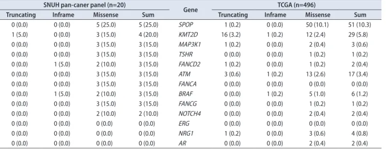
4. Comparison with the TCGA database
Among the 11 selected genes (SPOP, KMT2D, ATM, BRAF, FANCA, FANCD2, FANCG, MAP3K1, TSHR, NOTCH4, and AR), 10 genes were expressed more frequently
in our data than in TCGA, with the exception of AR (Table 2). SPOP and KMT2D mutations showed a >5% incidence rate in the TCGA database. In contrast, MAP3K1, FANCD2, TSHR, ATM, FANCA, BRAF, FANCG, and NOTCH4 were detected at a <5% incidence rate. Among these mutations, MAP3K1 (N749K), FANCD2 (V941A), TSHR (R310H), FAN- CA (G917R, R1011C, A816V), BRAF (K601E), and FANCG (P187Q) were likely to be pathogenic mutations with a Com- bined Annotation Dependent Depletion (CADD) score of over 20. The BRAF mutation was the only confirmed mutation present in other platforms, such as the Clinvar and OncoKB databases.
5. Univariate and multivariate analysis with clini- cal parameters
Clinical and pathologic features, as well as genetic al- teration data from the pan-cancer panel, of locally advanced prostate cancer were analyzed for their contribution to bio- chemical recurrence. The results of the univariate analysis are shown in Table 3. The Kaplan–Meier curve with a log- rank test of BRAF mutation is presented in Fig. 3. The multivariate Cox regression analysis revealed that prostate Fig. 1. Specific genetic alteration counts of each patient. A minimum of 1 to a maximum of 13 mutations per patient were found by multi-cancer panel analysis. Blue bars show SNP and Indel mutations and orange bars show number of structural variations.
SCP3R_R201 14 12 10 8 6 4 2 0
SCP3R_R203SCP3R_R204SCP3R_R206SCP3R_R207SCP3R_R21 1
SCP3R_R216
SCP3R_R202_1SCP3R_R205_1SCP3R_R208SCP3R_R209SCP3R_R210SCP3R_R212SCP3R_R213SCP3R_R214SCP3R_R215SCP3R_R217SCP3R_R218SCP3R_R219SCP3R_R220 Genetic alteration counts
Mutation count Fusion count Table 1. Baseline demographic and clinical characteristics of the pa-
tients (n=20)
Characteristic Value
Age (y) 69 (55–83)
Body mass index (kg/m2) 24.7 (18.7–31.0) Initial prostate-specific antigen (ng/dL) 66.3 (4.1–771.6) Biopsy Gleason grade group
2 2 (10)
3 4 (20)
4 10 (50)
5 4 (20)
Mean prostate volume (mL) 42.1 (10.0–87.6) Pelvic lymph node dissection 17 (85)
Positive surgical margin 11 (55)
Pathologic Gleason grade group
3 11 (55)
4 5 (25)
5 4 (20)
Seminal vesicle invasion 11 (55)
Perineural invasion 15 (75)
Intraductal carcinoma 4 (20)
Pathologic T stage
3a 11 (55)
3b 9 (45)
Pathologic N stage
0 13 (65)
1 5 (25)
x 2 (10)
Values are presented as mean (range) or number (%).
volume (hazard ratio [HR], 0.12; 95% confidence interval [CI], 0.01–0.74; p=0.02), pathological Gleason grade group (HR, 4.11;
95% CI, 1.14–14.88; p=0.03), and BRAF mutation (HR, 9.84;
95% CI, 2.27–149.18; p=0.03) showed a statistically significant association with biochemical recurrence (Table 4).
DISCUSSION
In this study, we successfully performed a pan-cancer panel analysis in patients with advanced prostate cancer. We achieved an average depth of over 500, coupled with a high sensitivity that allowed detection of a low allele frequency mutation (TP53, allele frequency=0.02, missense mutation).
The SNUH pan-cancer panel detected less commonly report- Fig. 2. Integrative analysis of cancer panel analysis of 20 patients. Each grey column represents specific data for 1 of the 20 pa- tients in order. Genomic polymorphism of SNP/Indel mutation by truncating, in the frame, missense is noted by a color dot in a grey column with black, brown, and green.
Structural variation was found in 5 patients annotated by purple color; the most com- mon finding was ERG: TMPRSS2 fusion.
No CNV amplification was found by our targeted next-generation sequencing (NGS) panel. This oncoprint was obtained by use of The cBioPortal for Cancer Genomics (http://cbioportal.org) graphic visualization tool.
Genetic alteration SPOP ERG NRG1 KMT2D MAP3K1 TSHR FANCD2 ATM FANCA BRAF FANCG PDGFRA PIK3CA ERCC2 DPYD FANCC EZH2 FGFR1 BRCA2 MAP3K4 ARID1A ERBB3 APC FGFR4 TP53 NOTCH4 POLQ KDM6A NOTCH3 MET NTRK1
25%
20%
5%
20%
15%
15%
15%
15%
15%
15%
15%
10%
10%
10%
10%
5%
5%
5%
5%
5%
5%
10%
10%
10%
10%
10%
10%
10%
10%
10%
10%
PDGFRB SQSTM1 EGFR CDKN2A FANCL FGFR3 ALK MUTYH NOTCH2 NFE2L2 HRAS ERBB4 FANCI RAD50 DDR1 IGFBP3 ATR TSC1 TOP2A SETD2 NTRK3 BRIP1 PALB2 TERT POLE CDK12 INPP4B MSH2 CTNNB1 CSF1R NOTCH1 FOXA1 ERBB2 SMAD4 EP300
5%
5%
5%
5%
5%
5%
5%
5%
5%
5%
5%
5%
5%
5%
5%
5%
5%
5%
5%
5%
5%
5%
5%
5%
5%
5%
5%
5%
5%
5%
5%
5%
5%
5%
5%
No alterations Truncating mutation Inframe mutation Missense mutation
ed mutations compared with previous studies undertaken in Western countries, which may be due to racial differences.
Among these mutations, the BRAF mutation was found to be an independent prognostic factor for biochemical recur- rence of prostate cancer.
We successfully detected 99 genetic alterations, including structural variations using pan-cancer panel analysis. The general mutation expression profile was similar to that re- ported by another researcher. The SPOP mutation and ERG fusion were the most common findings and were mutually exclusive as reported previously [11]. The most common ERG fusion partner was TMPRSS2; two additional fusion part- ners, SLMAP and SETD4, were also detected, which were Table 2. Comparison of the incidence of top-ranked genetic alteration of this study with the TCGA database. Muse, mutect, somaticsniper, and varscan were used to determine true mutations
SNUH pan-caner panel (n=20)
Gene TCGA (n=496)
Truncating Inframe Missense Sum Truncating Inframe Missense Sum
0 (0.0) 0 (0.0) 5 (25.0) 5 (25.0) SPOP 1 (0.2) 0 (0.0) 50 (10.1) 51 (10.3)
1 (5.0) 0 (0.0) 3 (15.0) 4 (20.0) KMT2D 16 (3.2) 1 (0.2) 12 (2.4) 29 (5.8)
0 (0.0) 0 (0.0) 3 (15.0) 3 (15.0) MAP3K1 1 (0.2) 0 (0.0) 2 (0.4) 3 (0.6)
0 (0.0) 0 (0.0) 3 (15.0) 3 (15.0) TSHR 0 (0.0) 0 (0.0) 1 (0.2) 1 (0.2)
0 (0.0) 1 (5.0) 2 (10.0) 3 (15.0) FANCD2 1 (0.2) 0 (0.0) 1 (0.2) 2 (0.4)
0 (0.0) 0 (0.0) 3 (15.0) 3 (15.0) ATM 3 (0.6) 1 (0.2) 13 (2.6) 17 (3.4)
0 (0.0) 0 (0.0) 3 (15.0) 3 (15.0) FANCA 0 (0.0) 0 (0.0) 0 (0.0) 0 (0.0)
0 (0.0) 1 (5.0) 2 (10.0) 3 (15.0) BRAF 0 (0.0) 1 (0.2) 5 (1.0) 6 (1.2)
0 (0.0) 0 (0.0) 3 (15.0) 3 (15.0) FANCG 0 (0.0) 0 (0.0) 1 (0.2) 1 (0.2)
0 (0.0) 0 (0.0) 2 (10.0) 2 (10.0) NOTCH4 0 (0.0) 0 (0.0) 2 (0.4) 2 (0.4)
0 (0.0) 0 (0.0) 0 (0.0) 0 (0.0) ERG 0 (0.0) 0 (0.0) 0 (0.0) 0 (0.0)
0 (0.0) 0 (0.0) 0 (0.0) 0 (0.0) NRG1 1 (0.2) 0 (0.0) 3 (0.6) 4 (0.8)
0 (0.0) 0 (0.0) 0 (0.0) 0 (0.0) AR 0 (0.0) 0 (0.0) 2 (0.4) 2 (0.4)
Values are presented as number (%).
SNUH, Seoul National University Hospital; TCGA, The Cancer Genome Atlas.
Table 3. Univariate analysis of clinical characteristics, pathologic pa- rameters, and genetic alterations with biochemical recurrence
Characteristic Univariate analysis HR (95% CI) p-value
Age >65 years 1.45 (0.45-4.66) 0.53
iPSA >20 0.77 (0.26-2.24) 0.63
Prostate volume (>50 mL) 0.95 (0.04-0.63) 0.01
Pathologic Gleason grade group 0.11
3 Reference
4–5 2.44 (0.83-7.20)
Pathologic T stage 0.49
T3a Reference
T3b 1.46 (0.50-4.24)
Positive surgical margin 0.54 (0.18-1.64) 0.28
Pathologic N stage 0.79
Intraductal carcinoma 4.46 (0.78-25.41) 0.09 Perineural invasion 2.00 (0.54-7.39) 0.30
BRAF mutation 4.48 (1.32-29.09) 0.03
SPOP mutation 1.87 (0.59-5.95) 0.29
KMT2D mutation 1.14 (0.31-4.19) 0.84
ATM mutation 0.55 (0.07-4.37) 0.57
FANCA mutation 0.30 (0.04-2.30) 0.25
FANCD2 mutation 0.82 (0.22-3.04) 0.77
FANCG mutation 1.81 (0.46-7.10) 0.40
MAP3K1 mutation 0.41 (0.05-3.19) 0.40
TSHR mutation 0.57 (0.13-2.57) 0.47
ERG mutation 0.45 (0.10-2.04) 0.30
HR, hazard ratio; CI, confidence interval; iPSA, initial prostate-specific antigen.
Fig. 3. Kaplan–Meier curve of BRAF mutation on biochemical recurrence with the log-rank test. Positive BRAF mutation worsened biochemical- recurrence-free survival with statistically significant difference (p=0.03).
0 1.0
0.8
0.6
0.4
0.2
40
Cumulativesurvival
Biochemical recurrence free survival (mo) 0.0
10 20 30
p=0.03 Kaplan Meier curve
No Yes Censored Censored BRAF mutation
not reported previously. SLMAP is known to interact with serine/threonine-protein kinase 24. The mutation in this re- gion was related to the Brugada syndrome. SETD4 is a cod- ing gene related to histone modification. Additionally, some rare genetic alterations were frequently detected in our study. For example, SNPs or Indels in MAP3K1, FANCD2, TSHR, ATM, FANCA, BRAF, FANCG, and NOTCH4 genes occurred at <5% frequency in the TCGA database [11,17];
however, they were relatively common in our study with frequency ranging from 15% to 20%. Among these differ- entially expressed genetic alterations, six were potentially pathogenic according to their CADD score; however, only the BRAF mutation (K601E) was confirmed to be pathogenic as per the Clinvar and OncoKB databases. This discrepancy may be due to the small number of Asians studied during the preparation of the TCGA database.
Because the incidence and behavior of prostate cancer is quite different in Western and Asian patients, there could be predisposing genetic differences between the two patient groups. Prostate cancer is less frequently diagnosed but shows more aggressive features in Asians compared with Western population [21,22]. A recent large-scale genetic study, comprising 65 whole-genome and 145 targeted se- quences in Chinese patients with prostate cancer, observed considerable differences compared with TCGA databases [21]. This discrepancy suggests that there are underlying genetic differences among different races in prostate can- cer development and behavior. In our study, multivariate analysis showed that BRAF mutation (HR, 9.84; p=0.03), Gleason grade group (HR, 4.11; p=0.03), and prostate volume (HR, 0.12; p=0.02) demonstrated significant association with biochemical recurrence. BRAF is a serine/threonine kinase family member, and alterations in this gene are commonly reported in malignant melanoma [23] and papillary thyroid
cancer [24]. BRAF is a component of mitogen-activated pro- tein kinase (MAPK) pathways, which play a major role in tumorigenesis. The incidence of BRAF mutations in prostate cancer shows considerable racial differences. According to the genetic analysis data mainly collected from white popu- lations [25], BRAF mutations are rare in prostate cancer in Western populations. However, the incidence of alterations in the BRAF gene in the Asian population is relatively high.
Cho et al. [26] reported that approximately 10% of Korean patients have BRAF mutations in prostate cancer. Genetic studies performed in a Chinese population also detected a higher incidence of BRAF gene alterations [27]. In this study, we observed that 15% of patients had BRAF-mutation- positive prostate cancers, indicating that BRAF mutation is an independent prognostic factor for biochemical recurrence.
Owing to the different behavioral and pathological features of prostate cancer owing to racial/ethnic differences, we suggest that the BRAF mutation may contribute to this dis- crepancy to some extent.
We discovered five potential therapeutic targets in nine patients (45%); however, they were not FDA-approved in prostate cancer. Each of the ATM or BRAF mutations was present in 3 patients. ATM is one of the DNA repair-related genes, such as BRCA1, BRCA2, ATM, Fanconi anemia gene, and CHEK2, which are targets of poly-adenosine diphosphate ribose polymerase (PARP) inhibitor. Olaparib is a PARP inhibitor that has shown promising results in a patient with prostate cancer harboring a defective DNA repair gene in a Phase 2 clinical trial. BRAF mutation is a target for the FDA-approved drugs vemurafenib and dabrafenib in malig- nant melanoma. However, no studies to date have focused on prostate cancer treatment. Prostate cancer caused by BRAF mutation is classified as I to II, manifested by kinase activ- ity, Ras-dependency, and dimerization status. BRAF class is important to predict MAPK target therapy response. K601E mutation is a class II BRAF mutation, which is sensitive to MEK inhibitors and vemurafenib in malignant melanoma.
The pan-BRAF inhibitor, PLX8394, is a potential drug for targeting BRAF (K601) mutation.
This study had several limitations. One important limita- tion was the small sample size. Because of the small size, the statistical power of this study was not enough to establish clinical importance of the studied genes. Since we utilized tNGS for genetic analysis, genomic alterations in the regions of interest of our cancer panel could not be detected. There- fore, recently discovered genetic alterations associated with biochemical recurrence could not be assessed. In this study, we undertook retrospective analysis from a prospectively collected cohort, and this approach has a potential risk of Table 4. Multivariate analysis of clinical characteristics, pathologic pa-
rameters, and genetic alterations with biochemical recurrence Characteristic Multivariate analysis
HR (95% CI) p-value Prostate volume (>50 mL) 0.12 (0.01–0.74) 0.02
Pathologic Gleason grade group 0.03
3 Reference
4–5 4.11 (1.14–14.88)
Pathologic T stage 0.09
T3a Reference
T3b 2.81 (0.84–9.78)
Intraductal carcinoma 0.22
BRAF mutation 9.84 (2.27–149.18) 0.03
HR, hazard ratio; CI, confidence interval.
bias. The natural course of prostate cancer is relatively slow;
thus, a short follow-up period (25.6 months) is another limi- tation. Despite these limitations, we successfully conducted tNGS using a pan-cancer panel with excellent depth (>500).
CONCLUSIONS
In conclusion, a pan-cancer panel, comprising prostate cancer-related genes, is a useful tool for evaluating genetic alterations in patients with locally advanced prostate can- cers. Our results suggest that the BRAF mutation is associ- ated with biochemical recurrence in the Korean population.
Nevertheless, a well-designed large-scale investigation is needed for a better understanding of this mutation.
CONFLICTS OF INTEREST
The authors have nothing to disclose.
ACKNOWLEDGMENTS
The bio-specimens for this study were provided by the Seoul National University Hospital Cancer Tissue Bank. All samples were obtained with informed consent of patients under IRB-approved protocols.
This study was supported by grants from the National R&D Program for Cancer Control (HA17C0039) and the Korea Health Technology R&D Project (HI14C1277) through the Korea Health Industry Development Institute (KHIDI), funded by the Ministry of Health & Welfare, Republic of Korea. None of the sponsors had any access to the data or any influence on or access to the analysis plan, the results, or the manuscript.
AUTHORS’ CONTRIBUTIONS
Research conception and design: Chang Wook Jeong and Jungyo Suh. Data acquisition: Jungyo Suh and Seongmin Choi. Statistical analysis: Chang Wook Jeong and Jungyo Suh. Data analysis and interpretation: Jungyo Suh and Seongmin Choi. Drafting of the manuscript: Jungyo Suh.
Critical revision of the manuscript: Chang Wook Jeong, Ja Hyeon Ku, and Hyeon Hoe Kim. Obtaining funding: Chang Wook Jeong. Administrative, technical, or material support:
Seongmin Choi and Kwang Soo Kim. Supervision: Chang Wook Jeong, Ja Hyeon Ku, Hyeon Hoe Kim, and Cheol Kwak. Approval of the final manuscript: Cheol Kwak.
REFERENCES
1. Roberts JS, Gornick MC, Le LQ, Bartnik NJ, Zikmund-Fisher BJ, Chinnaiyan AM; MI-ONCOSEQ Study team. Next-gener- ation sequencing in precision oncology: patient understanding and expectations. Cancer Med 2019;8:227-37.
2. Luthra R, Patel KP, Routbort MJ, Broaddus RR, Yau J, Simien C, et al. A targeted high-throughput next-generation sequencing panel for clinical screening of mutations, gene amplifications, and fusions in solid tumors. J Mol Diagn 2017;19:255-64.
3. Kamps R, Brandão RD, Bosch BJ, Paulussen AD, Xanthoulea S, Blok MJ, et al. Next-generation sequencing in oncology: ge- netic diagnosis, risk prediction and cancer classification. Int J Mol Sci 2017;18:E308.
4. Cheng DT, Mitchell TN, Zehir A, Shah RH, Benayed R, Syed A, et al. Memorial sloan kettering-integrated mutation profiling of actionable cancer targets (MSK-IMPACT): a hybridization capture-based next-generation sequencing clinical assay for solid tumor molecular oncology. J Mol Diagn 2015;17:251-64.
5. Cheng DT, Prasad M, Chekaluk Y, Benayed R, Sadowska J, Ze- hir A, et al. Comprehensive detection of germline variants by MSK-IMPACT, a clinical diagnostic platform for solid tumor molecular oncology and concurrent cancer predisposition test- ing. BMC Med Genomics 2017;10:33.
6. Siegel RL, Miller KD, Jemal A. Cancer Statistics, 2017. CA Cancer J Clin 2017;67:7-30.
7. Ferlay J, Soerjomataram I, Dikshit R, Eser S, Mathers C, Rebelo M, et al. Cancer incidence and mortality worldwide: sources, methods and major patterns in GLOBOCAN 2012. Int J Can- cer 2015;136:E359-86.
8. Arnold M, Karim-Kos HE, Coebergh JW, Byrnes G, Antilla A, Ferlay J, et al. Recent trends in incidence of five common cancers in 26 European countries since 1988: analysis of the european cancer observatory. Eur J Cancer 2015;51:1164-87.
9. Benafif S, Kote-Jarai Z, Eeles RA; PRACTICAL Consortium.
A review of prostate cancer genome-wide association studies (GWAS). Cancer Epidemiol Biomarkers Prev 2018;27:845-57.
10. Schumacher FR, Al Olama AA, Berndt SI, Benlloch S, Ahmed M, Saunders EJ, et al.; Australian Prostate Cancer BioResource (APCB); IMPACT Study; Canary PASS Investigators; Breast and Prostate Cancer Cohort Consortium (BPC3); PRAC- TICAL (Prostate Cancer Association Group to Investigate Cancer-Associated Alterations in the Genome) Consortium;
Cancer of the Prostate in Sweden (CAPS); Prostate Cancer Genome-wide Association Study of Uncommon Susceptibility Loci (PEGASUS); Genetic Associations and Mechanisms in Oncology (GAME-ON)/Elucidating Loci Involved in Prostate Cancer Susceptibility (ELLIPSE) Consortium. Association analyses of more than 140,000 men identify 63 new prostate
cancer susceptibility loci. Nat Genet 2018;50:928-36.
11. Cancer Genome Atlas Research Network. The molecular tax- onomy of primary prostate cancer. Cell 2015;163:1011-25.
12. Kumar A, Coleman I, Morrissey C, Zhang X, True LD, Gulati R, et al. Substantial interindividual and limited intraindividual genomic diversity among tumors from men with metastatic prostate cancer. Nat Med 2016;22:369-78.
13. Marzec J, Mao X, Li M, Wang M, Feng N, Gou X, et al.; PRAC- TICAL Consortium; CHIPGECS Group. A genetic study and meta-analysis of the genetic predisposition of prostate cancer in a Chinese population. Oncotarget 2016;7:21393-403.
14. Wang M, Takahashi A, Liu F, Ye D, Ding Q, Qin C, et al. Large- scale association analysis in Asians identifies new susceptibility loci for prostate cancer. Nat Commun 2015;6:8469.
15. Jeong CW, Suh J, Yuk HD, Tae BS, Kim M, Keam B, et al. Es- tablishment of the Seoul National University Prospectively Enrolled Registry for Genitourinary Cancer (SUPER-GUC):
a prospective, multidisciplinary, bio-bank linked cohort and research platform. Investig Clin Urol 2019;60:235-43.
16. Fraser M, Sabelnykova VY, Yamaguchi TN, Heisler LE, Living- stone J, Huang V, et al. Genomic hallmarks of localized, non- indolent prostate cancer. Nature 2017;541:359-64.
17. Robinson D, Van Allen EM, Wu YM, Schultz N, Lonigro RJ, Mosquera JM, et al. Integrative clinical genomics of advanced prostate cancer. Cell 2015;161:1215-28.
18. Chakravarty D, Gao J, Phillips SM, Kundra R, Zhang H, Wang J, et al. OncoKB: a precision Oncology Knowledge Base. JCO Precis Oncol 2017;2017.
19. Carneiro A, Barbosa ÁRG, Takemura LS, Kayano PP, Moran NKS, Chen CK, et al. The role of immunohistochemical analy- sis as a tool for the diagnosis, prognostic evaluation and treat-
ment of prostate cancer: a systematic review of the literature.
Front Oncol 2018;8:377.
20. Lo Iacono M, Buttigliero C, Monica V, Bollito E, Garrou D, Cappia S, et al. Retrospective study testing next generation se- quencing of selected cancer-associated genes in resected pros- tate cancer. Oncotarget 2016;7:14394-404.
21. Ren S, Wei GH, Liu D, Wang L, Hou Y, Zhu S, et al. Whole- genome and transcriptome sequencing of prostate cancer iden- tify new genetic alterations driving disease progression. Eur Urol 2018;73:322-39.
22. Jung KW, Won YJ, Oh CM, Kong HJ, Cho H, Lee DH, et al.
Prediction of cancer incidence and mortality in Korea, 2015.
Cancer Res Treat 2015;47:142-8.
23. Davies H, Bignell GR, Cox C, Stephens P, Edkins S, Clegg S, et al. Mutations of the BRAF gene in human cancer. Nature 2002;417:949-54.
24. Namba H, Nakashima M, Hayashi T, Hayashida N, Maeda S, Rogounovitch TI, et al. Clinical implication of hot spot BRAF mutation, V599E, in papillary thyroid cancers. J Clin Endocri- nol Metab 2003;88:4393-7.
25. Liu T, Willmore-Payne C, Layfield LJ, Holden JA. Lack of BRAF activating mutations in prostate adenocarcinoma: a study of 93 cases. Appl Immunohistochem Mol Morphol 2009;17:121-5.
26. Cho NY, Choi M, Kim BH, Cho YM, Moon KC, Kang GH.
BRAF and KRAS mutations in prostatic adenocarcinoma. Int J Cancer 2006;119:1858-62.
27. Ren G, Liu X, Mao X, Zhang Y, Stankiewicz E, Hylands L, et al.
Identification of frequent BRAF copy number gain and altera- tions of RAF genes in Chinese prostate cancer. Genes Chromo- somes Cancer 2012;51:1014-23.