362 서 론
전립선특이항원 (prostate-specific antigen; PSA)은 직장수 지검사 및 경직장초음파검사와 더불어 전립선암의 진단에 가장 흔히 사용되고 있는 종양표식자로서, 1980년대 임상
적으로 처음 사용되기 시작한 이후 현재까지 전립선암의 진단뿐 아니라 호르몬요법 및 항암화학요법 등의 각종 치 료에 대한 반응의 예측 등 전립선암의 병리행태에 근거한 중요한 예후 인자로 사용이 되고 있다. 그러나 다양한 임상 적 경험을 토대로 PSA가 전립선암뿐 아니라 전립선의 단순 비대나 급성 염증 등의 양성 질환에 있어서도 증가할 수
추적관찰의 임상적 의의
Free PSA and the Free PSA to Total PSA Ratio as a Predictor of Response to Hormone Treatment for Metastatic Prostate Cancer
Cheol Yong Yoon
From the Department of Urology, Korea University College of Medicine, Seoul, Korea
Purpose: In this study we analyzed the changes of fPSA and f-PSA% and its prognostic significance in course of hormone treatment for metastatic prostate cancer.
Materials and Methods: We retrospectively analyzed 75 patients with metastatic prostate cancer who received maximal androgen deprivation therapy and in whom the fPSA and f-PSA% had been serially checked for at least 1 year. The patients were divided into two groups: those patients with biological recurrence within 1 year, and those patients show- ing sensitivity to hormone therapy for longer than one year. Changes of the fPSA and f-PSA% in each group were analyzed in correlation with such prognostic factors as the PSA level and the Gleason sum.
Results: The initial PSA levels in each group were 508.0±331.4ng/ml and 39.8±7.6ng/ml, respectively and the fPSA levels were 59.4±19.4ng/ml and 6.7±4.1ng/ml, respectively; the group with early biological recurrence had significantly higher intial PSA and fPSA levels. The initial f-PSA%
was relatively lower in the patients with early recurrence (0.123±0.41 vs 0.159±0.37, respectively), but the difference was not statistically signifi- cant. The PSA nadir and the fPSA nadir in the early recurrence group were 6.1±10.1ng/ml and 0.89±3.9ng/ml, respectively, and these were sig- nificantly higher compared to those values of the hormone sensitive group, i.e., 2.4±8.4ng/ml and 0.41±0.2ng/ml, respectively. In the early recur- rence group, the f-PSA% changed from 0.123 to 0.092 and it gradually decreased during treatment. On the contrary, in the hormone sensitive group, the f-PSA% continuously increased during treatment, from 0.159 to 0.172.
Conclusions: These findings suggest that fPSA and f-PSA% are influenced by hormone treatment and the pattern of changes in the fPSA and f-PSA%
are different according to the responsiveness to hormone treatment.
(Korean J Urol 2006;47:362-367) ꠏꠏꠏꠏꠏꠏꠏꠏꠏꠏꠏꠏꠏꠏꠏꠏꠏꠏꠏꠏ
Key Words: Prostate-specific antigen, Prostate cancer, Hormone replace- ment therapy
대한비뇨기과학회지 제 47 권 제 4 호 2006
고려대학교 의과대학 비뇨기과학교실
윤철용
접수일자:2006년 1월 26일 채택일자:2006년 3월 3일
교신저자: 윤철용
고려대학교 구로병원 비뇨기과 서울시 구로구 구로2동 80번지 ꂕ 152-703
TEL: 02-818-6781 FAX: 02-818-6177 E-Mail: yoonyong@
korea.ac.kr
있다는 사실이 알려지게 됨으로써 전립선암의 진단 및 예 후 판단에 있어서 PSA의 단독 사용에 따른 한계를 극복하 기 위한 방안에 대한 필요성이 부각되었다. 이는 결과적으 로 유리/총 전립선특이항원비 (F/T PSA ratio), 전립선특이 항원밀도 (PSA density; PSAD) 및 전립선특이항원 증가속 (PSA velocity; PSAV) 등의 PSA에 근거한 여러 보조적 인자 의 발견을 촉진하게 되었으며, 이러한 인자들과 함께 사용 함으로써 PSA를 이용한 전립선암의 진단 및 예후 예측의 정확성이 상대적으로 증가하게 되었다.1
유리 전립선특이항원 (free PSA; fPSA)은 PSA 중 복합체 를 형성하지 않는 비활성 형태를 말하는데 그 자체만으로 는 진단적 중요성을 가지고 있지는 않으나 전립선암의 경 우 정상 조직에 비하여 상대적으로 총 전립선특이항원 (tPSA)에 대한 비율 (percentile free PSA; f-PSA%)이 감소할 뿐 아니라 나아가 f-PSA%가 종양의 크기 및 분화도 등의 질환 자체의 예후와도 깊은 관련성을 가진다는 사실이 알 려짐으로써 1997년 미국식품의약청 (USFDA)이 전립선암 의 진단 및 예후 예측 인자로서의 그 사용을 공식적으로 허용한 바 있다. 그러나 전립선의 생검 및 수술 또는 호르몬 요법 등의 치료에 앞서 진단적 정확성의 증대 및 예후의 예측 인자로서의 fPSA 역할에 대한 다양한 연구가 이루어 져 온 반면 전립선암의 호르몬치료 과정에서의 fPSA 및 f-PSA%의 변화 및 그 임상적 중요성에 대한 연구는 매우 미미한 실정이다. 이에 본 연구에서는 전이 전립선암의 호 르몬치료 과정에서의 fPSA 및 f-PSA%의 변화 및 그 예후적 중요성에 대하여 조사하였다.
대상 및 방법
2002년 1월부터 2004년 4월까지 본원에서 경직장전립선 생검을 통하여 전립선암으로 진단 받은 후 전산화단층촬영 또는 골주사 검사에서 전이 전립선암의 진단하에 호르몬치 료를 받은 환자로 1년 이상 추적관찰이 가능하였고 동 기간 동안 최소 2-3개월 간격으로 PSA 및 fPSA를 함께 추적 관찰 하였던 75명을 대상으로 하였다. 호르몬치료로는 goserline acetate 또는 leuprolide 등의 LHRH 유사체나 고환적출술과 항남성호르몬인 flutamide 또는 bicalutamide 등을 함께 사용 한 최대 남성호르몬차단요법을 사용하였으며, 호르몬요법 시행 전에 근치적전립선적출술이나 방사선치료를 받은 경 우 또는 항남성호르몬 단독요법이나 기타 항암화학제를 사 용한 치료를 받은 경우는 대상에서 제외하였다. PSA 및 fPSA 측정에는 Chemiluminescent Microparticle Immunoassay (CMIA) 방식을 이용한 ARCHITECT i2000SRⓇ(Abbott Diag- nostics, Abbott Park, USA)를 사용하였으며 total PSA가 0.2
ng/ml 이하나 가장 낮은 수치까지 떨어진 경우를 PSA nadir 로 정의하였다. 또한 EAU 진료지침에 따라 혈중 테스토스 테론이 거세수준을 유지한 상태에서 2주 간격으로 시행한 검사에서 연속 3회 이상 PSA의 증가를 보인 경우를 생화학 적 재발로 정의하였으며, 이에 따라 1년 이내에 생화학적 재발을 보였던 환자 군 (group I)과 1년 이상 호르몬치료에 반응을 보였던 군 (group II)으로 구분하여 각각의 군에서의 치료 기간 중 PSA 및 fPSA, f-PSA%의 변화를 관찰하였다.
통계분석에는 Student's t-test를 사용하였으며 p값이 0.05 미 만인 경우를 통계적으로 유의한 것으로 판정하였다.
결 과
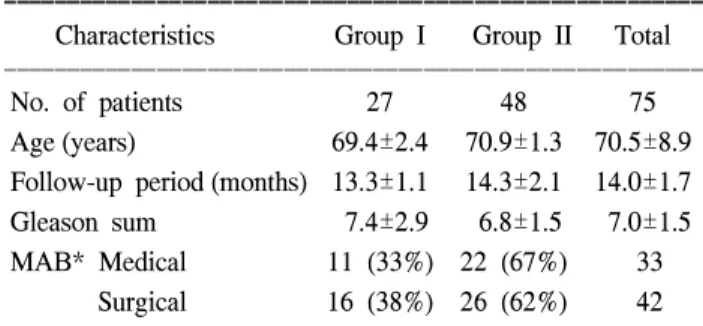
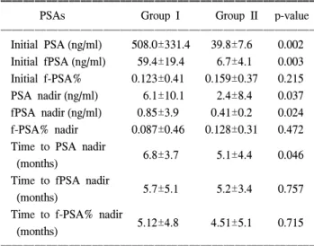
환자의 평균 연령은 70.5±8.9세였으며 1년 이내에 생화 학적 재발을 보인 경우는 27명으로 재발까지의 평균 소요 기간은 8.2±0.6개월이었다. 조기에 생화학적 재발을 보인 경우와 1년 이상 호르몬치료에 반응을 보였던 군의 Gleason sum은 각각 7.4±2.9와 6.8±1.5로 조기에 재발을 보인 경우 상대적으로 분화도가 나쁜 것으로 나타났으나 통계적으로 유의한 차이는 없었다 (Table 1). 두 군의 진단 당시의 PSA 는 각각 508.0±331.4ng/ml와 39.8±7.6ng/ml이었으며 fPSA 는 59.4±19.4ng/ml과 6.7±4.1ng/ml로 조기에 생화학적 재 발을 한 경우 모두 유의하게 높았다 (p=0.002 and 0.003 respectively). 진단 당시의 각 군의 f-PSA%는 0.123±0.41과 0.159±0.37로 조기에 생화학적 재발을 보인 경우 상대적으 로 낮았으나 유의한 차이는 없었다 (Table 2).
호르몬치료 후의 PSA nadir는 조기에 생화학적 재발을 보 인 경우 6.1±10.1ng/ml로 1년 이상 재발을 보이지 않았던 군의 2.4±8.4ng/ml에 비하여 유의하게 높았으며 (p=0.037), 각 군의 fPSA nadir는 0.85±3.9ng/ml와 0.41±0.2ng/ml로 역
Table 1. Characteristics of the patients
ꠚꠚꠚꠚꠚꠚꠚꠚꠚꠚꠚꠚꠚꠚꠚꠚꠚꠚꠚꠚꠚꠚꠚꠚꠚꠚꠚꠚꠚꠚꠚꠚꠚꠚꠚꠚꠚꠚꠚꠚꠚꠚꠚꠚꠚꠚꠚꠚꠚꠚꠚꠚꠚꠚꠚ Characteristics Group I Group II Total ꠏꠏꠏꠏꠏꠏꠏꠏꠏꠏꠏꠏꠏꠏꠏꠏꠏꠏꠏꠏꠏꠏꠏꠏꠏꠏꠏꠏꠏꠏꠏꠏꠏꠏꠏꠏꠏꠏꠏꠏꠏꠏꠏꠏꠏꠏꠏꠏꠏꠏꠏꠏꠏꠏꠏ
No. of patients 27 48 75 Age (years) 69.4±2.4 70.9±1.3 70.5±8.9 Follow-up period (months) 13.3±1.1 14.3±2.1 14.0±1.7 Gleason sum 7.4±2.9 6.8±1.5 7.0±1.5 MAB* Medical 11 (33%) 22 (67%) 33
Surgical 16 (38%) 26 (62%) 42 ꠏꠏꠏꠏꠏꠏꠏꠏꠏꠏꠏꠏꠏꠏꠏꠏꠏꠏꠏꠏꠏꠏꠏꠏꠏꠏꠏꠏꠏꠏꠏꠏꠏꠏꠏꠏꠏꠏꠏꠏꠏꠏꠏꠏꠏꠏꠏꠏꠏꠏꠏꠏꠏꠏꠏ
*maximal androgen blockade. Group I: patients with biological recurrence within 1 year of hormone treatment, Group II:
patients showing sensitivity to hormone treatment longer than 1 year
시 조기에 재발을 보인 군에서 유의하게 높았다 (p=0.024).
PSA nadir에 도달하기까지의 기간은 각각 6.8±3.7개월과 5.1±4.4개월로 조기에 재발을 한 경우 유의하게 길었으나 (p=0.046), fPSA nadir까지의 기간은 각각 5.7±5.1개월과 5.2±
3.4개월로 유의한 차이는 없었다 (Table 2).
f-PSA%는 1년 이상 호르몬치료에 지속적인 반응을 보였 던 경우, 기복이 있기는 하였으나 치료 기간 중 0.159에서 0.172까지 증가 소견을 보였던 반면, 조기에 생화학적 재발 을 한 경우 초기에 0.123에서 0.141까지 증가 소견을 보인 후 8-12개월에 0.092까지 점차 감소하는 양상을 보였다. 호 르몬치료 기간 중 1년 이상 호르몬치료에 반응을 보였던 군의 f-PSA%는 조기에 재발한 경우에 비하여 지속적으로 높았으며 8-12개월에 그 차이가 더욱 커졌으나 통계적인 유 의성은 없었다 (Table 3).
고 찰
1992년 혈청 PSA의 70-90%가 α1-antichymotripsin (ACT), α2-macroglobulin 또는 protein C inhibitor 등의 단백분해효 소억제제 (protease inhibitor)와 결합된 형태인 cPSA (com- plexd form PSA)로, 나머지 10-30%는 fPSA의 형태로 존재한 다는 사실이 밝혀짐과 동시에 전립선의 양성질환과 악성질 환에 있어서 이 두 가지 형태의 PSA의 발현 비율에 차이가 있다는 사실이 알려짐에 따라 이를 이용하여 전립선암 진 단에 있어서의 PSA의 낮은 민감도와 특이도에 동반된 문제 의 상당 부분을 극복할 수 있었다.2,3 예를 들어 조직학적으 로 전립선암으로 진단된 환자의 약 20%가 진단 시 4ng/ml 이하의 PSA치를 보이는 것으로 알려져 있으나 4ng/ ml의 PSA 절사값 (cutoff value)을 유지한 상태에서 10%의 f-PSA%
기준치를 함께 적용할 경우 민감도에는 특별한 영향을 주 지 않은 상태로 특이도를 3배 이상 증가시킬 수 있으며, 특 히 회색지대인 PSA 4-10ng/ml의 범위에서 전립선암의 진단 율을 약 25% 정도 증대시키는 것으로 알려져 있다.4-7 fPSA는 약 33kDa의 크기를 가지는 당단백으로서 단백분 해효소억제제와 비결합 형태로 존재함으로써 효소적으로 불활성 상태를 유지하며 최소한 PSA의 전구물 형태인 proPSA, “benign” PSA (BPSA), “intact” PSA의 3개의 상이한 형태가 존재한다. 이 중 proenzyme 또는 proPSA는 전립선암 과 BPSA는 양성 전립선비대증과 관련이 있는 것으로 보고 되고 있으나 정상 전립선 상피세포나 낮은 Gleason sum을 가지는 전립선암 조직에서 주로 분비가 되는 cPSA와 달리 fPSA 자체는 비대된 전립선 조직에서 주로 분비되는 것으 로 알려져 있다.8 전립선암과 전립선비대증에 있어서 fPSA 의 비율이 변하는 이유에 대하여서는 아직까지 명확하게 밝혀진 바가 없으나 전립선비대증의 경우 대표적인 PSA결
Table 3. Changes of PSA, fPSA and f-PSA% during hormone treatment
ꠚꠚꠚꠚꠚꠚꠚꠚꠚꠚꠚꠚꠚꠚꠚꠚꠚꠚꠚꠚꠚꠚꠚꠚꠚꠚꠚꠚꠚꠚꠚꠚꠚꠚꠚꠚꠚꠚꠚꠚꠚꠚꠚꠚꠚꠚꠚꠚꠚꠚꠚꠚꠚꠚꠚꠚꠚꠚꠚꠚꠚꠚꠚꠚꠚꠚꠚꠚꠚꠚꠚꠚꠚꠚꠚꠚꠚꠚꠚꠚꠚꠚꠚꠚꠚꠚꠚꠚꠚꠚꠚꠚꠚꠚꠚꠚꠚꠚꠚꠚꠚꠚꠚꠚꠚꠚꠚꠚꠚꠚꠚꠚꠚꠚꠚ Follow-up period (months)
PSAs Initial ꠏꠏꠏꠏꠏꠏꠏꠏꠏꠏꠏꠏꠏꠏꠏꠏꠏꠏꠏꠏꠏꠏꠏꠏꠏꠏꠏꠏꠏꠏꠏꠏꠏꠏꠏꠏꠏꠏꠏꠏꠏꠏꠏꠏꠏꠏꠏꠏꠏꠏꠏꠏꠏꠏꠏꠏꠏꠏꠏꠏꠏ
2-4 5-7 8-10 11-12
ꠏꠏꠏꠏꠏꠏꠏꠏꠏꠏꠏꠏꠏꠏꠏꠏꠏꠏꠏꠏꠏꠏꠏꠏꠏꠏꠏꠏꠏꠏꠏꠏꠏꠏꠏꠏꠏꠏꠏꠏꠏꠏꠏꠏꠏꠏꠏꠏꠏꠏꠏꠏꠏꠏꠏꠏꠏꠏꠏꠏꠏꠏꠏꠏꠏꠏꠏꠏꠏꠏꠏꠏꠏꠏꠏꠏꠏꠏꠏꠏꠏꠏꠏꠏꠏꠏꠏꠏꠏꠏꠏꠏꠏꠏꠏꠏꠏꠏꠏꠏꠏꠏꠏꠏꠏꠏꠏꠏꠏꠏꠏꠏꠏꠏꠏ
Group I PSA (ng/ml) 508.0 50.35 28.45 7.12 17.89
fPSA (ng/ml) 59.4 6.43 3.89 0.87 1.75
f-PSA% 0.123 0.127 0.141 0.113 0.092
Group II PSA (ng/ml) 39.8 3.89 2.81 2.92 3.21
fPSA (ng/ml) 6.7 0.56 0.43 0.47 0.55
f-PSA% 0.159 0.151 0.147 0.165 0.172
ꠏꠏꠏꠏꠏꠏꠏꠏꠏꠏꠏꠏꠏꠏꠏꠏꠏꠏꠏꠏꠏꠏꠏꠏꠏꠏꠏꠏꠏꠏꠏꠏꠏꠏꠏꠏꠏꠏꠏꠏꠏꠏꠏꠏꠏꠏꠏꠏꠏꠏꠏꠏꠏꠏꠏꠏꠏꠏꠏꠏꠏꠏꠏꠏꠏꠏꠏꠏꠏꠏꠏꠏꠏꠏꠏꠏꠏꠏꠏꠏꠏꠏꠏꠏꠏꠏꠏꠏꠏꠏꠏꠏꠏꠏꠏꠏꠏꠏꠏꠏꠏꠏꠏꠏꠏꠏꠏꠏꠏꠏꠏꠏꠏꠏꠏ PSA: prostate-specific antigen
Table 2. PSA, fPSA and f-PSA% in each group
ꠚꠚꠚꠚꠚꠚꠚꠚꠚꠚꠚꠚꠚꠚꠚꠚꠚꠚꠚꠚꠚꠚꠚꠚꠚꠚꠚꠚꠚꠚꠚꠚꠚꠚꠚꠚꠚꠚꠚꠚꠚꠚꠚꠚꠚꠚꠚꠚꠚꠚꠚꠚꠚꠚꠚ
PSAs Group I Group II p-value
ꠏꠏꠏꠏꠏꠏꠏꠏꠏꠏꠏꠏꠏꠏꠏꠏꠏꠏꠏꠏꠏꠏꠏꠏꠏꠏꠏꠏꠏꠏꠏꠏꠏꠏꠏꠏꠏꠏꠏꠏꠏꠏꠏꠏꠏꠏꠏꠏꠏꠏꠏꠏꠏꠏꠏ Initial PSA (ng/ml) 508.0±331.4 39.8±7.6 0.002 Initial fPSA (ng/ml) 59.4±19.4 6.7±4.1 0.003 Initial f-PSA% 0.123±0.41 0.159±0.37 0.215 PSA nadir (ng/ml) 6.1±10.1 2.4±8.4 0.037 fPSA nadir (ng/ml) 0.85±3.9 0.41±0.2 0.024
f-PSA% nadir 0.087±0.46 0.128±0.31 0.472
Time to PSA nadir
6.8±3.7 5.1±4.4 0.046 (months)
Time to fPSA nadir
5.7±5.1 5.2±3.4 0.757 (months)
Time to f-PSA% nadir
5.12±4.8 4.51±5.1 0.715 (months)
ꠏꠏꠏꠏꠏꠏꠏꠏꠏꠏꠏꠏꠏꠏꠏꠏꠏꠏꠏꠏꠏꠏꠏꠏꠏꠏꠏꠏꠏꠏꠏꠏꠏꠏꠏꠏꠏꠏꠏꠏꠏꠏꠏꠏꠏꠏꠏꠏꠏꠏꠏꠏꠏꠏꠏ PSA: prostate-specific antigen
합 단백분해효소억제제인 ACT의 합성이 상대적으로 감소 하기 때문에 전립선암에 비하여 상대적으로 비결합 형태인 fPSA의 비율이 증가하는 것으로 생각한다.9 따라서 이론적 으로는 fPSA에 비하여 전립선암 특이성을 가지는 cPSA를 전립선암의 진단에 사용하는 것이 더욱 타당한 것으로 보 이며, 실제 최근 여러 연구결과 cPSA와 PSA를 함께 사용한 C/T PSA ratio가 F/T PSA ratio에 비하여 전립선생검 결과를 더욱 정확하게 예측하는 것으로 보고되어 있다.2 그러나 1998년 ACT와 결합한 형태의 cPSA를 측정할 수 있는 단클 론항체 (monoclonal antibody)가 개발되기 전까지 임상적으 로 사용이 가능한 cPSA 측정법이 사실상 없었을 뿐 아니라 이후에도 다른 복합 형태의 영향 때문에 ACT와 결합한 형 태만을 측정하는 PSA-ACT 등의 cPSA 측정법의 이용에 많 은 어려움이 있어 최근까지도 fPSA에 대한 응용이 상대적 으로 활발히 이루어져 왔다.10,11
fPSA를 이용한 초기 이용 단계에서는 fPSA가 연령, 전립 선의 크기, 전립선비대증이나 전립선암에 대한 어떠한 치 료에도 영향을 받지 않는 것으로 생각하였으나 최근 여러 임상적 경험을 통하여 PSA와 마찬가지로 다양한 전립선 내 적, 외적 인자에 의해 영향을 받는다는 사실이 알려지게 되
었다.12,13 예를 들어 발열을 동반한 급성 요로감염 시 fPSA
는 PSA, cPSA와 더불어 증가하는 반면, 만성 전립선염의 경우 PSA는 증가하고 f-PSA%는 오히려 감소하는 것으로 알려져 있다.14,15 또한 전립선의 크기가 f-PSA%와 직접적인 연관성을 가지지 않는 전립선비대증의 경우와 달리 전립선 암의 경우 일반적으로 f-PSA%가 전립선의 크기와 밀접한 관련성을 가지는 것으로 알려져 있으나 Fujinami 등16은 전 립선의 크기가 30cm3 이상인 경우에만, 반면 Stephan 등17은 전립선의 크기가 40cm3 이하인 경우에만 이러한 관련성이 유의하다는 상이한 결과를 보고하고 있다.18
이러한 결과들은 fPSA와 f-PSA%가 단순히 전립선암의 진단을 위한 생검 여부를 결정하기 위해 사용되는 정적인 지표가 아니라 각종 검사 및 수술, 호르몬요법 등 전립선 자체 및 관련 질환에 연관된 다양한 인자들로부터의 영향 을 반영하는 역동적인 지표로서, PSA와 마찬가지로 각종 치료에 대한 반응 및 이에 따른 예후를 예측할 수 있게 하 는 인자로서의 가능성을 보여주는 것이다. 예를 들어 전립 선생검 전의 f-PSA%가 전립선암의 크기나 Gleason sum뿐 아니라 근치적 수술 시 피막 외 침범 여부, 변연 양성률 등 의 다양한 예후 인자와 밀접한 관련성을 가진다는 사실은 이미 잘 알려져 있으며, 나아가 Crook 등19은 방사선치료 후 전립선암의 재발에 대한 예측에 있어서 f-PSA%의 유용성 에 대하여 보고한 바 있다.20-25
최근의 연구 결과 호르몬치료 과정에서의 fPSA와 f-PSA%
의 변화는 전립선암과 전립선비대증에 있어서 차이가 있는 것으로 보고되고 있는데 예를 들어 전립선비대증에 대한 피 나스테리드 치료 시 PSA와 fPSA 그리고 PSA-ACT가 동일한 비율로 감소하기 때문에 결과적으로 C/T 또는 F/T PSA ratio 는 변화가 없는 것으로 알려져 있는 반면, Stein 등26은 전립 선암에 대한 LHRH 유사체치료 시 f-PSA%가 변화를 보이 게 되며 치료에 양호한 반응을 보인 경우 그렇지 않은 경우 에 비하여 치료 기간 중 f-PSA%가 상대적으로 더 많이 증 가하나 재발 시 다시 감소한다고 보고한 바 있다.27,28 Tanaka 등29도 f-PSA%가 전립선암의 진단 당시의 병기에는 영향을 받지 않지만 호르몬치료에 반응을 보이는 기간 동안 전립 선비대증 때와 비슷한 수준으로 상승하였다가 질환이 재발 됨에 따라 다시 서서히 치료 전의 수치까지 감소한다고 하 였다. 이러한 현상은 호르몬치료 시 전립선비대증의 경우 와 달리 전립선암과 직접적으로 관련된 cPSA의 감소가 fPSA의 감소보다 상대적으로 빨리 일어나기 때문인 것으로 풀이되고 있다.
본 연구에서는 전이 전립선암의 호르몬치료 과정에서의 fPSA 및 f-PSA%의 변화를 1년 이상 주기적으로 추적관찰 한 결과 조기에 생화학적재발을 보였던 군의 경우 치료 시 작 시점부터 첫 5-7개월 동안 fPSA%가 지속적으로 증가한 반면 이후 점차 감소하는 소견을 보였다. 전체 환자 중 생화 학적 재발이 확인이 되었던 27명에서 재발까지의 평균 기 간은 8.2±0.6개월이었던 점을 고려할 때 이러한 f-PSA%의 증감의 변화는 PSA와 마찬가지로 호르몬치료에 대한 반응 의 정도와 관련성이 있는 것으로 생각한다. 즉 조기에 생화 학적 재발을 보였던 군에서 PSA가 8-10개월에 최저치에 도 달하였다가 이후 점차 상승한 것과 유사하게 초기 5-7개월, 즉 호르몬치료에 반응을 보이던 시기까지는 f-PSA%가 점 차 증가하다가 PSA의 재상승 시기보다 2-3개월 빠른, 8-10 개월을 기점으로 점차 감소하기 시작하는 것으로 나타났 다. 반면 호르몬치료에 지속적으로 반응을 보인 군의 경우 치료 기간 중 일시적으로 f-PSA%가 감소하는 양상을 보였 으나 전반적으로 상승을 계속하였으며 통계적으로 유의하 지는 않았으나, f-PSA%는 초기 진단 시보다 생화학적 재발 시점인 8-10개월을 기준으로 두군간에 그 차이가 더욱 커지 는 현상을 보였다.
이러한 결과는 fPSA와 f-PSA%가 전립선암의 호르몬치료 에 영향을 받을 뿐 아니라, 호르몬치료에 대한 반응의 정도 에 따라 그 변화 양상이 다르다는 기존의 연구 결과에 상응 하는 것으로서 전이 전립선암의 호르몬치료 시 PSA와 함께 사용할 수 있는 보조적인 예후 인자로서의 중요성을 보여 주는 것이다. 따라서 향후 전이 전립선암의 호르몬치료 시 fPSA와 f-PSA%의 추적관찰의 임상적 응용 가능성에 대하
여 대규모 집단을 대상으로 한 추가적인 연구가 필요할 것 으로 생각한다.
결 론
본 연구 결과 전이 전립선암의 호르몬치료 과정에서 fPSA와 f-PSA%가 지속적으로 변하며 나아가 호르몬치료에 대한 반응 정도에 따라 그 변화 양상에 차이가 있다는 점을 알게 되었다. 더욱이 전립선암의 호르몬치료 시 예후 예측 인자로서 추적관찰에 가장 흔히 사용되는 PSA에 비하여 f-PSA% 변화가 호르몬 감수성의 변화를 조기에 반영하는 것으로 나타나, 호르몬치료 시 PSA에 기초한 추가적인 예 후 인자로서의 가능성을 보여주었다. 본 연구가 제한된 추 적관찰 기간 및 조사군을 대상으로 한 소규모 조사였음을 감안할 때, 향후 전이 전립선암의 호르몬치료 시 PSA와 fPSA, f-PSA%의 주기적 추적 관찰의 임상적 의의에 대한 대규모 군을 대상으로 한 장기간의 추가적 연구가 필요할 것으로 생각한다.
REFERENCES
1. Djavan B, Remzi M, Zlotta AR, Ravery V, Hammerer P, Reis- sigl A, et al. Complexed prostate-specific antigen, complexed prostate-specific antigen density of total and transition zone, complexed/total prostate-specific antigen ratio, free-to- total prostate-specific antigen ratio, density of total and transition zone prostate-specific antigen: results of the prospective multi- center European trial. Urology 2002;60:4-9
2. Mitchell ID, Croal BL, Dickie A, Cohen NP, Ross I. A pro- spective study to evaluate the role of complexed prostate spe- cific antigen and free/total prostate specific antigen ratio for the diagnosis of prostate cancer. J Urol 2001;165:1549-53 3. Catalona WJ. Clinical utility of measurements of free and total
prostate-specific antigen (PSA). Prostate 1996;7:64-9 4. Haese A, Dworschack RT, Partin AW. Percent free prostate
specific antigen in the total prostate specific antigen 2 to 4 ng./ml. range does not substantially increase the number of biopsies needed to detect clinically significant prostate cancer compared to the 4 to 10ng/ml. range. J Urol 2002;168:504-8 5. Lein M, Semjonow A, Graefen M, Kwiatkowski M, Abramjuk
C, Stephan C, et al. A multicenter clinical trial on the use of (-5, -7) pro prostate specific antigen. J Urol 2005;174:2150-3 6. Ito K, Yamamoto T, Ohi M, Kurokawa K, Suzuki K, Yama- naka H. Free/total PSA ratio is a powerful predictor of future prostate cancer morbidity in men with initial PSA levels of 4.1 to 10.0ng/ml. Urology 2003;61:760-4
7. Miele ME. Percent free PSA as an additional measure in a prostate cancer screen. Clin Lab Sci 2001;14:102-7
8. Khan MA, Sokoll LJ, Chan DW, Mangold LA, Mohr P, Mikolajczyk SD, et al. Clinical utility of proPSA and “benign”
PSA when percent free PSA is less than 15%. Urology 2004;
64:1160-4
9. Bjork T, Bjartell A, Abrahamsson PA, Hulkko S, di Sant' Agnese A, Lilja H. Alpha 1-antichymotrypsin production in PSA-producing cells is common in prostate cancer but rare in benign prostatic hyperplasia. Urology 1994;43:427-34 10. Jung K, Brux B, Knabich A, Lein M, Sinha P, Schnorr D,
et al. A gap between total prostate-specific antigen and the sum of free prostate-specific antigen plus alpha1-antichymo- trypsin-prostate-specific antigen in patients with prostate car- cinoma but not in those with benign prostate hyperplasia. Clin Chem 1999;45:422-4
11. Allard WJ, Zhou Z, Yeung KK. Novel immunoassay for the measurement of complexed prostate-specific antigen in serum.
Clin Chem 1998;44:1216-23
12. Meyer A, Jung K, Lein M, Rudolph B, Schnorr D, Loening SA. Factors influencing the ratio of free to total prostate-speci- fic antigen in serum. Int J Cancer 1997;74:630-6
13. Gregorakis AK, Malovrouvas D, Stefanakis S, Petraki K, Scorilas A. Free/total PSA (F/T ratio) kinetics in patients with clinically localized prostate cancer undergoing radical prosta- tectomy. Clin Chim Acta 2005;357:196-201
14. Zackrisson B, Ulleryd P, Aus G, Lilja H, Sandberg T, Hugos- son J. Evolution of free, complexed, and total serum prostate- specific antigen and their ratios during 1 year of follow-up of men with febrile urinary tract infection. Urology 2003;62:
278-81
15. Jung K, Meyer A, Lein M, Rudolph B, Schnorr D, Loening SA. Ratio of free-to-total prostate specific antigen in serum cannot distinguish patients with prostate cancer from those with chronic inflammation of the prostate. J Urol 1998;159:
1595-8
16. Fujinami K, Miura T, Takizawa A, Osada Y, Kawakami S.
Comparison of value of free-to total prostate specific antigen, prostate specific antigen density and prostate specific antigen density of transition zone for diagnosis of prostate cancer in patients with a PSA level of 4.1-10ng/ml. Nippon Hinyokika Gakkai Zasshi 2005;96:475-9
17. Stephan C, Lein M, Jung K, Schnorr D, Loening SA. The in- fluence of prostate volume on the ratio of free to total prostate specific antigen in serum of patients with prostate carcinoma and benign prostate hyperplasia. Cancer 1997;79:104-9 18. Haese A, Graefen M, Noldus J, Hammerer P, Huland E, Hul-
and H. Prostatic volume and ratio of free-to-total prostate spe- cific antigen in patients with prostatic cancer or benign pro- static hyperplasia. J Urol 1997;158:2188-92
19. Crook JM, Bunting PS. Percent free prostate-specific antigen after radiotherapy for prostate cancer. Urology 1998;52:100-5 20. Morote J, Encabo G, Lopez MA, De Torres IM. The free-to- total serum prostatic specific antigen ratio as a predictor of the
pathological features of prostate cancer. BJU Int 1999;83:
1003-6
21. Aus G, Becker C, Lilja H, Khatami A, Pihl CG, Hugosson J. Free-to-total prostate-specific antigen ratio as a predictor of non-organ-confined prostate cancer (stage pT3). Scand J Urol Nephrol 2003;37:466-70
22. Li W, Ren Y, Mee V, Wong PY. Prostate-specific antigen ratio correlates with aggressiveness of histology grades of prostate cancer. Clin Biochem 1999;32:31-7
23. Grossklaus DJ, Smith JA Jr, Shappell SB, Coffey CS, Chang SS, Cookson MS. The free/total prostate-specific antigen ratio (%fPSA) is the best predictor of tumor involvement in the radical prostatectomy specimen among men with an elevated PSA. Urol Oncol 2002;7:195-8
24. Wymenga LF, Duisterwinkel FJ, Groenier K, Visser-van Brum- men P, Marrink J, Mensink HJ. Clinical implications of free- to-total immunoreactive prostate-specific antigen ratios. Scand J Urol Nephrol 2000;34:181-7
25. Anfossi E, Rossi D, Grisoni V, Sauvan R, Bladou F, Serment G. What is the role of the correspondence of free PSA/total PSA in the staging of local prostate cancer? Series of 50 radi- cal prostatectomy cases. Prog Urol 1999;9:479-82
26. Stein A, Barak M, Mecz Y, Rubinov R, Lurie A. Serum free/
total prostatic-specific antigen in prostate cancer patients treat- ed with LH-RH agonists. Eur Urol 1997;32:64-8
27. Espana F, Martinez M, Royo M, Estelles A, Alapont JM, Navarro S, et al. Changes in molecular forms of prostate- specific antigen during treatment with finasteride. BJU Int 2002;90:672-7
28. Morote J, Lorente JA, Raventos CX, Lopez MA, Encabo G, De Torres I, et al. Effect of finasteride on the percentage of free PSA: implications in the early diagnosis of prostatic can- cer. Actas Urol Esp 1998;22:835-9
29. Tanaka M, Murakami S, Suzuki N, Shimazaki J. Change in the ratio of free-to-total prostate-specific antigen during pro- gression of advanced prostate cancer. Int J Urol 2000;7:83-7