Corresponding author: Michael F. Holick
Department of Medicine, Vitamin D, Skin and Bone Research Laboratory, Section of Endocrinology, Nutrition and Diabetes, Boston University Medical Center, 85 East Newton Street, M-1013, Boston, MA 02118, USA
Tel: +1-617-638-4546, Fax: +1-617-638-8882, E-mail: [email protected]
※ Supported in part by the UV Foundation and the Mushroom Council.
Vitamin D: A D-Lightful Vitamin for Health
Michael F. Holick
Vitamin D, Skin and Bone Research Laboratory, Section of Endocrinology, Department of Medicine, Nutrition and Diabetes, Boston University Medical Center, Boston, MA, USA
Vitamin D is a sunshine vitamin that has been produced on this earth for more than 500 million years. Because foods contain so lit- tle vitamin D most humans have always depended on sun exposure for their vitamin D requirement. Vitamin D deficiency has been defined as a serum 25-hydroxyvitamin D concentration < 20 ng/mL (50 nmol/L); vitamin D insufficiency as a serum 25-hydroxyvi- tamin D of 21-29 ng/mL and vitamin D sufficiency as a serum 25-hydroxyvitamin D of 30-100 ng/mL whereas toxicity is usually not seen until blood levels are above 150 ng/mL. Vitamin D deficiency is a global health problem that increases risk for metabolic bone diseases in children and adults as well as many chronic illnesses including autoimmune diseases, type 2 diabetes, cardiovascular disease, infectious disease, and cancer. The major causes of vitamin D deficiency are lack of adequate sensible exposure to sunlight, inadequate dietary intake and obesity. The United States Endocrine Society recommended that to prevent vitamin D deficiency in those at risk, children 1 year and older require 600-1,000 international unit (IU) of vitamin D daily and adults require 1,500-2,000 IU of vitamin D daily. Obese patients require 2-3 times more vitamin D to both treat and prevent vitamin D deficiency. (Endocrinol Metab 27:255-267, 2012)
Key Words: 25-hydroxyvitamin D, Autoimmune diseases, Communicable diseases, Neoplasms, Osteomalacia, Sunlight, Type 2 di- abetes mellitus, Vitamin D, Vitamin D deficiency
INTRODUCTION
Vitamin D, the sunshine vitamin, has received a lot of attention recently as a result of a meteoric rise in the number of publications that have related vitamin D deficiency with many acute and chronic illnesses not related to calcium metabolism including autoimmune diseases, some cancers, type 2 diabetes, cardiovascular disease, and infectious diseases. Vitamin D deficiency is now recognized as a global pandemic. The major cause for vitamin D deficiency is the lack of appreciation that sun exposure has been and continues to be the major source of vitamin D for children and adults of all ages.
Vitamin D plays a crucial role in the development and maintenance of a healthy skeleton throughout life. There remains some contro- versy regarding what blood level of 25-hydroxyvitamin D (25[OH]
D) should be attained both for bone health and reducing risk for vitamin D deficiency associated chronic diseases.
PREHISTORICAL PERSPECTIVE
Vitamin D is likely to be the oldest hormone that has been pho- tosynthesized for more than 500 million years in early life forms [1].
Emiliania huxleyi, a phytoplankton that has existed in the Sargasso Sea for more than 500 million years, was found to be able to pro- duce vitamin D2 after exposure to sunlight. Although the function for vitamin D is unknown in these primitive organisms it has been suggested that when vitamin D2 was produced in the plasma mem- brane of these early life forms it was then ejected out of the plasma membrane resulting in a transient opening of the membrane that permitted the transport of calcium into the cell [2]. As life forms evolved they maintained their ability to produce vitamin D during sun exposure. Three hundred and fifty million years ago the ocean dwelling vertebrates ventured onto land and maintained their abil- ity to produce vitamin D in their skin from sun exposure. It has been speculated that one of the causes for the demise of dinosaurs
This is an Open Access article distributed under the terms of the Creative Commons Attribution Non-Commercial License (http://creativecommons.org/licenses/by-nc/3.0/) which permits unrestricted non-commercial use, distribution, and reproduction in any medium, provided the original work is properly cited.
Copyright © 2012 Korean Endocrine Society
after the asteroid struck the earth was that the globe was enveloped in a dark cloud of ash which would have prevented land vertebrates from making any vitamin D. Therefore vitamin D deficiency could have contributed to the death of these giant vertebrates. It was the nocturnal rodent that survived this holocaust. They had adapted to their nighttime environment thereby needing little if any vitamin D to survive [2].
HISTORICAL PERSPECTIVE
The intimate relationship between sunlight and human health became evident with the industrialization of Northern Europe [3].
It was recognized in the mid-1600s that children living in London and Glasgow developed a devastating bone disease known as rick- ets. This bone deforming disease spread throughout Europe and Northeastern United States. In the mid-1800s cod liver oil was found to be effective in treating the disease. However by the turn of the 20th century upwards of 90% of children living in the inner cities in Northern Europe and Northeastern United States had evidence of rickets. Huldschinsky [4] was the first to report that children ex- posed to a mercury arc lamp could be cured of their disease. Hess and Unger [5] reported in 1921 that exposing children to sunlight in New York City was effective in treating rickets. This quickly led Steenbock [6] to introduce the concept of exposing various foods to ultraviolet radiation to impart antirachitic activity. This observation led to the fortification of milk with vitamin D. This simple fortifica- tion process resulted in the eradication of rickets as a health prob- lem for children living in the United States, Canada, and Europe.
In the early 1950s there were several reports of young children who had high blood calcium and altered facial features in Great Britain [7]. The experts concluded that this was due to vitamin D intoxication and believed that the intoxication was coming from the over fortification of milk with vitamin D. This caused great hysteria leading to laws being passed in Europe forbidding the for- tification of any food or consumer product with vitamin D.
Several investigations occurred but no one was able to find any product had contained an excess amount of vitamin D. It is more likely that these children had Williams syndrome which is known to be associated with birth defects especially regarding facial fea- tures with elfin faces, mental retardation, supravalvular aortic ste- nosis and that they are hypersensitive to vitamin D resulting in hy- percalcemia [8].
Unfortunately these laws remain operative today and most Euro-
Fig. 1. Schematic representation of the synthesis and metabolism of vitamin D for regulating calcium, phosphorus, and bone metabolism. During exposure to sunlight 7-dehydrocholesterol in the skin is converted to previtamin D3. Previta- min D3 immediately converts by a heat dependent process to vitamin D3. Exces- sive exposure to sunlight degrades previtamin D3 and vitamin D3 into inactive photoproducts. Vitamin D2 and vitamin D3 from dietary sources is incorporated into chylomicrons, transported by the lymphatic system into the venus circula- tion. Vitamin D (D represents D2 or D3) made in the skin or ingested in the diet can be stored in and then released from fat cells. Vitamin D in the circulation is bound to the vitamin D binding protein which transports it to the liver where vi- tamin D is converted by the vitamin D-25-hydroxylase to 25-hydroxyvitamin D [25(OH)D]. This is the major circulating form of vitamin D that is used by clinicians to measure vitamin D status (although most reference laboratories report the normal range to be 20-100 ng/mL, the preferred healthful range is 30-60 ng/mL).
It is biologically inactive and must be converted in the kidneys by the 25-hy- droxyvitamin D-1α-hydroxylase (1-OHase) to its biologically active form 1,25-di- hydroxyvitamin D [1,25(OH)2D]. Serum phosphorus, calcium fibroblast growth factors (FGF-23) and other factors can either increase (+) or decrease (-) the re- nal production of 1,25(OH)2D. A 1,25(OH)2D feedback regulates its own synthe- sis and decreases the synthesis and secretion of parathyroid hormone (PTH) in the parathyroid glands. A 1,25(OH)2D increases the expression of the 25-hy- droxyvitamin D-24-hydroxylase (24-OHase) to catabolize 1,25(OH)2D to the wa- ter soluble biologically inactive calcitroic acid which is excreted in the bile. A 1,25(OH)2D enhances intestinal calcium absorption in the small intestine by stim- ulating the expression of the epithelial calcium channel (ECaC) and the calbin- din 9K (calcium binding protein, CaBP). A 1,25(OH)2D is recognized by its recep- tor in osteoblasts causing an increase in the expression of receptor activator of nuclear factor kappa B ligand (RANKL). Its receptor RANK on the preosteoclast binds RANKL which induces the preosteoclast to become a mature osteoclast.
The mature osteoclast removes calcium and phosphorus from the bone to main- tain blood calcium and phosphorus levels. Adequate calcium and phosphorus levels promote the mineralization of the skeleton. Reprinted from Holick MF copy- right 2007 with permission. 7-DHC, 7-dehydrocholesterol; UVB, ultraviolet B.
Solar UVB radiation
Inactive photoproducts
Chylomicrons
Pi, Ca & other
Parathyroid glands factors +/-
Major circulating metabolite
< 20 ng/mL
< 150 ng/mL Intoxication
Deficient Reference
range 20-100 ng/mL
Ideal 30-60 ng/mL
pean countries with the exception of Sweden and Finland who re- cently permitted the fortification of milk and dairy products with vitamin D still forbid fortification of dairy products with vitamin D.
SOURCES OF VITAMIN D
The major source of vitamin D for humans is exposure to sun- light. During sunlight exposure the ultraviolet B radiation (290-315 nm) penetrates into the epidermis and is absorbed by 7-dehydro- cholesterol [9]. This absorption results in 7-dehydrocholesterol be- ing converted to previtamin D3 (Fig. 1). Once informed previtamin D3 which is thermally labile, undergoes a transformation of its dou- ble bonds to form vitamin D3. This process takes approximately 4
hours to complete [10]. As vitamin D is being produced it is ejected out of the cell membrane into the extravascular space and diffuses into the dermal capillary bed where it is bound to the vitamin D binding protein [11].
A variety of factors dramatically influence subcutaneous produc- tion of vitamin D3. A sunscreen with a sun protection factor of 30 absorbs approximately 98% of solar ultraviolet B (UVB) radiation and thereby reduces the production of vitamin D3 in the skin by approximately 98% [12]. Melanin pigmentation is a very effective sunscreen. Therefore people of color especially blacks require much longer exposures to sunlight to produce the same amount of vitamin D as a white person would require. One study reported that when a white adult was exposed to UVB radiation in a tanning
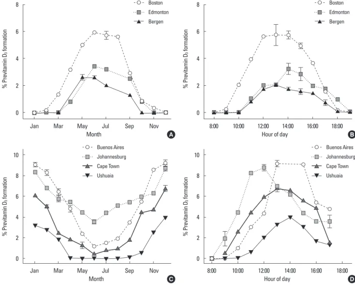
Fig. 2. (A, B) Influence of season, time of day, and latitude on the synthesis of previtamin D3 in Northern hemispheres. (C, D) Influence of season, time of day, and latitude on the synthesis of previtamin D3 in Southern hemispheres. The hour indicated in B and D is the end of the 1-hour exposure time. Reprinted from Holick MF copyright 1998 with permission.
% Previtamin D3 formation % Previtamin D3 formation
8
6
4
2
0
8
6
4
2
0
Jan Mar May Jul Sep Nov 8:00 10:00 12:00 14:00 16:00 18:00
Month Hour of day
Boston Edmonton Bergen
Boston Edmonton Bergen
A B
% Previtamin D3 formation
% Previtamin D3 formation
10
8
6
4
2
0 10
8
6
4
2
0
8:00 10:00 12:00 14:00 16:00 18:00
Jan Mar May Jul Sep Nov
Hour of day Month
Buenos Aires Johannesburg Cape Town Ushuaia Buenos Aires
Johannesburg Cape Town Ushuaia
D C
bed he was able to raise his blood level of vitamin D by 50 fold. A black adult exposed to the same amount of UVB radiation was un- able to raise his blood level of vitamin D. Exposure to six times more UVB radiation raised the blood level in the black adult by approximately 30 fold [13].
The zenith angle of the sun also has a dramatic influence on the ability of the skin to produce vitamin D3. Solar UVB radiation is ef- ficiently absorbed by the stratospheric ozone layer and no more than approximately 1% reaches the earth’s surface at the equator in the summer. Thus as the zenith angle of the sun increases the solar UVB radiation has a longer path length of ozone to pass through.
As a result essentially all UVB radiation is absorbed during the win-
ter at latitudes above and below approximately 32°. This is also the explanation for why even in the summer at the equator exposure to early morning and late afternoon sunlight will not result in any significant production of vitamin D3 in the skin (Fig. 2) [2].
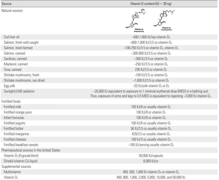
Very few foods naturally contain vitamin D [14]. Oily fish, includ- ing salmon, mackerel and herring, cod liver oil and sun exposed mushrooms naturally contain vitamin D (Table 1). Some countries including the United States and Canada, among others, permit for- tification of some foods with approximately 100 international units (IUs) per serving. For example in United States 8 oz of milk and some orange juices contain 100 IUs of vitamin D. In Europe some countries permit cereals and margarine to be fortified with vitamin
Table 1. Sources of vitamin D2 and vitamin D3
Source Vitamin D content (IU = 25 ng)
Natural sources
Cod liver oil ~400-1,000 IU/tsp vitamin D3
Salmon, fresh wild caught ~600-1,000 IU/3.5 oz vitamin D3
Salmon, fresh farmed ~100-250 IU/3.5 oz vitamin D3, vitamin D2
Salmon, canned ~300-600 IU/3.5 oz vitamin D3
Sardines, canned ~300 IU/3.5 oz vitamin D3
Mackerel, canned ~250 IU/3.5 oz vitamin D3
Tuna, canned 236 IU/3.5 oz vitamin D3
Shiitake mushrooms, fresh ~100 IU/3.5 oz vitamin D2
Shiitake mushrooms, sun dried ~1,600 IU/3.5 oz vitamin D2
Egg yolk ~20 IU/yolk vitamin D3 or D2
Sunlight/UVB radiation ~20,000 IU equivalent to exposure to 1 minimal erythemal dose (MED) in a bathing suit.
Thus, exposure of arms and legs to 0.5 MED is equivalent to ingesting ~3,000 IU vitamin D3. Fortified foods
Fortified milk 100 IU/8 oz usually vitamin D3
Fortified orange juice 100 IU/8 oz vitamin D3
Infant formulas 100 IU/8 oz vitamin D3
Fortified yogurts 100 IU/8 oz usually vitamin D3
Fortified butter 56 IU/3.5 oz usually vitamin D3
Fortified margarine 429/3.5 oz usually vitamin D3
Fortified cheeses 100 IU/3 oz usually vitamin D3
Fortified breakfast cereals ~100 IU/serving usually vitamin D3
Pharmaceutical sources in the United States
Vitamin D2 (Ergocalciferol) 50,000 IU/capsule
Drisdol (vitamin D2) liquid 8,000 IU/cc
Supplemental sources
Multivitamin 400, 500, 1,000 IU vitamin D3 or vitamin D2
Vitamin D3 400, 800, 1,000, 2,000, 5,000, 10,000, and 50,000 IU
Reprinted from Holick MF copyright 2007 with permission. Designated calciferol which usually means vitamin D2. IU, international unit.
D. This however is rarely practiced in Asian countries. Both vitamin D2, which is produced from the UV irradiation of yeast, and vita- min D3, which is chemically produced from cholesterol obtained from lanolin, are used in supplements and for food fortification.
Although there has been some controversy as to whether vitamin D3 is as effective as vitamin D2 in maintaining vitamin D status in humans most studies in children and adults have demonstrated that they are equally effective [15-17].
VITAMIN D METABOLISM FOR CALCIUM METABOLISM
Once vitamin D (D represents either D2 or D3) is ingested or pro- duced in the skin it travels on the vitamin D binding protein to the liver where it is converted to [1,25(OH)D]. A 25(OH)D is the major circulating form of vitamin D and is used by clinicians to determine a person’s vitamin D status. However 25(OH)D is biologically inert and requires a further hydroxylation in the kidneys on carbon one to form 1,25-dihydroxyvitamin D [1,25(OH)2D]. Once informed 1,25(OH)2D travels to the small intestine and interacts with the nu- clear vitamin D receptor (VDR) to increase expression of proteins that result in the enhancement of intestinal calcium absorption [14, 18]. A 1,25(OH)2D interacts with its receptor in osteoblasts resulting in the expression of receptor activator nuclear factor kappa B ligand (RANKL). The receptor RANK on the preosteoclasts interacts with RANKL inducing them to become mature osteoclasts. These ma- ture osteoclasts release HCl and collagenases which dissolve the mineral and matrix respectively to release the precious calcium into the circulation [14,19]. A variety of factors including serum calcium, phosphorus, parathyroid hormone (PTH) and fibroblast growth factor 23 regulate the renal production of 1,25(OH)2D (Fig. 1) [14].
EXTRARENAL PRODUCTION OF 1,25(OH)2D
It is now recognized that essentially all of the cells in the body express VDR. Thus essentially all cells and organs in the body in- cluding the brain, vascular smooth muscle, breast, prostate, pan- creas, skin, and macrophages are targets for 1,25(OH)2D. Remark- ably many of these organs and cells also have the capacity to con- vert 25(OH)D to 1,25(OH)2D [20,21]. Thus colon, prostate, brain, breast, skin, and immune cells including macrophages can locally produce 1,25(OH)2D (Fig. 3) [14,20-22].
It has been estimated that as many as 2,000 genes are directly or indirectly regulated by 1,25(OH)2D [23,24]. One example is that
Fig. 3. Metabolism of 25-hydroxyvitamin D [25(OH)D] to 1,25 dihydroxyvitamin D 1,25(OH)2D for non-skeletal functions. When a monocyte/macrophage is stim- ulated through its toll-like receptor 2/1 (TLR2/1) by an infective agent such as Mycobacterium tuberculosis (TB), or its lipopolysaccharide (LPS) the signal up- regulates the expression of vitamin D receptor (VDR) and the 25-hydroxyvitamin D-1-hydroxylase (1-OHase). The 25(OH)D levels > 30 ng/mL provides adequate substrate for the 1-OHase to convert it to 1,25(OH)2D. A 1,25(OH)2D returns to the nucleus where it increases the expression of cathelicidin (CD) which is a peptide capable of promoting innate immunity and inducing the destruction of infective agents such as TB. It is also likely that the 1,25(OH)2D produced in the monocytes/macrophage is released to act locally on activated T (AT) and acti- vated B (AB) lymphocytes which regulate cytokine and immunoglobulin synthe- sis respectively. When 25(OH)D levels are ~30 ng/mL, it reduces risk of many common cancers. It is believed that the local production of 1,25(OH)2D in the breast, colon, prostate, and other cells regulates a variety of genes that control proliferation including p21 and p27 as well as genes that inhibit angiogenesis and induced apoptosis. Once 1,25(OH)2D completes the task of maintaining normal cellular proliferation and differentiation, it induces the 25-hydroxyvita- min D-24-hydroxylase (24-OHase). The 24-OHase enhances the metabolism of 1,25(OH)2D to calcitroic acid which is biologically inert. Thus, the local produc- tion of 1,25(OH)2D does not enter the circulation and has no influence on calci- um metabolism. The parathyroid glands have 1-OHase activity and the local production of 1,25(OH)2D inhibits the expression and synthesis of PTH. The pro- duction of 1,25(OH)2D in the kidney enters the circulation and is able to down regulate renin production in the kidney and to stimulate insulin secretion in the β-islet cells of the pancreas. Reprinted from Holick MF copyright 2007 with permission.
BP, blood pressure; BS, blood sugar; PTH, parathyroid hormone; RXR, retinoic acid X receptor.
Breast, colon, prostate
when macrophages are infected with tuberculosis (TB) the toll like receptors become activated resulting in signal transduction to the nucleus resulting in an increase in the expression of VDR and the 25-hydroxyvitamin D-1-hydroxylase (cyp27B1) [25]. Once formed, 1,25(OH)2D enters the nucleus, binds to its VDR resulting in the expression of a cathelicidin a defensen protein that is capable of killing infective agents such as TB [25]. A 1,25(OH)2D has also been shown to inhibit cancer cell proliferation, induce cancer cell matu- ration and apoptosis and inhibit angiogenosis and may help explain the studies associating vitamin D deficiency with increased risk for many deadly cancers including colon and breast cancer [26-28]. A 1,25(OH)2D also down regulates renin production and enhances insulin secretion which may help explain the reported cardiovas- cular benefits of vitamin D (Fig. 3) [29-32].
DEFINITIONS OF VITAMIN D DEFICIENCY, INSUFFICIENCY, AND SUFFICIENCY
There is general agreement by the Institute of Medicine (IOM) and the Endocrine Society that vitamin D deficiency should be de- fined as a 25(OH)D < 20 ng/mL [33,34]. The IOM concluded that a blood level of 25(OH)D > 20 ng/mL will guarantee that more than 99% of adults will have no evidence of vitamin D deficiency bone disease [33]. However a reevaluation of the study by Priemel et al. [35]
revealed that 8.5% of otherwise healthy adult German motor vehi-
cle accident victims had evidence of vitamin D deficiency osteo- malacia [36]. The authors of the study concluded that to guarantee no evidence of vitamin D deficiency bone disease that a blood level of 25(OH)D should be > 30 ng/mL [35]. It has also been reported by several investigators that PTH levels plateau when 25(OH)D are approximately 30 ng/mL (Fig. 4) [37-39]. They also evaluated the literature relating vitamin D status, i.e., serum 25(OH)D, with skel- etal muscle function and falls and concluded that a blood level of 25(OH)D > 30 ng/mL reduced risk for falls and maximized muscle performance [34,40]. Based on this evidence the Endocrine Society recommended that to guarantee the maximum effect of vitamin D on bone and muscle health for adults the blood level of 25(OH)D should be at least 30 ng/mL. Furthermore a review of the literature demonstrated that vitamin D toxicity was not observed until blood levels of 25(OH)D were > 200 ng/mL. Therefore the Endocrine Soci- ety recommended that vitamin D deficiency should be defined as a 25(OH)D < 20 ng/mL. They defined a 25(OH)D of 21-29 ng/mL as vitamin D insufficiency and a blood level of 30 ng/mL and up to 100 ng/mL as vitamin D sufficiency. Because there are a variety of assays that have variable accuracy the Endocrine Society further recom- mended that the preferred range for 25(OH)D be 40-60 ng/mL [34].
VITAMIN D DEFICIENCY PANDEMIC
When defining vitamin D deficiency as a 25(OH)D < 20 ng/mL
Parathyroid hormone (pg/mL) Prevalence of secondary hyperparathyroidism
80 70 60 50 40 30 20 10 0
100 90 80 70 60 50 40 30 20 10
0-9 10-14 15-19 20-24 25-29 30-34 35-39 40-49 50-59 ≥ 60 0 0-9 10-14 15-19 20-24 25-29 30-34 35-39 ≥ 40
Serum 25(OH)D (ng/mL) Serum 25(OH)D
20
75 122
214
201 203 149 262
Fig. 4. (A) Mean ± SE serum parathyroid hormone (PTH; picograms per milliliter) by serum 25-hydroxyvitamin D (25[OH]D) subgroups. Subject PTH concentrations (picograms per milliliter) relative to serum 25(OH)D concentrations sorted by subgroups delineated by predefined cutoffs for analyses of 25(OH)D inadequacy. Serum PTH values began to increase with 25(OH)D concentrations less than 29.8 ng/mL. (B) Percent of subjects with secondary hyperparathyroidism by 25(OH)D level. The percent of subjects with secondary hyperparathyroidism (PTH > 40 pg/mL) sorted by subgroups with serum 25(OH)D concentrations delineated by predefined cutoffs for analyses of 25(OH)D inadequacy. Reprinted from Holick MF et al. J Clin Endocrinol Metab 90:3215-3224, 2005 [39] with permission.
A B
studies throughout the world have revealed that essentially all chil- dren and adults are at risk for vitamin D deficiency and its health consequences (Fig. 5). In United States the Centers for Disease Con- trol reported that 32% of children and adults were vitamin D defi- cient. Indeed children and adults whether they lived in countries near the equator or far north and south of the equator were at equally high risk for vitamin D deficiency (Fig. 3). Pregnant and lactating women are especially at high risk for vitamin D deficiency [41,42].
A study conducted in Boston reported that pregnant women who took 600 IUs of vitamin D daily at the time they gave birth 76%
of them and 81% of their newborns had a blood level of 25(OH)D
< 20 ng/mL [41].
Studies conducted in Korea have also confirmed widespread vita- min D deficiency especially in the younger generation [43]. Essen- tially all children 10 years and older and all adults through every
decade of life were found to have a blood level of 25(OH)D < 20 ng/
mL. Only 6.7% of all females and 13.2% of males were found to have a blood level of 25(OH)D > 30 ng/mL [43]. Similar to other studies, Korean adults were more likely to be vitamin D deficient if they worked indoors and during the winter months demonstrating the importance of sun exposure as their major source of vitamin D [43].
CONSEQUENCES OF THE VITAMIN D DEFICIENCY PANDEMIC
During pregnancy vitamin D deficiency has been associated with an increased risk for preeclampsia [44]. At the time of birthing vita- min D deficiency was associated with an increased risk for requir- ing a cesarean section [45]. Vitamin D deficiency during infancy has been linked to increased risk for wheezing disorders, asthma
11 22
24 26
24
28 27
9
31 61
45 75
56 74
90 98
56 60 92
36 19
22 28
24 25
24
% Population 25(OH)D < 20 ng/mL% Population 25(OH)D < 20 ng/mL Vitamin D (IUs) consumed per day
30
25
20
15
10
5
0
100 90 80 70 60 50 40 30 20 10 0
600
500
400
300
200
100
1-8 9-13 14-18 19-30 31-50 51-70 > 70 0
AU CA CH IN KR MA ME MO NZ NA NE US
1-3 9-13
9-13 19-30
19-30 51-70
51-70
4-8 14-18
14-18 31-50
31-50
≥ 71 ≥ 71
Age group (yr)
Age group (yr)
IOM recommended daily allowance
Both Male
Male Female
Diet, CSFII 1994-96, 1998 Diet, NHANES III Diet + Supplements, NHANES III
Female
Fig. 5. (A) Prevalence at risk of vitamin D deficiency defined as a 25-hy- droxyvitamin D < 12-20 ng/mL by age and sex: United States, 2001-2006.
(B) Mean intake of vitamin D (international unit, IU) from food and food plus dietary supplements from Continuing Survey of Food Intakes by Indi- viduals (CSFII) 1994-1996, 1998 and the Third National Health and Nutri- tion Examination Survey (NHANES III) 1988-1994. (C) Reported incidence of vitamin D deficiency defined as a 25-hydroxyvitamin D < 20 ng/mL around the globe including Australia (AU), Canada (CA), China (CH), India (IN), Korea (KR), Malaysia (MA), Middle East (ME), Mongolia (MO), New Zealand (NZ), North Africa (NA), Northern Europe (NE), and United States (US). Reprinted from Holick MF et al. J Clin Endocrinol Metab 97:1153-1158, 2012 [76] with permission.
A
C
B
and increased risk for upper respiratory tract infections as well as growth retardation and infantile rickets [46,47]. There have now been several reported cases of infants who were brought to the emergency department because of a suspected fracture due to min- imum trauma by the parents only to find out that upon further skel- etal survey that the infant has had multiple fractures in various stages of healing suggesting child abuse [48]. Often these children are removed from the parents and the parents are charged with child abuse. Radiologists who are suspicious that the child has been abused will over-interpret X-rays as having multiple fractures when in fact many of the skeletal X-ray findings that are reported as frac- tures, including transverse lucencies, flared ribs, and Loosers zones, are classic signs for infantile rickets and can look like a fracture to the uninformed eye [48,49].
Vitamin D also plays an important role in calcium homeostasis [14] and cardiovascular health [31,32]. A Korean child who presented with cardiomegaly, pulmonary congestion with an ejection fraction of 17% was found to be vitamin D deficient and hypocalcemic [50].
After 2 weeks of vitamin D therapy, the serum calcium levels re- turned to normal and the cardiomegaly and pulmonary congestion completely resolved and the ejection fraction increased to 66%.
A study conducted in Finland reported that infants during the first year of life who received 2,000 IUs of vitamin D daily during their first year of life reduced their risk of developing type 1 diabetes 31 years later by 88% [51]. Males born at far Northern and Southern lati- tudes had a 10-15 higher risk for developing type 1 diabetes [52].
Those who are born at a latitude below 35° N and live there for the first 10 years of their life have a 100% increased risk for developing multiple sclerosis for the rest of their life no matter where they live after the first 10 years [53]. Nurses who had the highest intake of vi- tamin D had a more than 40% reduced risk for developing multiple sclerosis [54]. Women who had a highest intake of vitamin D also reduced their risk of developing rheumatoid arthritis by 44% [55].
Living at higher latitudes was associated with increased risk for developing colorectal cancer, prostate cancer and breast cancer [26-28]. In the Harvard Nurses’ Health Study it was observed that nurses who had on average a blood level of 25(OH)D to 45 ng/mL had a 50% lower risk of developing breast cancer [27]. It was also concluded that increasing vitamin D intake to 1,000 IUs daily re- duced risk of colorectal cancer by 50% [56]. Although 400 IU of vi- tamin D3 daily along with calcium supplementation was not found to reduce risk for colorectal cancer in the women participating in the Women’s Health Initiative it was reported that those women
who had a baseline 25(OH)D < 12 ng/mL had a 253% higher risk of developing colorectal cancer compared to women who had a baseline of 25(OH)D > 24 ng/mL [57]. This observation is consis- tent with the recent report of a 34% reduced risk of colorectal ade- noma in Japanese women who had a blood level of 31-34 ng/mL when compared to women who had a blood level of 25(OH)D of 14-19 ng/mL [58]. A study in postmenopausal women who received 1,200 mg of calcium and 1,100 IUs of vitamin D3 daily for 4 years reduced their risk of developing all cancers by more than 60% [59].
VDRs exist in cardiovascular tissue and it is estimated that more than 200 genes may be directly or indirectly regulated by 1,25(OH)2D.
This may be the explanation for the observation that vitamin D de- ficiency was associated with a 50% increased risk for developing a myocardial infarction [60]. A blood level of 25(OH)D < 30 ng/mL was associated with an 80% increased risk for developing periph- eral vascular disease [61]. A meta-analysis of mortality studies re- vealed that men and women with the highest blood levels of 25(OH) D had on average a 15% reduced risk for mortality [62,63].
Vitamin D deficiency has been associated with type 2 diabetes.
Recent prospective studies have reported that men and women with the lowest blood levels of 25(OH)D were at higher risk for de- veloping type 2 diabetes or accelerating from pretype 2 diabetes to type 2 diabetes [64,65]. Vitamin D deficiency has also been associ- ated with insulin resistance [30].
Vitamin D plays a critical role in immunomodulation [20]. Acti- vated T and B lymphocytes have a VDR and 1,25(OH)2D is known to alter cytokine production and antibody production. It was docu- mented more than 100 years ago that children with rickets were at higher risk for upper respiratory tract infections and mortality from them [3,66]. A study conducted in Japanese school children who received 1,200 IUs of vitamin D3 daily during the winter reduced their risk of developing influenza A infection by 42% [67]. A study done at Yale revealed that healthy adults reduced their risk of de- veloping upper respiratory tract viral infections by more than two- fold when their blood level for 25(OH)D was 38 ng/mL [68].
In the United States a recent survey of adolescents revealed that more than 50 million were at risk for vitamin D deficiency and had a 2.4 fold higher risk for having high blood pressure, 2.5 fold higher risk for an elevated blood sugar and a 4 fold increased risk for pre- type 2 diabetes (metabolic syndrome) [69,70]. A study conducted on African American teenage boys and girls who received 2,000 IUs of vitamin D3 daily for 4 months and increased their blood level of 25(OH)D from 11-34 ng/mL had a significant reduction in arte-
rial wall stiffness compared to African American teenagers who had received 400 IUs of vitamin D3 daily during the same period of time and were only able to raise their blood level of 25(OH)D from 11-24 ng/mL [71].
STRATEGIES FOR TREATING AND PREVENTING VITAMIN D DEFICIENCY
Sensible sun exposure is a good source of vitamin D3. The capac- ity of the skin to produce vitamin D is extremely high. An adult in a bathing suit exposed to an amount of sunlight that causes a slight pinkness to the skin known as a minimal erythema dose is equiva-
lent to ingesting approximately 15,000-20,000 IUs of vitamin D (Fig.
6). Therefore exposing arms and legs and abdomen and back when appropriate can generate several thousand IUs of vitamin D3 that lasts 2-3 times longer than if the same dose was taken orally [11].
The IOM recommends that infants immediately receive 400 IUs of vitamin D daily and should remain on this amount during the first year of life. Children 1 year and older should receive 600 IUs of vitamin D daily. The recommendation for adults up to the age of 70 years is the same, i.e., 600 IUs daily and adults over 70 years should receive 800 IUs of vitamin D daily [33]. It has been estimated however that for every 100 IUs of vitamin D ingested the blood level of 25(OH)D increases by approximately 0.6-1 ng/mL [16,71].
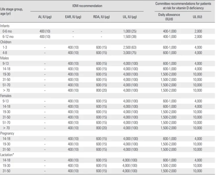
Table 2. Vitamin D intakes recommended by the IOM and the endocrine practice guidelines committee
Life stage group, age (yr)
IOM recommendation Committee recommendations for patients
at risk for vitamin D deficiency AI, IU (µg) EAR, IU (µg) RDA, IU (µg) UL, IU (µg) Daily allowance
(IU/d) UL (IU)
Infants
0-6 mo 400 (10) - - 1,000 (25) 400-1,000 2,000
6-12 mo 400 (10) - - 1,500 (38) 400-1,000 2,000
Children
1-3 - 400 (10) 600 (15) 2,500 (63) 600-1,000 4,000
4-8 - 400 (10) 600 (15) 3,000 (75) 600-1,000 4,000
Males
9-13 - 400 (10) 600 (15) 4,000 (100) 600-1,000 4,000
14-18 - 400 (10) 600 (15) 4,000 (100) 600-1,000 4,000
19-30 - 400 (10) 600 (15) 4,000 (100) 1,500-2,000 10,000
31-50 - 400 (10) 600 (15) 4,000 (100) 1,500-2,000 10,000
51-70 - 400 (10) 600 (15) 4,000 (100) 1,500-2,000 10,000
> 70 - 400 (10) 800 (20) 4,000 (100) 1,500-2,000 10,000
Females
9-13 - 400 (10) 600 (15) 4,000 (100) 600-1,000 4,000
14-18 - 400 (10) 600 (15) 4,000 (100) 600-1,000 4,000
19-30 - 400 (10) 600 (15) 4,000 (100) 1,500-2,000 10,000
31-50 - 400 (10) 600 (15) 4,000 (100) 1,500-2,000 10,000
51-70 - 400 (10) 600 (15) 4,000 (100) 1,500-2,000 10,000
> 70 - 400 (10) 800 (20) 4,000 (100) 1,500-2,000 10,000
Pregnancy
14-18 - 400 (10) 600 (15) 4,000 (100) 600-1,000 4,000
19-30 - 400 (10) 600 (15) 4,000 (100) 1,500-2,000 10,000
31-50 - 400 (10) 600 (15) 4,000 (100) 1,500-2,000 10,000
Lactation*
14-18 - 400 (10) 600 (15) 4,000 (100) 600-1,000 4,000
19-30 - 400 (10) 600 (15) 4,000 (100) 1,500-2,000 10,000
31-50 - 400 (10) 600 (15) 4,000 (100) 1,500-2,000 10,000
*Mother’s requirement 4,000-6,000 (mother’s intake for infant’s requirement if infant is not receiving 400 IU/d).
AI, adequate intake; EAR, estimated average requirement; IOM, Institute of Medicine; IU, international units; RDA, recommended dietary allowance; UL, tolerable upper intake level.
Although the amount of vitamin D recommended by the IOM will likely increase blood levels of 25(OH)D to 20 ng/mL this amount will not reach blood levels above 30 ng/mL. The Endocrine Soci- ety’s Practice Guidelines whose goal was to provide guidance to health care professionals on how to treat and prevent vitamin D deficiency recommended that children under one year should re- ceive 600 IUs of vitamin D daily and up to 1,000 IUs is safe. For all children over 1 year of age they should take 600 IUs of vitamin D daily and up to 2,000 IUs is safe. For adults it was recommended that they ingest 1,500-2,000 IUs of vitamin D daily and up to 10,000 IUs of vitamin D a day was considered to be safe (Table 2). How- ever for obese children and adults they may require at least 2-3 times more vitamin D because the vitamin D is sequestered in the body fat. Also patients with fat malabsorption syndromes or who are on medications such as glucococorticoids, anti-seizure medications, and AIDS medications that increase the destruction of 25(OH)D often require more vitamin D to satisfy their requirement [14,34].
An effective method to treat vitamin D deficiency is to give 50,000 IU vitamin D once a week for 8 weeks (equivalent to 7,000 IU/day).
To prevent recurrence 50,000 IU every 2 weeks is effective (equiv- alent to 3,300 IU/day). This treatment regimen has been effective for as long as 6 years without any toxicity [72].
Neither the IOM nor the Endocrine Society recommends screen- ing for 25(OH)D. Following guidelines for vitamin D supplementa- tion along with ingesting foods that contain vitamin D and sensible sun exposure should guarantee a healthy vitamin D status. How-
ever the Endocrine Society does recommend measuring serum 25(OH)D levels for patients at risk such as obese patients, patients on medications that enhance the catabolism of 25(OH)D as well as patients with a sensitivity to vitamin D including those with granu- lomatous disorders including sarcoidosis [24].
CONCLUSIONS
Humans have always depended on the sun to supply them with their vitamin D requirement. Evidence suggests that as our earliest ancestors migrated north and south of the equator it was necessary for them to have a mutation that resulted in the lack of skin pig- mentation in order for them to produce enough vitamin D for their skeletal health. Vitamin D deficiency in utero and during the first few years of life has devastating consequences for females because they have a flat pelvis and small pelvic outlet making child birthing difficult if not impossible. This would have been the driver in evo- lution for skin pigmentation to have devolved and why people liv- ing in Northern Europe are fair skinned.
The cause for the vitamin D deficiency pandemic is due to the misguided notion that sun exposure should be avoided from birth until death. Since there is very little vitamin D obtained from di- etary sources, sensible sun exposure is still the best and most reli- able source of vitamin D for children and adults. Concerns about
Serum vitamin D (ng/mL)
50
40
30
20
10
0 0 20 40 60
Time (hr)
Oral 25,000 IU Oral 10,000 IU 1 MED
Fig. 6. Comparison of serum vitamin D3 levels after a whole-body exposure (in a bathing suit; bikini for women) to 1 minimal erythemal dose (MED) of simulat- ed sunlight compared with a single oral dose of either 10,000 or 25,000 inter- national unit (IU) of vitamin D2. Reprinted from Holick MF copyright 1994 with permission.
Fig. 7. A schematic representation of the major causes for vitamin D deficiency and potential health consequences. Reprinted from Holick MF copyright 2010 with permission.
AODM, adult-onset diabetes mellitus; CHD, coronary heart disease; FEV1, forced expiratory volume in 1 second; HBP, high blood pressure; URI, upper respiatory infection; TB, tuberculosis; MS, multiple sclerosis; RA, rheumatoid arthritis.
sun exposure and skin cancer need to be put into perspective. Al- though nonmelanoma skin cancer is the most common cancer, it is easy to detect and easy to treat and not lethal if detected early [73].
These cancers occur because of chronic excessive exposure to sun- light which is why most of them occur on the most sun exposed areas including the face, back of the neck and top of the hands.
The recommendation for exposing arms, legs, abdomen and back when appropriate to suberythemal doses of sunlight 2-3 times a week during the spring, summer and fall is a good approach. Even the Australian Society of Dermatologists and the New Zealand Bone and Mineral Society now recognize that the abstinence message of avoiding all sun exposure has caused a vitamin D deficiency epi- demic in Australia and New Zealand and they now recommend that limited sun exposure can be a good source for vitamin D [74,75].
What is of course of most concern is the deadly skin cancer mel- anoma. It has been linked to sun exposure. Evidence suggests how- ever that most melanomas occur on the least sun exposed areas and occupational sun exposure decreases risk for melanoma [73].
The major risk factors include having a number of sun burning ex- periences as a child and young adult, large number of moles, being red headed and having a genetic predisposition. There is no evi- dence that sensible sun exposure increases risk for melanoma and may actually decrease risk.
The bottom-line is that everyone should be aware of their vita- min D status not only to maximize their bone health but also to re- duce risk of acute and chronic illnesses associated with vitamin D deficiency (Fig. 7). This can be accomplished by increasing intake of foods that naturally contain vitamin D or are fortified with vita- min D, taking a vitamin D supplement and obtaining a sensible amount of sun exposure at times of the day and season when the sun is able to produce vitamin D in the skin. As the Endocrine So- ciety recommends there is no downside to improving your vitamin D status. The only exceptions are people who have granulomatous disorders such as sarcoidosis and TB since they are more sensitive to vitamin D because of the macrophage converting 25(OH)D to 1,25(OH)2D in an unregulated fashion [34].
REFERENCES
1. Holick MF: Phylogenetic and evolutionary aspects of vitamin D from phy- toplankton to humans. In: Pang PK, Schreibman MP. Vertebrate Endocri- nology: Fundamentals and Biomedical Implications. pp7-43, San Diego, Academic, 1989
2. Holick MF: Vitamin D: A millenium perspective. J Cell Biochem 88:296-
307, 2003
3. Holick MF: Resurrection of vitamin D deficiency and rickets. J Clin Invest 116:2062-2072, 2006
4. Huldschinsky K: Heilung von Rachitis durch Kunstliche Hohensonne.
Deutsche Med Wochenschr 45:712-713, 1919
5. Hess AF, Unger LJ: The cure of infantile rickets by sunlight. JAMA 77:
39-41, 1921
6. Steenbock H: The induction of growth promoting and calcifying proper- ties in a ration by exposure to light. Science 60:224-225, 1924
7. Samuel HS: Infantile hypercalcaemia, nutritional rickets, and infantile scurvy in great Britain. A British Paediatric Association report. Br Med J 1:1659-1661, 1964
8. Taylor AB, Stern PH, Bell NH: Abnormal regulation of circulating 25- hydroxyvitamin D in the Williams syndrome. N Engl J Med 306:972- 975, 1982
9. Holick MF, MacLaughlin JA, Clark MB, Holick SA, Potts JT Jr, Ander- son RR, Blank IH, Parrish JA, Elias P: Photosynthesis of previtamin D3 in human skin and the physiologic consequences. Science 210:203-205, 1980
10. Tian XQ, Chen TC, Matsuoka LY, Wortsman J, Holick MF: Kinetic and thermodynamic studies of the conversion of previtamin D3 to vitamin D3 in human skin. J Biol Chem 268:14888-14892, 1993
11. Haddad JG, Matsuoka LY, Hollis BW, Hu YZ, Wortsman J: Human plasma transport of vitamin D after its endogenous synthesis. J Clin Invest 91:2552-2555, 1993
12. Matsuoka LY, Ide L, Wortsman J, MacLaughlin JA, Holick MF: Sunscreens suppress cutaneous vitamin D3 synthesis. J Clin Endocrinol Metab 64:
1165-1168, 1987
13. Clemens TL, Adams JS, Henderson SL, Holick MF: Increased skin pig- ment reduces the capacity of skin to synthesise vitamin D3. Lancet 1:74- 76, 1982
14. Holick MF: Vitamin D deficiency. N Engl J Med 357:266-281, 2007 15. Heaney RP, Recker RR, Grote J, Horst RL, Armas LA: Vitamin D(3) is
more potent than vitamin D(2) in humans. J Clin Endocrinol Metab 96:
E447-E452, 2011
16. Holick MF, Biancuzzo RM, Chen TC, Klein EK, Young A, Bibuld D, Re- itz R, Salameh W, Ameri A, Tannenbaum AD: Vitamin D2 is as effective as vitamin D3 in maintaining circulating concentrations of 25-hydroxyvi- tamin D. J Clin Endocrinol Metab 93:677-681, 2008
17. Gordon CM, Williams AL, Feldman HA, May J, Sinclair L, Vasquez A, Cox JE: Treatment of hypovitaminosis D in infants and toddlers. J Clin Endocrinol Metab 93:2716-2721, 2008
18. Christakos S, Dhawan P, Liu Y, Peng X, Porta A: New insights into the mechanisms of vitamin D action. J Cell Biochem 88:695-705, 2003 19. Holick MF: The D-lightful vitamin D for child health. JPEN J Parenter
Enteral Nutr 36(1 Suppl):9S-19S, 2012
20. Adams JS, Hewison M: Update in vitamin D. J Clin Endocrinol Metab 95:471-478, 2010
21. Bikle D: Nonclassic actions of vitamin D. J Clin Endocrinol Metab 94:26- 34, 2009
22. Holick MF: Vitamin D and health: evolution, biologic functions, and rec- ommended dietary intakes for vitamin D. Clin Rev Bone Miner Metab 7:2-19, 2009
23. Nagpal S, Na S, Rathnachalam R: Noncalcemic actions of vitamin D re-
ceptor ligands. Endocr Rev 26:662-687, 2005
24. Wang TT, Tavera-Mendoza LE, Laperriere D, Libby E, MacLeod NB, Na- gai Y, Bourdeau V, Konstorum A, Lallemant B, Zhang R, Mader S, White JH: Large-scale in silico and microarray-based identification of direct 1,25- dihydroxyvitamin D3 target genes. Mol Endocrinol 19:2685-2695, 2005 25. Liu PT, Stenger S, Li H, Wenzel L, Tan BH, Krutzik SR, Ochoa MT,
Schauber J, Wu K, Meinken C, Kamen DL, Wagner M, Bals R, Stein- meyer A, Zügel U, Gallo RL, Eisenberg D, Hewison M, Hollis BW, Ad- ams JS, Bloom BR, Modlin RL: Toll-like receptor triggering of a vitamin D-mediated human antimicrobial response. Science 311:1770-1773, 2006 26. Garland C, Shekelle RB, Barrett-Connor E, Criqui MH, Rossof AH, Paul O: Dietary vitamin D and calcium and risk of colorectal cancer: a 19-year prospective study in men. Lancet 1:307-309, 1985
27. Garland CF, Gorham ED, Mohr SB, Grant WB, Giovannucci EL, Lipkin M, Newmark H, Holick MF, Garland FC: Vitamin D and prevention of breast cancer: pooled analysis. J Steroid Biochem Mol Biol 103:708-711, 2007
28. Grant WB: An estimate of premature cancer mortality in the U.S. due to inadequate doses of solar ultraviolet-B radiation. Cancer 94:1867-1875, 2002
29. Li YC: Vitamin D regulation of the renin-angiotensin system. J Cell Bio- chem 88:327-331, 2003
30. Chiu KC, Chu A, Go VL, Saad MF: Hypovitaminosis D is associated with insulin resistance and beta cell dysfunction. Am J Clin Nutr 79:820-825, 2004
31. Holick MF: Vitamin D, sunlight and cancer connection. Anticancer Agents Med Chem. In press 2012
32. Lee JH, O’Keefe JH, Bell D, Hensrud DD, Holick MF: Vitamin D defi- ciency an important, common, and easily treatable cardiovascular risk fac- tor? J Am Coll Cardiol 52:1949-1956, 2008
33. Ross AC; Institute of Medicine (U.S.), Committee to Review Dietary Ref- erence Intakes for Vitamin D and Calcium: Dietary reference intakes for calcium and vitamin D. Washington DC, National Academies Press, 2011 34. Holick MF, Binkley NC, Bischoff-Ferrari HA, Gordon CM, Hanley DA,
Heaney RP, Murad MH, Weaver CM; Endocrine Society: Evaluation, treat- ment, and prevention of vitamin D deficiency: an Endocrine Society clini- cal practice guideline. J Clin Endocrinol Metab 96:1911-1930, 2011 35. Priemel M, von Domarus C, Klatte TO, Kessler S, Schlie J, Meier S, Proksch
N, Pastor F, Netter C, Streichert T, Püschel K, Amling M: Bone mineraliza- tion defects and vitamin D deficiency: histomorphometric analysis of iliac crest bone biopsies and circulating 25-hydroxyvitamin D in 675 patients.
J Bone Miner Res 25:305-312, 2010
36. Maxmen A: Nutrition advice: the vitamin D-lemma. Nature 475:23-25, 2011
37. Chapuy MC, Schott AM, Garnero P, Hans D, Delmas PD, Meunier PJ:
Healthy elderly French women living at home have secondary hyperpara- thyroidism and high bone turnover in winter. EPIDOS Study Group. J Clin Endocrinol Metab 81:1129-1133, 1996
38. Thomas MK, Lloyd-Jones DM, Thadhani RI, Shaw AC, Deraska DJ, Kitch BT, Vamvakas EC, Dick IM, Prince RL, Finkelstein JS: Hypovitaminosis D in medical inpatients. N Engl J Med 338:777-783, 1998
39. Holick MF, Siris ES, Binkley N, Beard MK, Khan A, Katzer JT, Petruschke RA, Chen E, de Papp AE: Prevalence of vitamin D inadequacy among postmenopausal North American women receiving osteoporosis therapy. J
Clin Endocrinol Metab 90:3215-3224, 2005
40. Murad MH, Elamin KB, Abu Elnour NO, Elamin, MB, Alkatib AA, Fatourechi MM, Almandoz JP, Mullan RJ, Lane, MA, Liu H, Erwin PJ, Hensrud DD, Montori VM: Interventions to raise vitamin D level and functional outcomes: a systematic review and meta-analysis. Poster session presented at: The Americal College of Preventive Medicine Annual Meet- ing, 2010 Feb 17-20, Arlington, VA.
41. Lee JM, Smith JR, Philipp BL, Chen TC, Mathieu J, Holick MF: Vitamin D deficiency in a healthy group of mothers and newborn infants. Clin Pe- diatr (Phila) 46:42-44, 2007
42. Hollis BW: Vitamin D requirement during pregnancy and lactation. J Bone Miner Res 22 Suppl 2:V39-V44, 2007
43. Choi HS, Oh HJ, Choi H, Choi WH, Kim JG, Kim KM, Kim KJ, Rhee Y, Lim SK: Vitamin D insufficiency in Korea: a greater threat to younger generation: the Korea National Health and Nutrition Examination Survey (KNHANES) 2008. J Clin Endocrinol Metab 96:643-651, 2011 44. Bodnar LM, Catov JM, Simhan HN, Holick MF, Powers RW, Roberts JM:
Maternal vitamin D deficiency increases the risk of preeclampsia. J Clin Endocrinol Metab 92:3517-3522, 2007
45. Merewood A, Mehta SD, Chen TC, Bauchner H, Holick MF: Association between vitamin D deficiency and primary cesarean section. J Clin Endo- crinol Metab 94:940-945, 2009
46. Camargo CA Jr, Rifas-Shiman SL, Litonjua AA, Rich-Edwards JW, Weiss ST, Gold DR, Kleinman K, Gillman MW: Maternal intake of vitamin D during pregnancy and risk of recurrent wheeze in children at 3 y of age.
Am J Clin Nutr 85:788-795, 2007
47. Holick MF, Lim R, Dighe AS: Case records of the Massachusetts General Hospital. Case 3-2009. A 9-month-old boy with seizures. N Engl J Med 360:398-407, 2009
48. Keller KA, Barnes PD: Rickets vs. abuse: a national and international epi- demic. Pediatr Radiol 38:1210-1216, 2008
49. Bishop NJ, Plotkin H: When is a fracture child abuse? Bone 23(5 Suppl 1):S458, 1998
50. Kim BG, Chang SK, Kim SM, Hwang JS, Jung JW: Dilated cardiomyop- athy in a 2 month-old infant: a severe form of hypocalcemia with vitamin d deficient rickets. Korean Circ J 40:201-203, 2010
51. Hyppönen E, Läärä E, Reunanen A, Järvelin MR, Virtanen SM: Intake of vitamin D and risk of type 1 diabetes: a birth-cohort study. Lancet 358:
1500-1503, 2001
52. Mohr SB, Garland CF, Gorham ED, Garland FC: The association between ultraviolet B irradiance, vitamin D status and incidence rates of type 1 dia- betes in 51 regions worldwide. Diabetologia 51:1391-1398, 2008 53. Ponsonby AL, McMichael A, van der Mei I: Ultraviolet radiation and au-
toimmune disease: insights from epidemiological research. Toxicology 181- 182:71-78, 2002
54. Munger KL, Levin LI, Hollis BW, Howard NS, Ascherio A: Serum 25-hy- droxyvitamin D levels and risk of multiple sclerosis. JAMA 296:2832- 2838, 2006
55. Merlino LA, Curtis J, Mikuls TR, Cerhan JR, Criswell LA, Saag KG; Iowa Women’s Health Study: Vitamin D intake is inversely associated with rheu- matoid arthritis: results from the Iowa Women’s Health Study. Arthritis Rheum 50:72-77, 2004
56. Garland CF, Garland FC, Gorham ED, Lipkin M, Newmark H, Mohr SB, Holick MF: The role of vitamin D in cancer prevention. Am J Public
Health 96:252-261, 2006
57. Holick MF: Calcium plus vitamin D and the risk of colorectal cancer. N Engl J Med 354:2287-2288, 2006
58. Yamaji T, Iwasaki M, Sasazuki S, Sakamoto H, Yoshida T, Tsugane S: As- sociation between plasma 25-hydroxyvitamin D and colorectal adenoma according to dietary calcium intake and vitamin D receptor polymorphism.
Am J Epidemiol 175:236-244, 2012
59. Lappe JM, Travers-Gustafson D, Davies KM, Recker RR, Heaney RP: Vi- tamin D and calcium supplementation reduces cancer risk: results of a ran- domized trial. Am J Clin Nutr 85:1586-1591, 2007
60. Wang TJ, Pencina MJ, Booth SL, Jacques PF, Ingelsson E, Lanier K, Ben- jamin EJ, D’Agostino RB, Wolf M, Vasan RS: Vitamin D deficiency and risk of cardiovascular disease. Circulation 117:503-511, 2008
61. Melamed ML, Muntner P, Michos ED, Uribarri J, Weber C, Sharma J, Raggi P: Serum 25-hydroxyvitamin D levels and the prevalence of periph- eral arterial disease: results from NHANES 2001 to 2004. Arterioscler Thromb Vasc Biol 28:1179-1185, 2008
62. Melamed ML, Michos ED, Post W, Astor B: 25-hydroxyvitamin D levels and the risk of mortality in the general population. Arch Intern Med 168:
1629-1637, 2008
63. Thomas GN, ó Hartaigh B, Bosch JA, Pilz S, Loerbroks A, Kleber ME, Fischer JE, Grammer TB, Böhm BO, März W: Vitamin D levels predict all-cause and cardiovascular disease mortality in subjects with the meta- bolic syndrome: the Ludwigshafen Risk and Cardiovascular Health (LU- RIC) Study. Diabetes Care 35:1158-1164, 2012
64. Gagnon C, Lu ZX, Magliano DJ, Dunstan DW, Shaw JE, Zimmet PZ, Si- karis K, Ebeling PR, Daly RM: Low serum 25-hydroxyvitamin D is asso- ciated with increased risk of the development of the metabolic syndrome at five years: results from a national, population-based prospective study (The Australian Diabetes, Obesity and Lifestyle Study: AusDiab). J Clin Endocrinol Metab 97:1953-1961, 2012
65. Deleskog A, Hilding A, Brismar K, Hamsten A, Efendic S, Östenson CG:
Low serum 25-hydroxyvitamin D level predicts progression to type 2 dia- betes in individuals with prediabetes but not with normal glucose toler- ance. Diabetologia 55:1668-1678, 2012
66. Hess AH: Collected writings. Vol 1. pp669-719, Springfield, Charles C.
Thomas, 1936
67. Urashima M, Segawa T, Okazaki M, Kurihara M, Wada Y, Ida H: Ran- domized trial of vitamin D supplementation to prevent seasonal influenza A in schoolchildren. Am J Clin Nutr 91:1255-1260, 2010
68. Sabetta JR, DePetrillo P, Cipriani RJ, Smardin J, Burns LA, Landry ML:
Serum 25-hydroxyvitamin d and the incidence of acute viral respiratory tract infections in healthy adults. PLoS One 5:e11088, 2010
69. Reis JP, D. vM, Miller ER 3rd, Michos ED, Appel LJ: Vitamin D status and cardiometabolic risk factors in the United States adolescent popula- tion. Pediatrics 124:e371-379, 2009
70. Kumar J, Muntner P, Kaskel FJ, Hailpern SM, Melamed ML: Prevalence and associations of 25-hydroxyvitamin D deficiency in US children:
NHANES 2001-2004. Pediatrics 124:e362-e370, 2009
71. Heaney RP, Davies KM, Chen TC, Holick MF, Barger-Lux MJ: Human serum 25-hydroxycholecalciferol response to extended oral dosing with cholecalciferol. Am J Clin Nutr 77:204-210, 2003
72. Pietras SM, Obayan BK, Cai MH, Holick MF: Vitamin D2 treatment for vitamin D deficiency and insufficiency for up to 6 years. Arch Intern Med 169:1806-1808, 2009
73. Kennedy C, Bajdik CD, Willemze R, De Gruijl FR, Bouwes Bavinck JN;
Leiden Skin Cancer Study: The influence of painful sunburns and lifetime sun exposure on the risk of actinic keratoses, seborrheic warts, melanocytic nevi, atypical nevi, and skin cancer. J Invest Dermatol 120:1087-1093, 2003
74. New Zealand Ministry of Health, Cancer Society of New Zealand, Acci- dent Compensation Corporation (N.Z.): Consensus statement on vitamin D and sun exposure in New Zealand. Wellington, Ministry of Health, 2012
75. Daly RM, Gagnon C, Lu ZX, Magliano DJ, Dunstan DW, Sikaris KA, Zimmet PZ, Ebeling PR, Shaw JE: Prevalence of vitamin D deficiency and its determinants in Australian adults aged 25 years and older: a na- tional, population-based study. Clin Endocrinol (Oxf) 77:26-35, 2012 76. Holick MF, Binkley NC, Bischoff-Ferrari HA, Gordon CM, Hanley DA,
Heaney RP, Murad MH, Weaver CM: Guidelines for preventing and treat- ing vitamin D deficiency and insufficiency revisited. J Clin Endocrinol Metab 97:1153-1158, 2012

![Fig. 3. Metabolism of 25-hydroxyvitamin D [25(OH)D] to 1,25 dihydroxyvitamin D 1,25(OH)2D for non-skeletal functions. When a monocyte/macrophage is stim-ulated through its toll-like receptor 2/1 (TLR2/1) by an infective agent such as Mycobacterium tube](https://thumb-ap.123doks.com/thumbv2/123dokinfo/5226707.125246/5.892.459.822.114.553/metabolism-hydroxyvitamin-dihydroxyvitamin-skeletal-functions-macrophage-infective-mycobacterium.webp)