대한빙사선의학회지 1997: 36: 451-453
Paraquat중독 폐 병변의 장기간 추적 HRCT소견 :
1 예 보고l
김 영 통 · 홍세용 2 . 김 일영
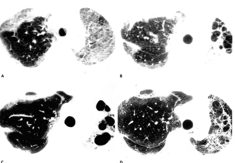
치 사량의 Paraquat를 음독한 35세 여 자 환자에 서 16개 월 동안의 임 상소견과 함께 폐 병 변 의 HRCT소견의 변화를 보고하고자 한다. HRCT소견은 음독 7일후는 양측에 마쇄음영이 보였고, 음독 6주후에는 마쇄음영을 보였던 부위가 폐경화와 냥성변화를 보였고, 음독 5개 월후는 폐용적이 더 감소하였고, 음독 16개월후는 폐용적의 증가와 정상 폐음영이 보였고,
전에 보이던 낭성병변은 줄었다.
Paraquat (Gramoxone
@,1,1
’-dimethyl-
4,4’강ipyridylium dichloride) 는 세계에서 가장 흔히 사용되는 제초제이다.Paraquat는 20%용액 10ml정도로 인체에 치명적이며, 폐, 간,
신장, 심근과 부신 펴질에 손상을 일으킨다. Paraquat에 의한 사망 원인은 저산소증에 의한 호흡 부전이 약 70%로 가장 많 기 때문에 (1), 흉부단순촬영과 흉부 HRCT소견은 환자의 예 후에 중요하다. Paraquat 에 의한 시간 변화에 따른 HRCT소 견이 기술된바 있으나 (2) , 한 환자에서 HRCT소견의 연속적인 변화는 보고 된 바 없다. 이에 저자들은 치사량의 Paraquat를 음독한 환자에서 16개월 동안 4차례의 추적 HRCT를 경험하 였기에 임상소견과함께 보고하는바이다.
증례보고
35세 여자 환자가 자살 목적으로
20%
Paraquat용액을 여섯 모금을 음독한후 2시간 후에 본원 응급실로 도착하였다. 가족 력과 과거력상 특이병력이 없었다. 이학적소견상 급성병색이었 고, 의식은 명료하였다. 호흡음은 정상이었고, 싱잡음은 없었다.혈압은
130
/80mmHg, 맥박은92/ min
, 호흡은 25/min이었 다. 내원당시 시행한 뇨 Paraquat의 측정상 강양성(+++)
을 보였으며, 혈중
Paraquat
농도는 2133ng/ml
였다. 입원 당 시 동액 가스분석은PH 7.210
,P02 78.9mmHg
,PC02 28.2 mmHg , HC03 1
1.3mMol
/L , 0
2 sat 93.3%였다. 한달 후의 동백 가스분석은PH 7.359
,P02 45.3mmHg
,PC02 45.3 mmHg
,HC03 24.9mMol
/L
,0
2 sat 79.3% 였고, 폐기능검사 는vital capacit
y (VC) 가0.81L
(예측치 : 2.90L) 였다.Vital
capacity는 음독 8개월후는 1.3 1L였고, 음독 167H 월후는 1.
93L
로 증가하였다. HRCT소견은 음독 7일후는 양측폐야에 마쇄음
l 순천향대 학교 의 과대 학 진단방사선과학교실 2순천향대학교 의과대학 내과학교실
이 논문은 1996년 10월 22일 접수하여 1996년 12월 7일에 채택되었음
영이 보였고(Fig.
1A)
, 음독 6주와 5개월후는 폐용척의 감소와 함께 마쇄음영을 보였던 부위가 폐경화와 낭성변화를 보였고(F
ig.1B
,lC)
, 음독 167H 월에는 좌측 폐용적의 증가와 정상 폐읍영이 보였고, 이전에 보이던 낭성병변은 줄었다 (Fig. 1D).
환자는 흡입산소분압을 낮추면서, 혈액투석
(hemoperfusion)
과 항산화제(Antioxidant)
인Superoxide dismutase
등을 병 행하면서 치료하였다. 환자는 8주간 업원 치료후 계속 외래로 추적 중이며, 환자의 증상은 많이 호전되 었으나, 운동시 호흡곤 란은아직도남아있다.고 찰
우리 나라성인의 사망원인중사고와중독에 의한사망은순 환기 질환, 신생물에 이어, 3번째 사언으로 이중
Paraquat
중 독에 의한 사망은 단일 질환으로 매우 높은 사망 원인이다 (3).우리 나라에서는 Paraquat는 Gramoxone으로 알려져 있고,
24.5%의 푸른 용액으로 시판된다. Paraquat의 초기 중독 증세 는소화기 계통점막의 부식 효과및 간손상,특히 신세뇨관손 상과 더불어 요독증세가 나타나고 점진적으로 폐손상이 있어 호흡 부전에 이르는 유독한 약물로
20
% 용액 한 모금 흑은 그 이하에서도 치명적인 결과가 올 수 있다. Paraquat는 호기성 생물체내에서 용해되어nicotinamide
-adenine dinucleotide(NADP) 의 환원을 감소시켜
superoxide
및peroxide radical
을 형성하여 세포막 파괴를 일으키고 조직에 변화를 초래하며,
산소 분압이 높고 활성화된 산소들의 제거 효소계가 상대적으 로 적은 폐포세포가 가장 심한 비가역적 손상을 받는다 (4, 5).
그래서 Paraquat중독의 최근 치료방법으로
free
radical의 생 성을 억제시키기 위해 폐포세포내의superoxide dismutase
농 도를 높이는 항산화제를 사용하여 좋은 효과를 보았다고 한다(6).
Paraquat에 의한 폐병변의 병리 소견은 초기에는 폐포내 혹은 간질에 부종과 출혈,hyaline
막 형성, 그리고 폐포세포의껴 잉
A
C
•
김영통 오I
:
Paraquat중독 펴|병변의 장기간 추적 HRCT소견B
증식 등이 고 점 차 섬 유화가 악화된다 (4 ,
5
,7).
Paraquat에 의 한 폐병변의 방사선학적 소견은 음독후 7일 이내 폐 경화를 보 이고, 그후 7일 동안 낭성 흑은 선상의 음영을 보이고, 음독 l달 후에 별집 모양 (honeycombing) 이 보인다고 하였다 (8). Lee등 (2) 도 음독 7일 이내에 마쇄음영을 보이고, 이 병변은 기관지 확장증과 불규칙한 선상 음영을 동반한 폐경화로 변화한다고 보고하였다. 저자들의 증례도 음독후 일주일 내에 폐포내 혹은 간질에 부종과출혈에 의한마쇄음영을보였고,이 부위가폐경 화를 거쳐 냥성변화와 폐용적의 감소등의 폐섬유화소견을 보였 다. 그러나 음독 167~월후에 폐용척의 증가와 낭성변화의 호전 을 보였고, 폐기능검사도 호전되였다.결론적으로 Paraquat에 의한 폐병변의 HRCT소견은 음독 일주일에 마쇄음영으로 보였고, 이 부위가 폐경화, 낭성변화와
7
% 4
D
Fig. 1. A. HRCT scan obtained 7days after Paraquat intake at the 1eve1 of aortic arch shows patchy areas of ground-g1ass opacity in both 1ungs.
B. Follow-up HRCT scan obtained 6weeks after Paraquat intake at the same 1eve1 to (A), shows initia1 areas of ground-g1ass opacity in the 1eft upper 10be have changed into areas of consolidation and irregu1ar lines with multip1e cystic air spaces. Vo1ume of 1eft 1ung decreased marked1y. Note patchy areas of consolidation in right upper 1obe.
c.
Follow-up CT scan obtained 5 months after Paraquat intake at the same 1eve1 shows that cystic air spaces are 1arger and the vo1ume of the 1eft 1ung is smaller.D. Follow-up HRCT scan obtained 16 months after Paraquat intake at the same 1eve1 shows that vo1ume of 1eft 1ung have increased. Extent of cystic 1esions is smaller than (C) with multip1e irregu1ar lines.
폐용적의 감소를 점차 보이다가, 음독 16개월에는 폐용적의 증 가와 이전에 보이던 낭성병변이 감소하였다. 시간경과에 따른 폐용적의 증가와 낭성병변의 호전은 Paraquat에 의한 폐병변 이 과연 비가역적 인가하는 의문점을 갖게 하며, 최근 들어 치료 가 발달되어 과거에는 사망했을 환자들이 생존함에 따라 (6) , 더 많은 보고와 연구가 되어야할것으로 사료된다.
~ 71 무등4
口 •
l 이재석, 정미경, 김태준등 Paraquat( Gramoxone@) 중독환자의 임상적 고찰 대한내과학회지 1994; 47: 93-100
2. Lee SH, Lee KS, Ahn 1M, Kim SH, Hong SY. Paraquat poisoning of the lung: thin-section CT findings. Radiology 1995; 195 ‘ 271-274
대한밤사선의학회지
1997: 36 : 451-453
3 경제기획원 조사통계국: 1988년 사망원인 통계 연보-인구동 태 신고에 의한 집계 경제기획원 조사통계국, 1989
4. 이경수, 김의한, 이병호, 검건상 가토폐의 Paraquat중독 고해 상 전산화단층촬영 소견과 병리소견의 비교연구 대한방사선
Scavenger를 이용한 치료 경험 대한내과학회지 1996;51:
99-107
학회지 1992; 28: 865-874
7. Smith LL. Mechanism of paraquat toxicity in lung and its rel- evance to treatment. Human Toxico/ 1987: 6; 31-36
8. 1m JG, Lee KS, Han MC, Kim SJ, Kim IO. paraquat poisoning Findings on chest radiography and CT in 42 patients. AJR 1991;
157: 687-701 5. Thurlbeck WM, Thurlbeck SM. Pulmonary effects of Paraquat
poisoning. Chesl 1976; 69 : 276-280
6 홍세 용, 양동호, 김 영 통 Paraquat 중독 환자에 서 Free Radical
J
Korean Radiol Soc 1997: 36 : 451 - 453
Paraquat-Induced Pulmonary Lesions:
HRCT Findings in Long-Term Follow-up: A Case Repore
Young Tong Kim
,M.D.
,Sae Yong Hong
,M.D. 2
,Il Young Kim
,M.D.
1 Department of Diagnostic Radiology, College of Medi다nζ Soonchunhyang University 2Department of Internal Medicine, College of Medicine, Soonchunhyang University
We illustrate serial HRCT findings over a 16-month period in a 35-year-old woman who had ingested paraquat. Initial areas of ground-glass opacity changed into areas of multiple air cysts on follow up scan obtained five months after ingestion. A further follow-up scan obtained 16 months after ingestion showed improvement
,with increased lung volume and normalized lung architecture.
Index Words : Paraquat Lung
,CT
Computed tomograph y(CT) , high-resolution
Address reprint requests to: Young Tong Kim, M.D., Department of Radiology, Soonchunhyang University HospitaL 23-20, Bongmyung-Dong, 330-100, Chunan, Korea Tel. 82-417-559-2101 Fax. 82-417-574-6265
…
m
국제 학술대회 일정표[ 1 J
• 3rd International Conference on Magnetic Reson- ance Imaging (1997/03/12-16)
venue: The Regent Hotel Melbourne, Australia
contact: Ms. Probati Milton, B. Sc., Convention Professionals, P.O. Box 4031, Balwyn East, Victoria 3129, Australia (tel: 61 - 3 -98990368; fax: 61 -3 -98990368)
• Annual Meeting American Institute of Ultrasound in Medicine (1997/03/23-26)
venue: San Diego, USA
contact: Jenny Clark, AIUM, Suite 100,
14750 Sweitzer Lane, Laurel, MD 20707-5906, USA‘ (tel: 1- 301-4984100; fax: 1-301-4984450)
• Second Congress Asian & Ocenian Soc. of Neuro-/
Head & Neck Radiology (1997/03/24- 27)
venue: Taipei Veterans Gen. Hosp Taipei, Taiwan, R. O. C contact: Dept. of Radiology, Veterans Gen. Hospital, 201,
Sec. 2, Shih-Pai Rd., Taipei, Taiwan 11217, R. O. C.
(tel : 886 -2 - 8757357; fax: 886 - 2 - 8733643)
• Course: Neuroradiology Update 1997 (1997/03/24-28)
venue: undetermined, USA‘
contact‘ Janice Ford, Hosp. of the Univ. of PA,
3400 Spruce Street, Philadelphia, PA 19104, USA (tel: 1 -215 -6626904; fax: 1 -215 -3495925)
• Course: Radiology for MRCP (1997/04/00-00) venue: Wolfson Conference Centre London, United
Kingdom
contact 、>,IolfsonConference Centre, Hammersmith Hospital, Du Cane Road, London WI2 ONN, United Kingdom‘ (tel:44- 181-7403245; fax:44-181-7404950)
• Course on Radiation Protection (1997/04/00-00) venue: Wolfson Conference Centre London, United
Kingdom
contact: Wolfson Conference Centre, Hammersmith Hospital, Du Cane Road, London WI2 ONN, United Kingdom.
(tel: 44 -181 - 7403245; fax’44-181-7404950)
• 8th Annual Meeting European Society of Pediatric Urology (1997/04/03-05)
venue: Pontificia Univ. Urban. Rome, Italy
contact: ESPU Meetings, Central Secretariat, 42 Devonshire Road, Cambridge CBI 2BL, United Kingdom (tel: 44 - 1223 -323437; fax: 44 -1223 -460396)
• 56th Soc. Assembly of Japan Radiological Soc./53rd Soc. Assembly of JSRT (1997/04/03- 06)
venue: Pacifico Yokohama Yokohama city, Japan contact: JMCP, Kitaotemachi Bldg., 1-7-6,
Chiyoda-ku, Tokyo 101, Japan
(tel: 81 - 3 - 52810456; fax: 81 -3 - 52810457)
• 3rd International Conference of Nuclear Cardiology (1997/04/06 - 09)
venue: Florence, Italy
contact: OIC, Via A. La Marmora 24, Firenze, Italy 50121
(tel: ; fax: 39 - 55 - 570227)
• 3rd Asian-Pacific Congress of Cardiovascular and Interventional Radiology (1997/04/06 -11)
venue: World Congress Centre Melbourne, Australia contact: Mrs .. Davies, APCCVIR, Radiology Department,
The Royal Melbourne Hosp., Parkville, Victoria 3050, Australia. (tel: 61 - 3 -93427293; fax: 61 -3 -93428369)
• 5th Scientific Meeting and Exh. Int. Soc. For Mang. Resonance in Medicine (1997/04/12-18)
venue: Vancouver Trade & Conv. Ctr. Vancouver, BC, Canada
contact: ISMRM Central Office, 2118 Milvia Street, Suite 201, Berkeley, CA 94704, USA (tel: 1 - 510-8411899; fax: 1 -510-8412340)
• 20th Annual Meeting of the Society for Computed Body Tomography and MR (1997/04/14-19)
venue: Grand Hyatt Washington, DC, USA contact: SCBT/MR, c/o Matrix Meetings,
P.O. Box 1103, Rochester, MN 55903-1026, USA (tel: 1 - 507 - 2885620; fax: 1 - 507 - 2880014)
• Sao Paulo Radiology Meeting (1997/04/18 -21) venue: Anhembi Convention Centre Sao Paulo, Brazil contact: Regina Carvalho, Soc. Paulista Radiologia,
Av. Paulista 491,40 andar, Cjs. 41 e 42, CEP 01311-909 Sao Paulo, Brazil.
(tel: 55 -11 -2843988; fax ‘ 55 -11 -2843152)
• 2nd Annual Angio/lnterventional Review Course (1997/04/19 - 20)
venue: Orlando, Florida, USA contact: Ryals & AssQciates, Inc.,
P.O. Box 1925, Roswell, GA 30077-1925, USA (tel: 1-770-6419773; fax’ 1 -770 - 5529859)
• 9th Annual Radiology Review Course: What You Need to Know (1997/04/20- 25)
venue: Orlando, Florida, USA contact: Ryals & Associates, Inc.,
P.O. Box 1925, Roswell, GA 30077-1925, USA.
(tel: 1- 770- 6419773; fax: 1-770-5529859)
• 2nd Annual Mammography Review Course (1997/04/25-27)
venue: The Buena Vista Palace Orlando, Florida, USA contact: Ryals & Associates, Inc.,
P.O. Box 1925, Roswell, GA 30077-1925, USA (tel ‘ 1-770- 6419773; fax‘ 1 -770 -5529859)