대한소화기학회지 1999;34:50 - 55
6)
Introduction
In human beings, cholestasis is observed in vari- ous diseases such as late viral hepatitis, carcinoma of
접수: 1998년 5월 29일, 승인: 1998년 11월 29일 연락처: Kyo-Cheol Mun, M.D., Ph.D., Department of Bio chemistry, Keimyung University School of Medicine, 194 Dong San Dong, Taegu, 700-712, Korea
Tel: (053) 250-7786, Fax: (053) 252-1605
the bile duct, gallstones in the bile duct, primary biliary cirrhosis, sclerosing cholangitis, biliary atresia, alcoholic hepatitis, and drug-induced liver disease.1 In rats, cholestasis can be induced by common bile duct (CBD) ligation which causes biochemical and morphological abnormalities in the liver such as inflammation, necrosis, fatty change, biliary hyperplasia, fibrosis, and cirrhosis.2-4
Most of ethanol absorbed in the body is metabo- lized in the liver;5,6 it is oxidized to acetaldehyde and
A ct iv it ie s of H e p a t ic E n zy m e s f or E t h a n ol M e t a b olis m in C h ole s t a t ic Ra t s w it h A c u t e E t h a n ol In t ox ica t ion
Kyo Cheol Mun, M.D., Chun Sik Kwak, Ph.D. and Yong Ho Jo, M.D.*
Department of Biochemistry, Keimyung University School of Medicine, Taegu;
Department of Biochemistry*, Kyung Hee University College of Medicine, Seoul, Korea
담관결찰 흰쥐에서 E tha nol 투여가 간의 E than ol 대사효소들의 활성도에 미치는 영향
계명대학교 의과대학 생화학교실, 경희대학교 의과대학 생화학교실*
문교철ㆍ곽춘식ㆍ조용호*
목적: 간질환이 있는 경우에 음주를 한다면 ethanol을 분해하는 각종 효소의 변동이 초래되어 ethanol 대 사 경로의 변동이 초래될 것이다. 이때 관여하는 ethanol 대사 관계 효소의 변화를 파악하고자 하였다 대상 및 방법: 흰쥐에게 담관을 결찰하여 담즙울체를 야기시킨 후 ethnol을 투여하고 간에서 ethanol 대 사에 관여한다고 알려진 alcohol dehydrogenase (ADH)와 microsomal ethanol oxidizing system (MEOS) 그 리고 catalase의 활성을 측정하였다. 결과: 정상 쥐에게 ethanol을 투여했을 경우는 간에서 ADH만 증가를 보였다. 담관을 결찰했을 경우에는 ADH와 catalase의 활성은 감소되고 MEOS의 활성은 증가되었다. 담 관을 결찰한 후 ethanol을 투여하였을 경우에는 담관만 결찰한 경우보다는 ADH와 catalase가 증가되었으 나 ADH의 경우는 정상군보다 활성이 낮았고 MEOS는 담관만 결찰한 군과 비슷한 활성을 보여주었다 결론: 정상 간에서 ethanol의 대사는 주로 ADH에 유도되어 대사되며, 담즙울체가 있는 경우에 ethanol이 투여되면, 간에서의 MEOS의 유도에는 한계가 있으며 또한 ADH의 유도만으로는 ethanol을 모두 대사시 킬 수 없어 부대사 경로의 하나인 catalase가 유도된다고 생각된다. (대한소화기학회지 1999;34:50 - 55) 색인단어: 담관결찰, Alcohol dehydrogenase, Catalase, Microsomal ethanol oxidizing system
54 The Korean Journal of Gastroenterology : Vol. 34, No. 1, 1999
then, metabolized further to acetate or excreted.5,6 The process of ethanol oxidation in the liver is mediated by alcohol dehydrogenase (alcohol: NAD+
oxidoreductase, EC 1.1.1.1, ADH), microsomal ethanol oxidizing system (MEOS) and catalase (hydrogen peroxide oxidoreductase, EC 1.11.1.6).5,7,8 The activi- ties of these alcohol-metabolizing enzymes can be changed in case of extrahepatic cholestasis. There fore, alcohol drinking under cholestasis can induce alteration in the activities of alcohol metabolizing enzymes.
To investigate the effects of alcohol ingestion on the alcohol-metabolizing enzymes in cholestatic liver, the activities of ADH, MEOS, and catalase were measured in rats by means of acute ethanol intoxi cation after CBD ligation.
S ubje ct a nd Me thods
1. A n im a ls
Thirty normal male rats of the Sprague-Dawley strain, weighing between 280 and 320 grams, were used in this experiment. All 30 rats were equally divided into 6 groups (group 1, group 2, group 3A and 3B group 4A and 4B). The group 1 were norma controls. The group 2 was CBD ligation group. The rats in this group were sacrificed at 14th day after CBD ligation. In group 3A and 3B, ethanol intoxication was performed. The rats were intoxicated with 4 g of ethanol per kg of body weight according to the method of Liu et al.9 Then, they were sacrificed 1.5 hours (Group 3A) and 24 hours (Group 3B) after acute ethanol intoxication. In the remaining two groups (group 4A and 4B), both CBD ligation and ethanol intoxication were carried out. At the 14th day after CBD ligation, the rats were intoxicated with 4 g of ethanol per kg of body weight according to the method of Liu et al.9 Then, they were sacrificed 1.5 hours (Group 4A) and 24 hours (Group 4B) after acute ethanol intoxication. All animals were main-
tained on commercial pellets purchased from Sam Yang Food Co.
The rats were fasted for 12 hours prior to sacrifice and they were anesthetized lightly with ether for surgery or being sacrificed. The CBD was exposed through a middle line incision. After double ligation of CBD, the mid point was cut.
2. C he m ica ls
-Nicotinamide adenine dinucleotide ( -NAD+, from yeast, grade III), -nicotinamide adenine dinu- cleotide reduced form ( -NADH, from yeast, grade III), N,N-dimethyl-p-nitrosoaniline, glycine, n-buta- nol, alcohol dehydrogenase of equine liver, -nicoti- namide adenine dinucleotide phosphate (NADP+, from yeast, Sigma grade), nicotinamide, glucose-6-phos- phate, semicarbazide-HCl, catalase of bovine liver, and bovine albumin were purchased from Sigma Chemical Co. (U.S.A.). Ethanol (99-100%) was pur- chased from E Merk Co. All other chemicals were of the highest purity available commercially.
3. E n zy m e s a m ple pr e pa r a t ion s
The rats were anesthetized lightly with ether, blood was collected from abdominal aorta, and liver was perfused through the portal vein with 0.25 M sucrose. The liver obtained was rinsed in 0.25 M sucrose. Then, the surface was wiped and dried.
Enzyme samples were obtained by centrifugation using density gradient made of sucrose.10
All the procedures described above were per- formed at 2-4℃.
4. E n zy m e a s s a y s
The assay of cytosolic ADH activity was carried out with ethanol and NAD+ as substrates according to the method of Koivula et al.11 Cytosolic ADH activity was expressed as nmol of NADH produced per minute per mg of protein.
The assay of serum ADH activity was carried out
문교철 외 2인. 담관결찰 후 Ethanol 투여시 Ethanol 대사효소들의 활성도 55
using n-butanol and N, N-dimethyl-p-nitrosoaniline as substrates according to the method of Skursky et al.12 Serum ADH activity was expressed as μmol of butyraldehyde produced per minute per mg of protein.
The assay of MEOS activity was carried out according to the method of Lieber and DeCarli.13 MEOS activity was expressed as nmol of acetal- dehyde produced per minute per mg of protein.
The assay of catalase activity was carried out using hydrogen peroxide as a substrate according to the method of Nelson and Kiesow.14 Catalase activity was expressed as μmol of hydrogen peroxide re- duced per minute per mg of protein.
5. S t a t is t ica l a n a ly s is
The values were expressed as mean±S.D. Sta- tistical evaluation of significant difference between mean values was performed with the Student' s t-test.
P values less than 0.05 were considered significant.
The PC-SAS version 6.04 program was used.
Re s ults
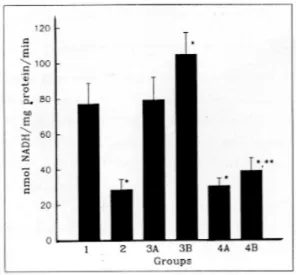
Hepatic ADH activity was lower in group 2 than in group 1 (Fig. 1). It was higher in groups 3A and 3B than in group 1, and also higher in group 4B than in group 2 (Fig. 1).
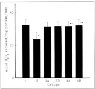
Hepatic MEOS activity was higher in group 2 than in group 1 (Fig. 2). There was no significant dif ference in the activities between group 4A, group 4B and group 2 (Fig. 2).
Hepatic catalase activity was lower in group 2 than in group 1 (Fig. 3). Hepatic catalase activity was higher in groups 4A and 4B than in group 2 (Fig. 3).
Serum ADH activity was higher in group 2 than in
Fig. 1. Hepatic ADH activity. Values are mean±S.D. of 5 rats in each group. group 1, Normal rats; group 2, CBD ligated rats; groups 3A and 3B, The rats were sacrificed at the 1.5 hours (group 3A) and 24 hours (group 3B) after acute ethanol intoxication; group 4A and 4B, The rats were sacrificed at the 1.5 hours (group 4A) and 24 hours (group 4B) after acute ethanol intoxication with CBD ligation.
* p<0.001 compared to group 1.
** p<0.05 compared to group 2.
Fig. 2. Hepatic MEOS activity. Values are mean±S.D. of 5 rats in each group. group 1, Normal rats; group 2, CBD ligated rats; groups 3A and 3B; The rats were sacrificed at the 1.5 hours (group 3A) and 24 hours (group 3B) after acute ethanol intoxication; group 4A and 4B, The rats were sacrificed at the 1.5 hours (group 4A) and 24 hours (group 4B) after acute ethanol intoxication with CBD ligation.
* p<0.05 compared to group 1.
56 대한소화기학회지 : 제 34 권 제 1 호 1999
group 1 (Fig. 4). It was higher in groups 4A and 4B than in group 2 (Fig. 4).
Dis cus s ion
It is well known that 98 percent of ethanol absor bed in normal human body is metabolized in the liver and alcohol dehydrogenase plays a role in this procebs.5,6 In our experiment, only the activity of ADH among the three ethanol metabolizing enzymes was increased after the acute ethanol intoxication in normal rats (groups 3A and 3B, Fig. 1). This result indicates that ethanol is metabolized mainly by ADH which is the major catalyst in the process of ethano oxidation5,6 in case of the normal subject who drinks alcoholic liquors.
CBD ligation in rats causes morphological abnor- malities in the liver such as inflammation, necrosis,
fatty change, biliary hyperplasia, fibrosis, cirrhosis,2-4 and also causes biochemical changes including enzy- me activities.15 In this experiment, the activity of MEOS among the three ethanol metabolizing enzy- mes was increased in the CBD ligated group (group 2, Fig. 2), but the activities of ADH and catalase were decreased (Fig. 1, 3). These results suggest that the homeostasis of ethanol metabolism under chole- stasis is maintained by induction of MEOS despite of decreased activities of ADH and catalase.
In the case of cholestasis combined with acute ethanol intoxication (groups 4A and 4B), the activi- ties of ADH and catalase were higher than in chole stasis group without ethanol intoxication (Fig. 1, 3).
However, cytosolic ADH activity in groups 4A and 4B was still lower than that in group 1. These results suggest that if the subject with cholestasis is exposed to a great quantities of alcoholic liquors, both ADH
Fig. 3. Hepatic catalase activity. Values are mean±S.D.
of 5 rats in each group. group 1, Normal rats; group 2, CBD ligated rats; groups 3A and 3B, The rats were sacrificed at the 1.5 hours (group 3A) and 24 hours (group 3B) after acute ethanol intoxication; group 4A and 4B, The rats were sacrificed at the 1.5 hours (group 4A) and 24 hours (group 4B) after acute ethanol intoxication with CBD ligation.
* p<0.01 compared to group 1.
** p<0.05 compared to group 2.
Fig. 4. Serum ADH activity. Values are mean±S.D. of 5 rats in each group. group 1, Normal rats; group 2, CBD ligated rats; groups 3A and 3B, The rats were sacrificed at the 1.5 hours (group 3A) and 24 hours (group 3B) after acute ethanol intoxication; group 4A and 4B, The rats were sacrificed at the 1.5 hours (group 4A) and 24 hours (group 4B) after acute ethanol intoxication with CBD ligation.
* p<0.001 compared to group 1.
** p<0.05 compared to group 2.
Mun et al. Activities of Hepatic Enzymes for Ethanol Metabolism in Cholestasis with Ethanol Intoxication 57
and catalase play roles in the process of ethanol oxidation. However, ADH can not metabolize all the alcoholic liquors absorbed under cholestasis, thus catalase is activated.
Increase of serum ADH activity was greater in group 4A and 4B than in group 2 (Fig. 4). These results suggest that large amount of ADH in the liver flows into the blood. Such flow is caused by in creased damage to the liver under the alcohol intoxi cation combined with cholestasis.
S umma ry
Background/Aims: The effects of alcohol inges- tion on the alcohol metabolizing enzymes of the liver under the cholestasis were investigated. Methods:
The activities of hepatic alcohol dehydrogenase (ADH), microsomal ethanol oxidizing system (MEOS), and catalase were measured in rats with extrahepatic cholestasis induced by common bile duct (CBD) ligation combined with acute ethanol intoxication.
Results: The activity of ADH was increased under the acute ethanol intoxication in normal rats. In the CBD ligated group, the activity of MEOS was in creased. When acute ethanol intoxication was com- bined with cholestasis, the activities of ADH and catalase were increased. Conclusions: These results suggest that if subjects with cholestasis drink a large quantity of ethanol, not only ADH but also catalase is activated because induction of MEOS activity is limited and ADH level is lower under the acute ethanol intoxication combined with cholestasis than under normal condition.
Key Words: Alcohol intoxication, Extrahepatic chole- stasis, Alcohol dehydrogenase, Cata- lase, Microsomal ethanol oxidizing system
Re fe re nce s
1. Eddleston ALWF. Liver and biliary tract disease. In Souhami RL, Moxham J, eds. Textbook of medicine 2nd ed. New York: Churchill Livingstone, 1994 615-656.
2. Kountouras J, Billing BH, Scheuer PJ. Prolonged bile duct obstruction: a new experimental model fo cirrhosis in the rats. Br J Exp Pathol 1984;65 305-311.
3. Chang DS, Kwak JS, Sohn TJ. An ultrastructura study on the proliferative changes of bile ductules after ligation of common bile duct. Kyungpook Univ Med J 1987;28:113-117.
4. Kim HS, Park JY, Kim EY, Kwak KS, Choi YH Chung JM. Morphologic change of hepatocytes induced by common bile duct ligation. Kor J Intern Med 1989;36:459-470.
5. Murray RK. Biochemical case histories. In: Murray RK, Granner DK, Mayer PA, Rodwell VW. eds Harper' s biochemistry. 24th ed. East Norwalk:
Appleton and Lange, 1996;814-828.
6. Chun BG. Pharmacological action of ethanol. J Korean Med Assoc 1998;41:6-14.
7. Seitz HK, Poschl G. The role of gastrointestina factors in alcohol metabolism. Alcohol Alcohol 1997;32:543-549.
8. Porodenko VA. The status of the ethanol oxidizing enzyme systems in fatal alcohol poisonings. Sud Med Ekspert 1997;40:15-18.
9. Liu SJ, Ramsey RK, Fallon HJ. Effect of ethanol o hepatic microsomal drug metabolizing enzymes in the rat. Biochem Pharmacol 1975;24:369-378.
10. Kwak CS, Kwak JS. Cell fractionation method o the rat liver. Keimyung Univ Med J 1986;5:45-53.
11. Koivula T, Koivusalo M, Lindros KO. Liver aldehyde and alcohol dehydrogenase activities in ra strains genetically selected for their ethanol pre ference. Biochem Pharmacol 1975;24:1807-1811.
12. Skursky L, Kovar J, Stachova M. A sensitive pho tometric assay for alcohol dehydrogenase activity in
58 The Korean Journal of Gastroenterology : Vol. 34, No. 1, 1999
blood serum. Anal Biochem 1979;99:65-71.
13. Lieber CS, DeCarli LM. Ethanol oxidation by hepatic microsomes: adaptive increase after ethanol feeding. Science 1968;162:917-918.
14. Nelson DP, Kiesow LA. Enthalpy of decomposition of hydrogen peroxide by catalase at 25℃ (with
molar extinction coefficients of H2O2 solutions in the UV). Anal Biochem 1972;49:474-478.
15. Mun KC, Kwak CS. Activities of hepatic antioxidan enzymes in bile duct ligated rats. Kor J Gast roenterol 1997;30:66-71.