Phenotype Difference between Familial and Sporadic Ankylosing Spondylitis in Korean Patients
Clustered occurrences of ankylosing spondylitis (AS) in family have been noticed. We evaluated patients with AS confirmed by the modified New York criteria for familial history of AS (one or more first to third degree relatives). The clinical characteristics and the recurrence risks (number of AS patients/number of familial members) of the familial AS compared to sporadic AS were investigated. Out of a total of 204 AS patients, 38 patients (18.6%) reported that they had a familial history of AS. The recurrence risks in the familial AS patients for first, second and third degree family members were 14.5%, 5.2%, and 4.4% respectively. Erythrocyte sedimentation rate (ESR) (22.6 ± 22.2 vs 35.4 ± 34.4, P = 0.029) and C-reactive protein (CRP) (1.24 ± 1.7 vs 2.43 ± 3.3, P = 0.003) at diagnosis, body mass index (21.9 ± 2.7 vs 23.7 ± 3.3, P = 0.002) and frequency of oligoarthritis (13.2% vs 33.7%, P = 0.021) were significantly lower in the familial form.
The presence of HLA-B27 (97.4% vs 83.1%, P = 0.044) was significantly higher in familial AS. In conclusion, Korean familial AS patients show a lower frequency of oligoarthritis, lower BMI, lower ESR and CRP at diagnosis and higher presence of HLA-B27.
Keywords: Spondylitis, Ankylosing; Familial; Sporadic; Phenotype; Recurrence Risk Hye Won Kim,1 Hye Rim Choe,2
Su Bin Lee,2 Won Ik Chang,2 Hyun Jun Chae,2 Jin Young Moon,2 Jisue Kang,3 Sungim Lee,4
Yeong Wook Song,2 and Eun Young Lee2
1Department of Internal Medicine, Eulji University College of Medicine, Seoul; 2Department of Internal Medicine, Seoul National University College of Medicine, Seoul; 3Biomedical Research Institute, Seoul National University Hospital, Seoul;
4Department of Applied Statistics, Dankook University, Yongin, Korea
Received: 4 September 2013 Accepted: 1 April 2014 Address for Correspondence:
Eun Young Lee, MD
Department of Internal Medicine, Seoul National University Hospital, 101 Daehak-ro, Jongno-gu, Seoul 110-799, Korea Tel: +82.2-2072-0852, Fax: +82.2-762-9662
E-mail: [email protected]
This work was supported by grants from by Eisai Korea Inc.
(SNUH grant No. 0620133330), 2013.
http://dx.doi.org/10.3346/jkms.2014.29.6.782 • J Korean Med Sci 2014; 29: 782-787
INTRODUCTION
Familial disease refers to the increased risk of disease experi- enced by relatives of patients (1). Familial disease is an epide- miologic concept where members of a family share genetic and environmental factors. Prediction of familial risk profiles relies on the presence of susceptibility genes, early age at onset or a family member with the same disease (1). The familial risk as- sessment provides a practical risk management plan for certain diseases based on the family history (2). For example, familial breast cancer is characterized by several features such as male involvement, bilateral or recurrent breast cancer at the same side and familial history of breast cancer or ovarian cancer and the BRCA gene mutation. By thorough inspection of the famil- ial degree, prophylactic mastectomy or oophorectomy may be recommended, which may reduce the breast cancer risk signif- icantly if there is a strong family history (3, 4).
Aggregation of ankylosing spondylitis (AS) or clustered inci- dence in the family has been reported previously. The recurrence risk was high in familial cohorts than sporadic cohorts or the general population (0.2%-0.9%), and was generally higher in first degree family members (5.9%-15%) than relatives with other degrees, and higher in monozygotic twins than dizygotic
twins (5, 6). About 10%-40% of AS patients were reported to be familial in the literature (7-9).
Phenotype studies on familial AS, comparing it with sporadic form of AS or spondyloarthropathy, have been published. These studies reported not only increased incidence of AS in the fa- milial cohort but also the distinct features of the disease. Said- Nahal et al. (10) reported that familial spondyloarthropathy showed clustering of arthritis and uveitis. Subjective outcomes such as pain, disability and objective radiographic changes were influenced by familiality (10). A UK study reported that fa- milial AS was a milder disease than the sporadic disease in terms of spinal mobility, physical, emotional and social well-being (11). According to a Spainish study, being female and having a young age at symptom onset, longer disease duration, uveitis, HLA-B27, higher pain visual analogue scale (VAS), higher Bath Ankylosing Spondylitis Disease Activity Index (BASDAI), and a good response to non-steroidal anti-inflammatory drugs (NS- AIDs) were reported to be associated with familiality (12). How- ever, there are some discrepancies between the studies in the characteristics associated with familiality and severity, and the treatment response and disease course have not been explored in association with familiality in some of these studies.
Familial history is one of the conditions of spondyloarthrop- Immunology, Allergic Disorders & Rheumatology
athy to the extent that it is included in the classification criteria of spondyloarthropathy (13). The contributions of HLA-B27 and familial aggregation in AS have been regarded as evidence of the genetic influence in that disease (14). An enormous paper about the HLA-B27 gene being the most important genetic fac- tor in the susceptibility of AS has been published (7, 8). The high risk of AS in HLA-B27 positive first degree relatives has been re- ported previously (9). However, the presence of HLA-B27 can- not discriminate between familial disease and sporadic disease since HLA-B27 is present in as many as 90%-100% of AS patients.
Furthermore, HLA-B27 negative familial AS has also been re- ported (10). Therefore, it is worthwhile to identify the shared fea- tures other than HLA-B27 that occur in the familial forms of AS.
The aim of this study was to explore the incidence of familial AS in Korea, and to compare the phenotype of the disease be- tween the familial disease and sporadic disease.
MATERIALS AND METHODS Study population
AS patients in the outpatient clinic of the Seoul National Uni- versity Hospital were consecutively enrolled from June 2012 to December 2012. Out of a total of 215 patients investigated, 11 patients who did not meet the 1984 modified New York criteria were excluded (15).
Index patients had a face-to-face interview with well-trained researchers to elicit their familial pedigree tree. For more infor- mation, telephone interviews were conducted with a member of the family as to whether he/she was a candidate for AS. If nec- essary, the candidates were invited to the clinic for evaluation and confirmation of AS. Demographic data including age, sex, body weight, height, smoking, familial history, current or prior clinical features (advanced features in radiography, psoriasis, inflammatory bowel disease, arthritis, enthesitis and uveitis), presence of HLA-B27, erythrocyte sedimentation rate (ESR), C- reactive protein (CRP) and treatment used were obtained by questionnaire surveys and a retrospective review of the medical report.
Definition of familiality
AS patients with a positive one or more first to third degree rela- tives with AS, defined by the 1984 modified New York criteria, were classified as having familial AS. Familial pedigrees were drawn for the first to third degree family members including the parents, children, siblings, grandparents, grandchildren, nephew, aunts, uncles and cousins. Patients were considered to have sporadic AS when they denied having relatives with AS.
Statistical analysis
Recurrence risks were defined as the ratio of the number of AS family patients/familial members in relatives of affected AS sub-
jects. Demographic and clinical characteristics of the study sample are described separately for familial AS patients and sporadic AS patients and according to gender and disease du- ration among familial AS patients. Characteristics of familial AS compared to sporadic AS were analyzed using chi-square tests or Fisher’s exact tests to compare categorical variables and Stu- dent’s t-test or Mann-Whitney test to compare continuous vari- ables, as appropriate. All statistical analyses were performed with SPSS 19.0 for Windows (SPSS 19.0 Inc., IBM Company, Chi- cago, IL, USA).
Ethics statement
This study was approved by the institutional review board of Seoul National University Hospital (IRB No. 1208-072-422). In- formed consent was submitted by all subjected patients.
RESULTS
Characteristics of the study population
A total of 204 AS patients (165 males and 39 females, mean age 37.0 ± 13.1 yr, mean disease duration 10.8 ± 7.7 months) were evaluated. Percentage of patients who were positive for HLA- B27 was 85.8%. Thirty-eight (18.6%) of 204 proband AS patients had a familial history. Of 2,412 family members investigated, 50 (33 males and 17 females) familial patients were identified.
Recurrence risk of AS
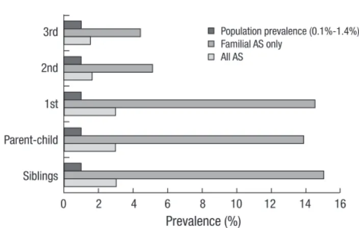
Recurrence risk in all AS patients for first, second, and third fa- milial degrees was 3.0%, 1.7%, and 1.5%. When restricted to the familial AS group, the recurrence risks increased to 14.6%, 5.2%, and 4.4%. If the relationship was close to the proband, the risks were higher in a dose dependent manner. In first degree families, siblings rather than the parents or children were more prone to have familiality (Fig. 1).
Prevalence (%)
0 2 4 6 8 10 12 14 16
3rd
2nd
1st
Parent-child
Siblings
Population prevalence (0.1%-1.4%) Familial AS only
All AS
Fig. 1. Recurrence risk of ankylosing spondylitis according to different type of familial degree. Population prevalence as a reference bar (0.1%-1.4%) was derived from the literature.
Characteristics of the familial AS compared to sporadic AS Significantly lower ESR and CRP were observed in familial AS patients compared to their sporadic AS counterparts (ESR, 22.6
± 22.2 vs 35.4 ± 34.4, P = 0.029; CRP, 1.24 ± 1.7 vs 2.43 ± 3.3, P = 0.003) (Table 1). A significantly lower body mass index (BMI) was noticed in the familial group (21.9 ± 2.7 vs 23.7 ± 3.3, P = 0.002). Familial AS was distributed in the quadrant of low CRP/
low BMI (Fig. 2). HLA-B27 positivity was significantly higher in familial AS (97.4% vs 83.1%, P = 0.044). Axial symptoms and the presence of sacroiliitis proven by plain radiography and advanc- ed radiographic findings such as syndesmophytes or sacroiliac ankylosis were not different between the two groups. Assess- ment of the pattern of arthritis revealed that oligoarthritis was significantly less common in the familial group, which also had less asymmetry. Particular parts of any joint disease were not
different (data not shown). Peripheral enthesitis including dac- tylitis, Achilles tendinitis and plantar fasciitis were less com- monly observed in familial AS. Extraarticular manifestations including psoriasis, uveitis and inflammatory bowel disease were not affected by familiality.
Familial AS patients according to age, sex and disease duration
There was no difference by sex in familial patients (Table 2). The formation of syndesmophytes was more seen in female patients, statistically not significant, though. Earlier disease onset is re- garded as a feature of familial disease in various diseases such as familial breast cancer and familial colon polyposis. There- fore, we investigated the age at onset of the disease and divided the familial AS patients by short (< 10 yr) or long disease (≥ 10 yr) duration to explore the differences (Table 3).
Age of disease onset seemed to be earlier and the percentage of female patients was lower in the familial AS group; however, both these findings did not reach statistical significance (Table Table 1. Characteristics of the familial form of ankylosing spondylitis (AS) compared
to sporadic AS
Parameters Familial AS
(n = 38) Sporadic AS (n = 166) P value Age (yr, mean ± SD) 33.7 ± 12.4 37.8 ± 13.1 0.081
Female, No. (%) 11 (28.9) 28 (16.9) 0.139
Onset age (yr, mean ± SD) 23.5 ± 9.9 26.8 ± 12.7 0.131 Duration (months, mean ± SD) 10.2 ± 6.9 10.9 ± 7.9 0.581 Diagnostic delay (days,
mean ± SD)
133.1 ± 166.6 136.3 ± 202.0 0.927 ESR (mm/hr, mean ± SD) 22.6 ± 22.2 35.4 ± 34.4 0.029† CRP (mg/dL, mean ± SD) 1.24 ± 1.7 2.43 ± 3.3 0.003‡ BMI (kg/m2, mean ± SD) 21.9 ± 2.7 23.7 ± 3.3 0.002‡ Smoking status
No. (%) Current smokers Ex-smokers Pack-years*
19 (50.0) 9 (26.3) 10 (26.3) 5.0 ± 8.4
65 (41.7) 46 (29.5) 45 (28.8) 6.3 ± 12.4
0.631
0.551
HLA-B27, No. (%) 37 (97.4) 138 (83.1) 0.044
Axial symptom Inflammatory back pain
Buttock pain 27 (71.1)
29 (76.3) 132 (80.0)
106 (64.2) 0.323 0.218
Syndesmophyte on X ray 9 (23.7) 42 (25.5) 1.000
Sacroiliitis by X ray Unilateral Bilateral Ankylosis
5 (13.2) 33 (86.8) 8 (21.1)
18 (10.8) 148 (89.2) 41 (24.7)
0.902 0.902 0.792 Peripheral enthesitis
Dactylitis Achilles tendinitis Heel pain
3 (7.9) 2 (5.3) 3 (7.9)
32 (19.3) 15 (9.0) 34 (20.5)
0.150 0.665 0.113 Peripheral arthritis
Monoarthritis Oligoarthritis Polyarthritis Symmetric Asymmetric
7 (18.4) 5 (13.2) 4 (10.5) 14 (36.8) 1 (2.6)
18 (10.8) 56 (33.7) 16 (9.6) 59 (35.5) 30 (18.1)
0.312 0.021 1.000 1.000 0.032
Psoriasis 2 (5.3) 7 (4.2) 1.000
Uveitis 9 (23.7) 38 (22.9) 1.000
Inflammatory bowel disease 1 (2.6) 1 (0.6) 0.816
*Pack-years is calculated based on the smoking history of current and ex-smokers;
†Significant difference (P < 0.05); ‡Significant difference (P < 0.01). SD, standard deviation; ESR, erythrocyte sedimentation rate; CRP, C-reactive protein; BMI, body mass index.
Body mas index (kg/m2 )
C-reactive protein (mg/dL)
0 2 4 6 8 10 12 14 16 18 20 36
34 32 30 28 26 24 22 20 18 16
Familial AS Sporadic AS
Fig. 2. Distribution of familial ankylosing spondylitis (AS) and sporadic AS according to BMI (body mass index) and CRP (C-reactive protein). Familial AS patients are dis- tributed in low CRP/low BMI quadrant. CRP and BMI are significantly lower in the fa- milial AS compared to sporadic AS.
Table 2. Variations in demographic, radiographic, and clinical features in familial an- kylosing spondylitis patients according to sex
Clinical features Female (n = 11) Male (n = 27) P value*
Syndesmophyte, No. (%) 8 (29.6) 1 (0.1) 0.237
Peripheral enthesitis, No. (%) 2 (18.2) 8 (29.6) 0.690 Peripheral arthritis, No. (%) 4 (36.4) 11 (40.7) 1.000
Uveitis, No. (%) 2 (18.2) 7 (25.9) 1.000
Psoriasis, No. (%) 1 (9.1) 1 (3.7) 0.501
Inflammatory bowel disease, No. (%) 1 (9.1) 0 (0.0) 0.290
*Fisher’s exact test for association between gender and clinical features.
1). In the graph directly comparing age at disease onset (Fig. 3), the two groups showed no differences and demonstrated the same peak of development in the 20s. In the familial AS patients, disease duration was not a factor that showed any difference between the short and long duration of the AS groups. Howev- er, higher female involvement and more peripheral arthritis were observed in the early AS with a disease duration of less than 10 yr group, whereas more peripheral arthritis and more uveitis were present in established AS with a disease duration of more than 10 yr, but these differences did not reach statistical significance.
DISCUSSION
The current report describes the increased occurrence of AS within families. The different phenotypes of familial AS com- pared to sporadic AS were explored. Lower inflammatory con- ditions in spite of the higher prevalence of HLA-B27 in the fa- milial AS group were remarkable in this report. This is similar to the findings from a study that showed that familial disease was milder in metrological measures than sporadic disease (11). To the best of our knowledge, this study is the first to examine fa- milial AS and its clinical manifestations in Asia.
Familial studies have been published primarily in Europe (10-12, 16, 17). Although higher familial recurrence has consis- tently been recognized, discrepancies have been found among the studies. Calin et al. (11) found that there were milder condi- tions in familial AS as assessed by spinal mobility, physical so- cial and pain score. In contrast, Paardt et al. (16) did not find difference in phenotype expression. In a study from Spain, fa- milial AS patients had a poorer pain visual activity score com- pared to sporadic AS patients. Young age at symptom onset and female dominance, higher frequency of uveitis, and HLA-B27 prevalence and a better response to NSAIDs in familial AS have also been addressed (12). In the Belgian study, psoriasis and uveitis were significantly higher (17). Dissimilarly, clustering of arthritis and uveitis in familial disease were observed and no relationships between familiality and psoriasis and inflamma- tory bowel disease were found in a report from France (10). How- ever, the definition of familiality and the extent of proband pa-
tients were different among the studies. We enrolled Korean AS patients and familial AS patients who were both defined by the 1984 modified New York Criteria for AS (15).
Strikingly, acute phase reactants, generally responsible for disease activity in AS, were lower in familial patients than spo- radic AS patients. It is a finding that supports that of previous report of Calin et al., in which familial AS was found to be a mild- er disease than sporadic disease in terms of spinal mobility, and physical, emotional and social well-being. One study from Spain reported that ESR and CRP were not different at the time of study (11), although, these cannot reflect genuine activity of the disease or predict the disease phenotype without the concomi- tant treatment such as TNF inhibitor or DMARDs being adjust- ed. Our study reported the ESR and CRP at diagnosis and found lower inflammatory conditions in familial AS than in sporadic disease. One study reported that the disease severity as assessed by BASDAI and BASFI was demonstrated to be correlated with familiality, leading one to consider that the severity of the dis- ease is genetically determined (18). At-risk families may have had a susceptibility gene rather than a severity gene (11). How- ever, evaluation of disease burden and activity requires more than serum ESR and CRP, these data should be interpreted with caution.
Lower BMI in the familial group was observed. Obesity is a condition in which inflammatory cytokines are increased (19).
This suggests that lower inflammatory conditions along with lower ESR and CRP in familial AS are noticeable features. The response to infliximab in AS was poorer in obese patients (20) although this cannot entirely explain the outcomes of familial AS. Therefore, it would be worthwhile to evaluate the treatment Table 3. Variations in demographic, radiographic, and clinical features in familial an-
kylosing spondylitis patients according to disease duration
Parameters Disease duration (yr)
P value*
< 10 (n = 23) ≥ 10 (n = 15)
Sex (female), No. (%) 8 (34.8) 3 (20.0) 0.470
Peripheral enthesitis, No. (%) 7 (30.4) 3 (20.0) 0.709 Peripheral arthritis, No. (%) 9 (54.2) 6 (40.0) 1.000
Uveitis, No. (%) 3 (13.0) 6 (40.0) 0.115
Psoriasis, No. (%) 1 (4.3) 1 (6.7) 1.000
Inflammatory bowel disease, No. (%) 1 (4.3) 0 (0.0) 1.000
*Fisher’s exact test for association between clinical features and disease duration.
Fig. 3. Age at disease onset in familial ankylosing spondylitis (AS) and sporadic AS.
Age at disease onset is not significantly different in familial AS and sporadic AS and the peak age of disease development are within the 20s in both group.
Number of patients
Age at disease onset (yr)
0 10 20 30 40 50 60 50
40
30
20
10
0
Familial AS Sporadic AS
response according to BMI in the familial disease in the future.
In our study, early disease onset or diagnostic delay were not observed to be associated with familial AS. This differs from a previous report, in which the relatives of probands who were younger had a higher risk of having AS (11, 21). Recognition of the AS history in family members may raise alertness to the symptoms of spondyloarthropathy and may encourage earlier inspection for the presence of HLA-B27 or the development of the disease. However, Korean families of AS sufferers did not show any difference in the age of symptom onset or the diag- nostic time-point. Similarly, age of onset was not a phenotype of familiality in the French study (10), suggesting that the age of disease onset is primarily determined by nongenetic causes (18). Furthermore, radiographic advancity in the axial or sacro- iliac joint was not different between familial AS and sporadic AS, probably because the disease duration was not different in both groups.
The sex ratio in our patients was not different between famil- ial and sporadic AS (28.9% vs 16.9%, P = 0.139). However, there was a trend for female predominance. A larger cohort may show a difference between the two groups in the sex ratio in the future study. In the recent report from Spain, female patients were predominant in the familial cohort (12). In contrast, a Belgian study reported the same sex prevalence (17). The importance of female sex in the familial disease has been previously describ- ed, with the young female index case having a higher familial recurrence among children and siblings (22). Generally, females are thought to be more resistant to getting spondyloarthritis than male. Therefore, an interpretation was given that female sex within a family cohort contributes to susceptibility rather than severity by females more easily passing the disease to their offsprings than males (23). However, research on the X chro- mosome failed to show any genetic evidence for susceptibility (24), and other genes responsible for susceptibility are still to be elucidated.
Regarding the differences between the sexes in familial AS, female patients from a French familial cohort showed signifi- cantly less radiographic sacroiliitis than male patients, although they had a similar incidence of symptomatic buttock pain (10).
In addition, the longer the disease duration, the higher the male/
female ratio in the French cohort (10). This is a supportive evi- dence that the female factor is important for predicting suscep- tibility to the disease rather than severity (22). Our familial pa- tients did not show a difference between the sexes, possibly due to the small numbers of the patients.
A familial risk model study revealed that heritability depends on genetic factors rather than environmental factor, and if the genetic factors are implicated, fewer than 5 genes are involved (5, 18). However, the GWAS study did not find any difference between the familial group and sporadic group (25). Although a significantly higher prevalence of HLA-B27 was demonstrated
in familial AS patients in this study, the presence of HLA-B27 still cannot predict or discriminate familial disease in the AS cohort. However, we witnessed again the high recurrence of AS in the familial group in this Korean patients as in the European patients, and the closer the relationships, the higher the recur- rence risk, indicating higher susceptibility in the familial group.
Clustering of certain phenotypes (e.g. arthritis and uveitis) in the familial AS group, suggesting a specific genetic or environ- mental factor contribution to specific phenotypes, has been re- ported . The current study suggests that further studies of the genetic determinations of such phenotypes are indicated, and that understanding the familial disease by phenotypes may fur- ther expand knowledge about the pathophysiology of AS. Prac- tically, this study also indicates that identification of the familial history may bring a distinct approach to patients with AS and prognosis of the disease.
The questionnaire or telephone inquiry based acquisition of information regarding the familial history in the study may have caused errors in data sourcing. Therefore, potential familial dis- ease patients could have been misclassified into sporadic dis- ease or vice versa. To avoid error, we enrolled consecutive out- patient patients and performed investigations pertinaciously.
In conclusion, we report that Korean AS patients with a fa- milial history have a higher presence of HLA-B27 than sporadic AS. The recurrence risk in the familial AS is highest in the first degree relatives, indicating higher susceptibility to the disease.
Distinct phenotypes of familial AS are lower frequency of oligo- arthritis, lower ESR and CRP at diagnosis and lower body mass index.
DISCLOSURE
The authors have no conflicts of interest to disclose.
ORCID
Hye Won Kim http://orcid.org/0000-0001-9450-3626 Hye Rim Choe http://orcid.org/0000-0003-0676-3546 Su Bin Lee http://orcid.org/0000-0003-0394-1749 Won Ik Chang http://orcid.org/0000-0002-5268-290X Hyun Jun Chae http://orcid.org/0000-0002-9113-6733 Jin Young Moon http://orcid.org/0000-0002-8688-2205 Jisue Kang http://orcid.org/0000-0002-7854-285X Sungim Lee http://orcid.org/0000-0001-6636-1077 Yeong Wook Song http://orcid.org/0000-0002-5384-3437 Eun Young Lee http://orcid.org/0000-0001-6975-8627 REFERENCES
1. Hopper JL. Disease-specific prospective family study cohorts enriched for familial risk. Epidemiol Perspect Innov 2011; 8: 2.
2. O’Neill SM, Rubinstein WS, Wang C, Yoon PW, Acheson LS, Rothrock N, Starzyk EJ, Beaumont JL, Galliher JM, Ruffin MT 4th. Familial risk for common diseases in primary care: the Family Healthware Impact Trial.
Am J Prev Med 2009; 36: 506-14.
3. Kramer JL, Velazquez IA, Chen BE, Rosenberg PS, Struewing JP, Greene MH. Prophylactic oophorectomy reduces breast cancer penetrance dur- ing prospective, long-term follow-up of BRCA1 mutation carriers. J Clin Oncol 2005; 23: 8629-35.
4. Hartmann LC, Schaid DJ, Woods JE, Crotty TP, Myers JL, Arnold PG, Petty PM, Sellers TA, Johnson JL, McDonnell SK, et al. Efficacy of bilat- eral prophylactic mastectomy in women with a family history of breast cancer. N Engl J Med 1999; 340: 77-84.
5. Brown MA, Laval SH, Brophy S, Calin A. Recurrence risk modelling of the genetic susceptibility to ankylosing spondylitis. Ann Rheum Dis 2000;
59: 883-6.
6. Brown MA, Kennedy LG, MacGregor AJ, Darke C, Duncan E, Shatford JL, Taylor A, Calin A, Wordsworth P. Susceptibility to ankylosing spon- dylitis in twins: the role of genes, HLA, and the environment. Arthritis Rheum 1997; 40: 1823-8.
7. Brewerton DA, Hart FD, Nicholls A, Caffrey M, James DC, Sturrock RD.
Ankylosing spondylitis and HL-A 27. Lancet 1973; 1: 904-7.
8. Schlosstein L, Terasaki PI, Bluestone R, Pearson CM. High association of an HL-A antigen, W27, with ankylosing spondylitis. N Engl J Med 1973;
288: 704-6.
9. Van der Linden SM, Valkenburg HA, de Jongh BM, Cats A. The risk of developing ankylosing spondylitis in HLA-B27 positive individuals: a comparison of relatives of spondylitis patients with the general popula- tion. Arthritis Rheum 1984; 27: 241-9.
10. Said-Nahal R, Miceli-Richard C, Berthelot JM, Duché A, Dernis-Labous E, Le Blévec G, Saraux A, Perdriger A, Guis S, Claudepierre P, et al. The familial form of spondylarthropathy: a clinical study of 115 multiplex families: Groupe Français d’Etude Génétique des Spondylarthropathies.
Arthritis Rheum 2000; 43: 1356-65.
11. Calin A, Kennedy LG, Edmunds L, Will R. Familial versus sporadic an- kylosing spondylitis: two different diseases? Arthritis Rheum 1993; 36:
676-81.
12. Almodóvar R, Font P, Zarco-Montejo P, Collantes E, Mulero J, Gratacós J, Juanola X, Ariza R. Phenotypic differences between familial versus spo- radic ankylosing spondylitis: a cross-sectional Spanish registry of spon- dyloarthropathies (REGISPONSER). Clin Exp Rheumatol 2011; 29: 822-7.
13. Dougados M, van der Linden S, Juhlin R, Huitfeldt B, Amor B, Calin A,
Cats A, Dijkmans B, Olivieri I, Pasero G, et al. The European Spondylar- thropathy Study Group preliminary criteria for the classification of spon- dylarthropathy. Arthritis Rheum 1991; 34: 1218-27.
14. Chou CT, Lin KC, Wei JC, Tsai WC, Ho HH, Hwang CM, Cherng JM, Hsu CM, Yu DT. Study of undifferentiated spondyloarthropathy among first- degree relatives of ankylosing spondylitis probands. Rheumatology (Ox- ford) 2005; 44: 662-5.
15. Van der Linden S, Valkenburg HA, Cats A. Evaluation of diagnostic cri- teria for ankylosing spondylitis: a proposal for modification of the New York criteria. Arthritis Rheum 1984; 27: 361-8.
16. Paardt Mv, Dijkmans B, Giltay E, van der Horst-Bruinsma I. Dutch pa- tients with familial and sporadic ankylosing spondylitis do not differ in disease phenotype. J Rheumatol 2002; 29: 2583-4.
17. De Vlam K, Vastesaeger N, Brasseur JP, Boonen A, Lenaerts J, Lesaffre E, Mielants H, Steinfeld S, Ribbens C, Leroi H, et al. Belgian patients with familial and sporadic ankylosing spondylitis differ in disease phenotype.
Arthritis Rheum 2006; 54: S717-8.
18. Hamersma J, Cardon LR, Bradbury L, Brophy S, van der Horst-Bruins- ma I, Calin A, Brown MA. Is disease severity in ankylosing spondylitis genetically determined? Arthritis Rheum 2001; 44: 1396-400.
19. Bastard JP, Maachi M, Lagathu C, Kim MJ, Caron M, Vidal H, Capeau J, Feve B. Recent advances in the relationship between obesity, inflamma- tion, and insulin resistance. Eur Cytokine Netw 2006; 17: 4-12.
20. Ottaviani S, Allanore Y, Tubach F, Forien M, Gardette A, Pasquet B, Pala- zzo E, Meunier M, Hayem G, Job-Deslandre C, et al. Body mass index influences the response to infliximab in ankylosing spondylitis. Arthritis Res Ther 2012; 14: R115.
21. Miceli-Richard C, Said-Nahal R, Breban M. Impact of sex on inheritance of ankylosing spondylitis. Lancet 2000; 355: 1097-8.
22. Calin A, Brophy S, Blake D. Impact of sex on inheritance of ankylosing spondylitis: a cohort study. Lancet 1999; 354: 1687-90.
23. Brown MA, Crane AM, Wordsworth BP. Genetic aspects of susceptibility, severity, and clinical expression in ankylosing spondylitis. Curr Opin Rheumatol 2002; 14: 354-60.
24. Hoyle E, Laval SH, Calin A, Wordsworth BP, Brown MA. The X-chromo- some and susceptibility to ankylosing spondylitis. Arthritis Rheum 2000;
43: 1353-5.
25. Joshi R, Reveille JD, Brown MA, Weisman MH, Ward MM, Gensler LS, Wordsworth BP, Evans DM, Assassi S. Is there a higher genetic load of susceptibility loci in familial ankylosing spondylitis? Arthritis Care Res (Hoboken) 2012; 64: 780-4.