Treatment Algorithm of Complications after Filler Injection:
Based on Wound Healing Process
Soft tissue filler injection has been a very common procedure worldwide since filler injection was first introduced for soft tissue augmentation. Currently, filler is used in various medical fields with satisfactory results, but the number of complications is increasing due to the increased use of filler. The complications after filler injection can occur at any time after the procedure, early and delayed, and they range from minor to severe. In this review, based on our experience and previously published other articles, we suggest a treatment algorithm to help wound healing and tissue regeneration and generate good aesthetic results with early treatment in response to the side effects of filler.
Familiarity with the treatment of these rare complications is essential for achieving the best possible outcome.
Keywords: Soft Tissue Filler; Injectable; Stem Cell; Filler Complications; Treatment Algorithm
Joo Hyun Kim, Duk Kyun Ahn, Hii Sun Jeong, and In Suck Suh
Department of Plastic and Reconstructive Surgery, Kangnam Sacred Heart Hospital, Hallym University Medical Center, Seoul, Korea
Received: 18 June 2014 Accepted: 28 July 2014 Address for Correspondence:
In Suck Suh, MD
Department of Plastic and Reconstructive Surgery, KangNam Sacred Heart Hospital, College of Medicine, Hallym University, 1 Singil-ro, Yongdeongpo-gu, Seoul 150-071, Korea Tel: +82.2-829-5182, Fax: +82.2-847-5183 E-mail: sismdps@chol.com
http://dx.doi.org/10.3346/jkms.2014.29.S3.S176 • J Korean Med Sci 2014; 29: S176-182
INTRODUCTION
Various managements for restoring volume to the body or reju- venation of facial wrinkles with injectable materials have been described over the last 100 yr. Autogenous fat graft was firstly performed for the correction of facial soft tissue defect at 1893 (1). In the beginning of 20th century, liquid paraffin was inject- ed for the facial contouring in Vienna (1). In spite of substantial complications and public backlash, alternative injectable sub- stances were explored.
Liquid silicone was used to improve body contouring in Swit- zerland and Japan during the 1940s and facial injections of liq- uid silicone was generalized and common procedure for reju- venation in the 1960s, particularly with the microdroplet injec- tion technique (2).
There was a great development of injectable fillers in 1981 with initial U.S. Food and Drug Administration (FDA)’s approv- al of bovine collagen fillers (Zyderm, Zyplast; Inamed Aesthet- ics, Santa Barbara, California) for cosmetic use (1). After that, bovine collagen became the standard for injectable materials.
The new era of injection with synthetic selective bioactive fillers began in 2003, when a Restylane (Medicis, Scottsdale, Arizona) became FDA approved product. Since then, the FDA has ap- proved a variety of fillers for cosmetic use, mostly in the Euro- pean market. This has led to a boom in the use of injectable fill- ers (1). Many filler types have been developed, resulting in ex- pectations for reduced complications. However, variable com- plications have been reported.
IDEAL MATERIAL
Ideal injectable fillers should possess the following characteris- tics. We evaluated the safety and efficacy to decide whether a material is ideal or not. The injectable filler materials should be safe, biocompatible, resistant to infection, and fixed to the sur- rounding tissue as well as should maintain its volume for wrin- kles and body contouring. It would be better induce a minimal foreign body reaction, including granuloma formation. And ideal materials for injection should not be teratogenic, carcino- genic and allergenic; not require pretesting; be painless, and in- expensive; and stable when stored at room temperature. And easy removal of filler is also necessary in case of complications (3, 4). Although the ideal filler has not yet been developed, re- searchers are still searching for fillers that meet these demand- ing criteria (4).
CATEGORIES OF FILLER
There are several methods for categorizing dermal fillers; we focus on the following three types in this article: natural, syn- thetic, mixed. Natural materials may be extract preparations of human or animal tissues. Natural material consists of autolo- gous fat and collagen, and the collagen is subdivided into por- cine collagen, bovine collagen and human collagen, which are xenogeneic and allogeneic collagens, respectively. Autologous fat is usually extracted from the abdomen or thigh. Autogenous dermofat graft has been used successfully for several decades, while the use of fat injection for body contouring began popu-
lar than 30 yr ago. Bioynthetic materials are chemical materials or biologic organic products, such as hyaluronic acid, silicone, calcium hydroxylapatite, polylactic acid, and polyacrylamide gel (4). One of the most commonly used fillers is hyaluronic acid, which consists of linear polymeric dimers of N-acetyl glu- cosamine and glucuronic acid (5). If the hyaluronic acid fillers are over injected or misinjected, they can be corrected or remov- ed by hyaluronidase immediately (1, 6). Some products, such as Artecoll (Artes Medical, San Diego, California), exhibit spe- cial characteristics because of an inner methylmethacrylate covered with a collagen barrier (4). The FDA has approved 22 fillers (Table 1).
INDICATIONS & CONTRAINDICATIONS
Currently, fillers are used in a variety of situations. Their main indications are for the filling of superficial or deep wrinkles as well as correction of congenital or acquired soft tissue depres- sion. Increasingly, fillers are used for volume replacement and enhancement procedures (7), including facial contouring, low- er eyelid wrinkle correction, and hand rejuvenation as well as the correction of facial asymmetry and congenital bony and soft-tissue defects (5, 8). Fillers are also used in patients suffer- ing from scleroderma, Romberg’s disease, or facial lipodystro- phy secondary to immunodeficiency syndrome treatment (9).
Another indications are lip augmentation and soft palate aug- mentation in secondary cleft lip and palate deformity, unilater- al paralysis of the vocal cords, correction of anophthalmic orbit syndrome and enophthalmos (8, 10-12).
By contrast, contraindications to the use of dermal fillers in- clude hypersensitivity to product components, bleeding disor- ders and a history of severe allergies and anaphylactic shock.
Polymethylmethacrylate injection is contraindicated into the mucosa of the lip. Polymethylmethacrylate and poly-L-lactic acid should not be used in patients who are suffered from ke- loid or hypertrophic scar (13).
FILLER COMPLICATIONS: TREATMENT ALGORITHM
As the number of filler injections increase, the cases of compli- cations also increase. After filler injection, risk of both short-du- ration and long-duration complications is always existence.
While most complications are mild and transient, more severe complications can occur, leaving patients with long lasting, func- tional and aesthetic problems. Some adverse events occur im- mediately after filler injection, while others don’t. The types of complications according to time of onset are illustrated in Table 2 (5).
Early complications
Swelling, ecchymosis and erythema
Swelling and ecchymosis may develop at the time of injection and they usually resolve spontaneously (14-16). Cold compres- sion and applying pressure helps to treat the symptoms. Imme- diately after injection, skin erythema is also transient and nor- mal. If erythema lasts for more than several days, it is likely to be a hypersensitivity reaction. However, it should be differenti- ated from infection after checking for accompanying fever. Man- agement for erythema with steroids can be useful. Vitamin K cream is also effective in the resolution of reddish swelling (5, 17). Patients with rosacea have a higher risk of developing ery- thema after injection and should be warned of this fact prior to injecting (5).
Lumps, nodules
Lumps or nodules usually appear to the cystic, edematous or sclerosing types shortly after treatment in the form of well-con- fined palpable lesions, which can result from injection in areas of thin soft-tissue coverage (e.g. eyelids, nasojugal region, and lip), injection of too much material, clumping of the filler, or dislocation by movement of the muscles (18-20). The lips are an area of high mobility with thin mucosa. Measures to avoid visibility of the implanted material include firm massage and meticulous placement of filler in the deep supraperiosteal plane (21, 22). Relatively short-term fillers such as hyaluronic acid products are preferable for these high-risk regions. An addi- tional benefit of using hyaluronic acid in these areas is that ir- Table 1. Fillers approved by the FDA
C ompo- nent
Polymethyl- methacrylate
(PMMA)
Hydroxyl- apatite
Poly-L- lactic acid
(PLLA)
Hyaluronic acid Collagen
Name Artefill Radiesse Sculptra Restylane-L
Restylane Zyderm
Zyplast Perlane
Belotero balance
Cosmoderm Cosmoplast Hyalform
Hyalform plus Evolence Juvederm 30
Juvederm volumna XC Juvederm 30 HV Juvederm 24 HV Captique
Fibrel
Elevess Prevelle
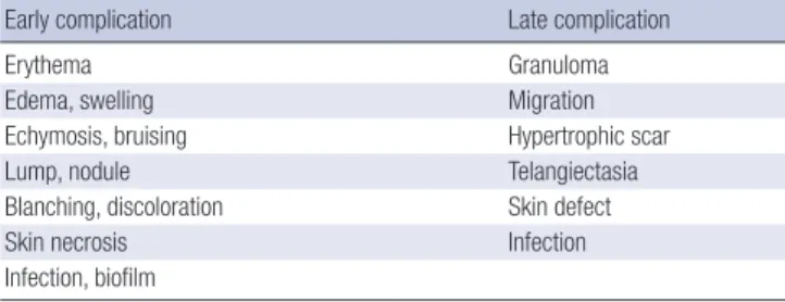
Table 2.Type of filler complication according to the time of onset
Early complication Late complication
Erythema Granuloma
Edema, swelling Migration
Echymosis, bruising Hypertrophic scar
Lump, nodule Telangiectasia
Blanching, discoloration Skin defect
Skin necrosis Infection
Infection, biofilm
regularities can be reversed with hyaluronidase. Semi-perma- nent fillers, such as poly-L-lactic acid, are longer lasting than hyaluronic acid. However, in cases of overcorrection, irregulari- ty and nodule formations are more persistent and difficult to treat. The management of resulting nodule as the cystic type is a simple puncture (incision and drainage) with a blade or sharp needle. If it is impossible to puncture the lump in the edema- tous or sclerosing types, a more invasive procedure, such as di- rect excision will be required or observation may be necessary until the product is absorbed (23-26). Lumps caused by poly-L- lactic acid or polymethylmethacrylate respond well to intrale- sional steroid injections, but steroids are less effective for calci- um hydroxylapatite (18).
Infection, erythematous nodule
As with any procedure that penetrates the skin, soft-tissue filler injections are also always associated with infection (5). Because chronic inflammation or infection leading to the formation of a granuloma can interfere with the wound healing process, infec- tion control is a significant process (27). Moreover, wound in- fections are often associated with aesthetically and functionally unfavorable scarring (28). Erythematous nodules, multiple red and tender lumps that persist beyond the first few days of treat- ment, may be signs of inflammation (14, 19, 21). Additionally, there is a risk for infection with swelling following filler injec- tion. If a single abscess is formed, contamination through the skin likely occurred during injection (29). But, if a patient has multiple abscesses, contamination likely occurred in the sy- ringe before injection. Once a fluctuant abscess is suspected, abscess culture and antibiotics are necessary. However, abscess- es should not be treated with antibiotics alone; they should be treated with incision and drainage (I & D) in the absence of cel- lulitis surrounding tissues. If the abscess is non-fluctuant, we can use antibiotics and steroids as the first line treatment (Fig.
1). In the early stage of treatment, hyaluronidase should not be used because of the risk of spreading the infected material dif- fusely into the surrounding tissues in the case of active cellulitis
(29). If these treatments fail, one should suspect biofilm, methi- cillin resistant staphylococcus aureus (MRSA) or non-typical tuberculosis (TB) and culture the infected material. In such a case, the patient should be given a TB test and treatments such as with quinolone and a third-generation macrolide, hyaluroni- dase, 5-fluorouracil (FU) or excision. The infection should be controlled with I & D at first, followed by hyaluronidase if nec- essary (29). We will discuss the biofilm theory later in this article.
Discoloration, blanching, and necrosis
Vascular-related events are the major, immediate complica- tions that are most likely to result in permanent sequelae. They can occur from intravascular embolism of the injected material, direct needle injury to the vessels, or external compression of the adjacent vasculature secondary to the hydrophilic proper- ties of the product (5). Inadvertent injections of the angular, dorsal nasal or supratrochlear artery are likely to lead to isch- emic responses that result in necrosis (15, 22, 30). Localized color changes in the affected areas should increase the index of suspicion about vascular compromise. The pathophysiology of vascular occlusion begins with immediate changes that are vis- ible in the vascular system, including initial blanching, which is followed by mottled discoloration called livedo reticularis. This is accompanied by pain unless there is a nerve block or local anesthetic blocking the pain pathways. The resulting ischemia produces a dusky discoloration that is associated with sluggish or absent capillary refill after digital compression as well as pos- sible loss of function. The final stage of vascular compromise is skin necrosis.
Appropriate treatment should be initiated immediately upon suspicion of vascular compromise. Injection should be stopped, and the area of injection should be massaged immediately as well as warm compresses applied to increase vasodilatation (5, 14, 21). Utilization of nitroglycerine paste and hyaluronidase is also advocated for cases that present early. Other treatments include systemic or topical steroids to reduce the associated in- flammation, mitigating the degree of injury. Although aspirin
A B
Fig. 1. A 32-yr old woman, with filler (Restylane; Medicis, Scottsdale, Arizona) injected into the glabella, alar and dorsum of the nose. She was referred to our department 3 days after filler injection. She complained of erythema and a pustule on nose with pain resulting from inflammation. Seven days after using antibiotics and steroids, the lesions were completely healed. (A) After injection of the dermal filler, erythema and a pustule are shown at the alar and dorsum of the nose. (B) After steroid and antibiotic treatment, the symptoms improved.
and intravenous (IV) prostaglandins have been suggested, their efficacy has not been proven (31). Other options with unproven efficacy are filler removal via puncture and low-molecular-wei- ght heparin. The aim for treatment is dissolving the product, fa- cilitating blood flow, and promoting vasodilation. Dayan et al.
(32) have suggested the use of hyaluronidase in all cases of vas- cular compromise, independent of the filler type, because of its edema-reducing benefits and theoretical advantage in reduc- ing the occluding vessel pressure. Because of vascular compro- mise and the resultant necrosis, debridement and wound care are required to minimize scarring. The treatment options are antibiotics and I&D as well as topical wound care, and adipose- derived stem cell injection to boost the healing process and tis- sue regeneration, which usually takes several weeks for granu- lation and reepithelialization once an eschar develops. Stem cells are a unique population of undifferentiated biological cells (33). They promote angiogenic processes by secreting angioge- nic factors. They also can stimulate cells that helpful in wound healing and differentiate cells that contribute to neovascular formation. However, exact mechanisms are not yet come out into the open (Fig. 2) (34). Many articles on human trials of adi- pose-derived stem cells are phase I safety trials and case reports (35).
Late complications
Adverse events from fillers can be sorted according to the tim- ing of symptom onset. Fourteen days after the procedure, late complications may occur.
Telangiectasia
Erythema and, occasionally, permanent telangiectasia may oc- cur at the injection site. In cases with erythema, prolonged in- tralesional or topical steroid therapy should be avoided because they can induce telangiectasia (5). And, in patients with telangi- ectasia, injection of the filler could worsen their appearance and size. Treatment with intense pulsed light therapy or pulsed dye laser can be helpful (18, 19).
Migration
Migration is thought to result from muscle- or gravity-induced displacement of the filler material (18). When the filler is locat- ed too superficially or in mobile anatomic areas, such as lips or lid area, sometimes migration can occur. This complication is strongly associated with injection of calcium hydroxylapatite into the lip. While speaking or eating and so on, the superficial and deep portion of the orbicularis oris muscle acts as a pump, which makes the material coalesce and results in nodule for- mation (26). To mechanically break up the product, intralesion- al steroid injections with massage should be performed. Alter- natively, the nodular formation site can be opened with a nee- dle for removal of the product. As an optional treatment, surgi- cal removal is also considered (36).
Hypertrophic scar
Superficial placement of fillers may be associated with hyper- trophic scarring. Superficial placement of fillers rarely resolves with permanent scar, consisting mainly of extracellular matrix components such as collagen, fibroblasts, and small vessels (37). For softening the hypertrophic scar, a pulsed dye laser or intralesional steroids are helpful. After this treatment, if there is no improvement, scar revision can be considered.
Granuloma and biofilm
The enveloped filler material may resist degradation and remain sequestered in the macrophages. These macrophages secrete various cytokines and other inflammatory materials that attract other macrophages and blood monocytes. Each macrophage may increase in size (epithelioid histiocytes) or fuse to form multinucleated foreign body giant cells. During this process, granuloma is formed (5). The reported rate of granuloma for- mation is 0.01% to 1% (18, 38). Granulomas can occur with all soft-tissue fillers, regardless of type, and usually appear after a latent period, which can be several months to years after filler injection (5). Nodule and granuloma are different in terms. Pa- thological diagnosis is not available, we can use nodule as a de- scriptive term. However, the latter term should only be used
A B C D E
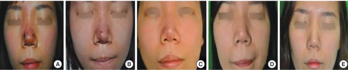
Fig. 2. A 25-yr old woman who had a filler (Artecoll; Artes Medical, San Diego, California) injected into her forehead and nose. She suffered pain that was accompanied by pus discharge and skin necrosis after 5 days. The adipose-derived stem cells were abstracted and injected into the damaged area on the nose tip. (A) The patient came to the clinic with a pustule and necrosis after filler injection. (B) Two days after adipose-cell derived stem cell injection. (C) Ten days after adipose-derived stem cell injection. The necrotic area shows nearly complete re-epithelization. (D) Six months after adipose-derived stem cell injection. The necrotic lesion was healed without scarring or pigmentation. (E) One year after stem cell injection. There were no demonstrable findings other than a small linear scar.
when the required pathological criteria for granulomas have been satisfied (29).
First treatment of choice for this complication is massage and intralesional corticosteroids. Granulomatous reactions af- ter hyaluronic acid injection can be cured with hyaluronidase (39). Patients who are not responsive to steroid alone can re- spond to the combination treatment of 5-FU and corticoste- roids, discouraging additional fibroblast activity and fibrosis. In the event of repeated failure of aforementioned therapies, sur- gical removal should be considered (5, 40). If these treatments fail, a biofilm is likely, consisting of MRSA or non-typical TB and culture of the infected material, TB test and treatments such as 2-drug with a quinolone and third-generation macrolide, hyal- uronidase, 5-FU or excision should be performed.
Recently, there has been discussion on the role of biofilms in causing delayed nodule formation, and it has not been precise- ly proven that biofilms are involved in granuloma formation.
However, many articles present arguments in favor of this hy- pothesis (16, 29, 41). Biofilms are accumulations of microor- ganisms within a self-developed matrix; they are irreversibly adherent to one another and to a variety of surfaces (42). In the biofilm, bacteria can safely avoid from immune defenses, so antibiotics have no effect. (29) Hyaluronidase helps break down the matrix, decreasing the biofilm mass (43). Other treatment options for biofilms are the prolonged use of antibiotics, admin- istration of intralesional 5-FU and intralesional laser therapy with a 532-nm or 808-nm laser (44). With respect to antimicro- bials, 2-drug therapy with a quinolone and a third-generation macrolide is recommended (16, 44).
Skin defects
There might be skin loss, scarring, and asymmetry, resulting in
skin defects in spite of active treatment after skin necrosis. In cases with skin defects, various reconstruction treatment op- tions can be used for the aesthetically and externally competent results. The primary usable methods of reconstruction are a full thickness skin graft (FTSG), local flap, composite graft and car- tilage graft. A FTSG usually involves performing a skin graft us- ing donor tissue from behind the ear, and composite and carti- lage grafts are also applied by the conchal cartilage of the ear, which is harvested according to the size of the defect. It is ad- vantageous that the scar from the donor site is invisible because it is usually harvested from ear, and the recipient site can achieve symmetry by supplying the site of skin loss defect (Fig. 3). And, in the case of a small defect, primary closure or local flaps are the best treatment of choice because they can offer similar skin color and texture like surrounding tissue (45).
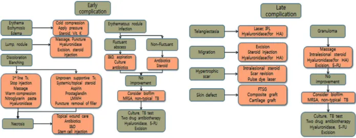
From these results, we constructed a treatment algorithm that incorporates our case and related articles (Fig. 4).
PREVENTIVE METHOD FOR FILLER INJECTION
Although proper treatment for complications is important, the prevention of the complications is crucial. The selection of ap- propriate injection techniques is important because it can help successful outcomes and reduce the risk of complications (36).
First, strategy for reducing the risk of infection is thorough clean- sing of the injection site. We should not inject the hydrophilic permanent filler materials through oral or nasal mucosa and not inject into the previous filler site or traumatized tissue (5).
There are several injection techniques that are associated with an increased risk of adverse events. Increasing the dissection of the subepidermal plane (i.e., a fan-like injection pattern), rapid injection, rapid flow rates, and higher volumes are included.
A B
E
C D
F
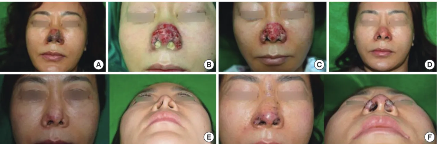
Fig. 3. A 53-yr old female who had a filler (Cutegel; Hyaluronic acid, BNC Korea Inc., Seoul) injected into her nasal tip. After 10 days, skin necrosis through the full skin layers of the nose developed. She underwent debridement of the necrotic tissue on the nose and injection of adipose-derived stem cells. One year after the operation, nasal deviation and asymmetry were shown due to scar contracture and the defect. Therefore, a composite graft on both alar rims with conchal cartilage was placed. (A) Skin necrosis occurred after filler injection. (B) In the operation, the necrotic area shows a raw surface after debridement and adipose-derived stem cell injection. (C) The day after the operation. Some areas show epithelization. (D) Fifteen days after the operation. Almost all of the area shows near re-epithelization except the small area of the nasal alar. (E) Deformity caused by the skin defect and scar contracture after total healing of the raw surface. (F) Asymmetry and deformity are improved after applying the composite graft.
(36). Specially, if a large volume bolus injection was adminis- tered, blood circulation defects could occur, which require at- tention. Large volume injections should only be performed when administered above the bone or into the dermis (5). Oth- er precautions include aspiration before injection and delivery of material at different points (21, 23). Some have advocated the use of small-caliber needles because they slow the speed of injection. The use of blunt needles is another method of reduc- ing complications. After filler injection, exposure to high and low temperatures should be avoided. For one day, filler recipi- ents should avoid touching or pressing on the site of the surgi- cal procedure and avoid contacting water to prevent inflamma- tion. For almost 1 week, drinking alcohol and smoking should be avoided.
CONCLUSION
Filler injection for soft tissue augmentation is a satisfactory pro- cedure with very modest results in spite of high expectations.
However, as the number of indications and performance in- creases, the number of complications also increases. It is im- portant for the physicians to know facial anatomy and high-risk regions. The experts of filler injection must be familiar with each filler material, the injection techniques, and the potential com- plications. If a complication does occur, the treatment algorithms of this article will be helpful for facilitating minimization of long- term sequelae. We summarized the key information about fill- ers and their complications, which can help clinicians, allowing them to successfully avoid and efficiently treat potential adverse events.
ORCID
Joo Hyun Kim http://orcid.org/0000-0001-8067-5814 Duk Kyun Ahn http://orcid.org/0000-0001-9232-8297
Hii Sun Jeong http://orcid.org/0000-0001-9408-8207 In Suck Suh http://orcid.org/0000-0003-4256-6398 REFERENCES
1. Kim JE, Sykes JM. Hyaluronic acid fillers: history and overview. Facial Plast Surg 2011; 27: 523-8.
2. Narins RS, Beer K. Liquid injectable silicone: a review of its history, im- munology, technical considerations, complications, and potential. Plast Reconstr Surg 2006; 118: 77s-84s.
3. Lemperle G, Morhenn V, Charrier U. Human histology and persistence of various injectable filler substances for soft tissue augmentation. Aes- thetic Plast Surg 2003; 27: 354-66; discussion 67.
4. Kinney BM, Hughes CE 3rd. Soft tissue fillers: an overview. Aesthet Surg J 2001; 21: 469-71.
5. Funt D, Pavicic T. Dermal fillers in aesthetics: an overview of adverse events and treatment approaches. Clin Cosmet Investig Dermatol 2013;
6: 295-316.
6. Vartanian AJ, Frankel AS, Rubin MG. Injected hyaluronidase reduces re- stylane-mediated cutaneous augmentation. Arch Facial Plast Surg 2005;
7: 231-7.
7. Goldberg DJ. Legal ramifications of off-label filler use. Clin Plast Surg 2006; 33: 597-601.
8. Cahill KV, Burns JA. Volume augmentation of the anophthalmic orbit with cross-linked collagen (Zyplast). Arch Ophthalmol 1989; 107: 1684-6.
9. Carr A, Miller J, Law M, Cooper DA. A syndrome of lipoatrophy, lactic acidaemia and liver dysfunction associated with HIV nucleoside ana- logue therapy: contribution to protease inhibitor-related lipodystrophy syndrome. AIDS 2000; 14: F25-32.
10. Chan RW, Titze IR. Viscosities of implantable biomaterials in vocal fold augmentation surgery. Laryngoscope 1998; 108: 725-31.
11. Flint PW, Corio RL, Cummings CW. Comparison of soft tissue response in rabbits following laryngeal implantation with hydroxylapatite, sili- cone rubber, and Teflon. Ann Otol Rhinol Laryngol 1997; 106: 399-407.
12. Hallen L, Johansson C, Laurent C. Cross-linked hyaluronan (Hylan B gel): a new injectable remedy for treatment of vocal fold insufficiency--an animal study. Acta Otolaryngol 1999; 119: 107-11.
Fig. 4. The treatment algorithm of the early and late complications.
13. Dayan SH. Complications from toxins and fillers in the dermatology clin- ic: recognition, prevention, and treatment. Facial Plast Surg Clin North Am 2013; 21: 663-73.
14. De Boulle K. Management of complications after implantation of fillers.
J Cosmet Dermatol 2004; 3: 2-15.
15. Jones D. Volumizing the face with soft tissue fillers. Clin Plast Surg 2011;
38: 379-90, v.
16. Rohrich RJ, Monheit G, Nguyen AT, Brown SA, Fagien S. Soft-tissue filler complications: the important role of biofilms. Plast Reconstr Surg 2010;
125: 1250-6.
17. Cohen JL, Bhatia AC. The role of topical vitamin K oxide gel in the reso- lution of postprocedural purpura. J Drugs Dermatol 2009; 8: 1020-4.
18. Lemperle G, Rullan PP, Gauthier-Hazan N. Avoiding and treating der- mal filler complications. Plast Reconstr Surg 2006; 118: 92s-107s.
19. Lemperle G, Duffy DM. Treatment options for dermal filler complica- tions. Aesthet Surg J 2006; 26: 356-64.
20. Alam M, Gladstone H, Kramer EM, Murphy JP Jr, Nouri K, Neuhaus IM, Spencer JM, Spenceri E, Van Dyke S, Ceilley RI, et al. ASDS guide- lines of care: injectable fillers. Dermatol Surg 2008; 34: S115-48.
21. Sclafani AP, Fagien S. Treatment of injectable soft tissue filler complica- tions. Dermatol Surg 2009; 35: 1672-80.
22. Alam M, Dover JS. Management of complications and sequelae with temporary injectable fillers. Plast Reconstr Surg 2007; 120: 98s-105s.
23. Cohen JL. Understanding, avoiding, and managing dermal filler com- plications. Dermatol Surg 2008; 34: S92-9.
24. Graivier MH, Bass LS, Busso M, Jasin ME, Narins RS, Tzikas TL. Calci- um hydroxylapatite (Radiesse) for correction of the mid- and lower face:
consensus recommendations. Plast Reconstr Surg 2007; 120: 55s-66s.
25. Beer KR. Radiesse nodule of the lips from a distant injection site: report of a case and consideration of etiology and management. J Drugs Der- matol 2007; 6: 846-7.
26. Berlin A, Cohen JL, Goldberg DJ. Calcium hydroxylapatite for facial re- juvenation. Semin Cutan Med Surg 2006; 25: 132-7.
27. Kang BS, Na YC, Jin YW. Comparison of the wound healing effect of cel- lulose and gelatin: an in vivo study. Arch Plast Surg 2012; 39: 317-21.
28. Mioton LM, Jordan SW, Hanwright PJ, Bilimoria KY, Kim JY. The rela- tionship between preoperative wound classification and postoperative infection: a multi-institutional analysis of 15,289 patients. Arch Plast Surg 2013; 40: 522-9.
29. DeLorenzi C. Complications of injectable fillers, part I. Aesthet Surg J 2013; 33: 561-75.
30. Kassir R, Kolluru A, Kassir M. Extensive necrosis after injection of hyal- uronic acid filler: case report and review of the literature. J Cosmet Der-
matol 2011; 10: 224-31.
31. Kim SG, Kim YJ, Lee SI, Lee CJ. Salvage of nasal skin in a case of venous compromise after hyaluronic acid filler injection using prostaglandin E.
Dermatol Surg 2011; 37: 1817-9.
32. Dayan SH, Arkins JP, Mathison CC. Management of impending necrosis associated with soft tissue filler injections. J Drugs Dermatol 2011; 10:
1007-12.
33. Salibian AA, Widgerow AD, Abrouk M, Evans GR. Stem cells in plastic surgery: a review of current clinical and translational applications. Arch Plast Surg 2013; 40: 666-75.
34. Sung HM, Suh IS, Lee HB, Tak KS, Moon KM, Jung MS. Case reports of adipose-derived stem cell therapy for nasal skin necrosis after filler injec- tion. Arch Plast Surg 2012; 39: 51-4.
35. Choi J, Minn KW, Chang H. The efficacy and safety of platelet-rich plas- ma and adipose-derived stem cells: an update. Arch Plast Surg 2012; 39:
585-92.
36. Bailey SH, Cohen JL, Kenkel JM. Etiology, prevention, and treatment of dermal filler complications. Aesthet Surg J 2011; 31: 110-21.
37. Ko WJ, Na YC, Suh BS, Kim HA, Heo WH, Choi GH, Lee SU. The effects of topical agent (kelo-cote or contractubex) massage on the thickness of post-burn scar tissue formed in rats. Arch Plast Surg 2013; 40: 697-704.
38. Carruthers A, Carruthers J. Non-animal-based hyaluronic acid fillers:
scientific and technical considerations. Plast Reconstr Surg 2007; 120:
33s-40s.
39. Brody HJ. Use of hyaluronidase in the treatment of granulomatous hyal- uronic acid reactions or unwanted hyaluronic acid misplacement. Der- matol Surg 2005; 31: 893-7.
40. Lemperle G, Gauthier-Hazan N. Foreign body granulomas after all in- jectable dermal fillers: part 2. Treatment options. Plast Reconstr Surg 2009;
123: 1864-73.
41. Christensen LH. Host tissue interaction, fate, and risks of degradable and nondegradable gel fillers. Dermatol Surg 2009; 35: 1612-9.
42. Goldman MP. Pressure-induced migration of a permanent soft tissue filler. Dermatol Surg 2009; 35: 403-5; discussion 5-6.
43. Pecharki D, Petersen FC, Scheie AA. Role of hyaluronidase in Strepto- coccus intermedius biofilm. Microbiology 2008; 154: 932-8.
44. Bjarnsholt T, Tolker-Nielsen T, Givskov M, Janssen M, Christensen LH.
Detection of bacteria by fluorescence in situ hybridization in culture-neg- ative soft tissue filler lesions. Dermatol Surg 2009; 35: 1620-4.
45. Yoon TH, Yun IS, Rha DK, Lee WJ. Reconstruction of various perinasal defects using facial artery perforator-based nasolabial island flaps. Arch Plast Surg 2013; 40: 754-60.