ABSTRACT
Purpose: Vitamin D is a potent immunomodulator. However, its role in the pathogenesis of allergic rhinitis is unclear.
Methods: The aim of this study was to evaluate the antiallergic effect of intranasally applied vitamin D in an allergic rhinitis mouse model. BALB/c mice were intraperitoneally sensitized with ovalbumin (OVA) and alum before they were intranasally challenged with OVA. Then, they were intranasally administered 1, 25-dihydroxyvitamin D3 (0.02 µg) or solvent. Allergic symptom scores, eosinophil infiltration, cytokine mRNA levels (interleukin [IL]-4, IL-5, IL-10, IL-13 and interferon-γ) in the nasal tissue, and serum total immunoglobulin E (IgE) and OVA-specific IgE, IgG1, and IgG2a were analyzed and compared with negative and positive control groups. Cervical lymph nodes (LNs) were harvested for flow cytometry analysis and cell proliferation assay.
Results: In the treatment group, allergic symptom scores, eosinophil infiltration, and mRNA levels of IL-4 and IL-13 were significantly lower in the nasal tissue than in the positive control group. The IL-5 mRNA level, serum total IgE, and OVA-specific IgE and IgG1 levels decreased in the treatment group; however, the difference was not significant. In the cervical LNs, CD86 expression had been down-regulated in CD11c+major histocompatibility complex II-high (MHCIIhigh) in the treatment group. Additionally, IL-4 secretion in the lymphocyte culture from cervical LNs significantly decreased.
Conclusions: The results confirm the antiallergic effect of intranasal 1,25-dihydroxyvitamin D3. It decreases CD 86 expression among CD11c+MHCIIhigh cells and T-helper type 2-mediated inflammation in the cervical LNs. Therefore, topically applied 1,25-dihydroxyvitamin D3 can be a future therapeutic agent for allergic rhinitis.
Keywords: Animal model; allergic rhinitis; dendritic cell; intranasal administration;
vitamin D; antiallergic agents
Original Article
Received: May 22, 2018 Revised: Sep 24, 2018 Accepted: Oct 10, 2018 Correspondence to Dong-Young Kim, MD, PhD
Department of Otorhinolaryngology-Head and Neck Surgery, Seoul National University Hospital, Seoul National University College of Medicine, 101 Daehak-ro, Jongno-gu, Seoul 03080, Korea.
E-mail: [email protected]
Copyright © 2019 The Korean Academy of Asthma, Allergy and Clinical Immunology • The Korean Academy of Pediatric Allergy and Respiratory Disease
This is an Open Access article distributed under the terms of the Creative Commons Attribution Non-Commercial License (https://
creativecommons.org/licenses/by-nc/4.0/) which permits unrestricted non-commercial use, distribution, and reproduction in any medium, provided the original work is properly cited.
ORCID iDs Sung-Woo Cho
https://orcid.org/0000-0003-0827-4471 Disclosure
There are no financial or other issues that might lead to conflict of interest.
Sung-Woo Cho ,1 Yu-Lian Zhang,1,2 Young Kyung Ko,3 Jae Min Shin,4 Jun Ho Lee,3 Chae-Seo Rhee,1,3 Dong-Young Kim3*
1 Department of Otorhinolaryngology-Head and Neck Surgery, Seoul National University Bundang Hospital, Seoul National University College of Medicine, Seongnam, Korea
2Center of Morphological Experiment, Medical College of Yanbian University, Yanji, China
3 Department of Otorhinolaryngology-Head and Neck Surgery, Seoul National University Hospital, Seoul National University College of Medicine, Seoul, Korea
4 Department of Otorhinolaryngology-Head and Neck Surgery, Soonchunhyang University Seoul Hospital, Soonchunhyang University College of Medicine, Seoul, Korea
Intranasal Treatment With 1,
25-Dihydroxyvitamin D3 Alleviates
Allergic Rhinitis Symptoms in a Mouse
Model
INTRODUCTION
Allergic rhinitis is an inflammatory, immunoglobulin E (IgE)-mediated disease characterized by allergic symptoms, such as nasal congestion, rhinorrhea, sneezing and nasal itching, after exposure to causative allergens. Although the exact pathogenesis remains unclear, it is known that T-helper type 2 (Th2)-driven immune response to allergens contributes to disease pathogenesis, and this is thought to occur after activation of mucosal epithelial and dendritic cells (DCs).1
Vitamin D has recently been discovered as a potent immune modulator. Deficiency in the serum level of vitamin D may contribute to the development of Th2-skewed allergic diseases, such as asthma, allergic rhinitis and atopic dermatitis.2,3 Previous studies have shown that vitamin D affects T cells, B cells, monocytes and macrophages.4 It also regulates the activity of DCs, which are key immune system cells. Vitamin D blocks the differentiation and maturation of DCs from monocytes and down regulates the expression of co-stimulatory molecules including CD80, CD83, and CD86.5,6 The down regulation of these co-stimulatory molecules reduces T cell activation, thereby leading to immune tolerance.7
Although vitamin D is known to regulate the activation threshold of DCs, most of these previous findings were from in vitro studies. Moreover, the exact role of vitamin D in allergic rhinitis is unclear, and especially little is known about the effect of topically applied vitamin D in allergic rhinitis. Therefore, in this study, we aimed to evaluate the antiallergic effect of intranasally applied 1,25-dihydroxyvitamin D3, a biologically active form of vitamin D, and its effect on DC activation in an allergic rhinitis mouse model.
MATERIALS AND METHODS
Animals
Four-week-old female BALB/c mice were used as experimental animals (YoungBio, Seongnam, Korea). Each mouse weighed 18–22 g and was maintained under specific pathogen-free conditions. All animal experiments in the present study followed the
guidelines and ethics of the Institutional Animal Care and Use Committee of the Biomedical Research Institute of Seoul National University Hospital (16-0106-S1A0[2]).
Induction and treatment of allergic rhinitis in mice
Mice were divided into 4 groups as follows: group 1 (n = 4) as a negative control group, group 2 (n = 6) as a positive control group, group 3 (n = 6) as a 1,25-dihydroxyvitamin D3 treatment group and group 4 (n = 4) as a sham treatment group. 1,25-dihydroxyvitamin D3 (Bonky® [Yuyu Pharma, Inc., Seoul, Korea] injection 1 µg) and its solvent were acquired from YuYu Pharma, Inc. The main ingredients of the solvent were polysorbate 20 (4.0 mg/
mL), NaCl (1.5 mg/mL), sodium L-ascorbate (10.0 mg/mL), disodium edetate (1.11 mg/
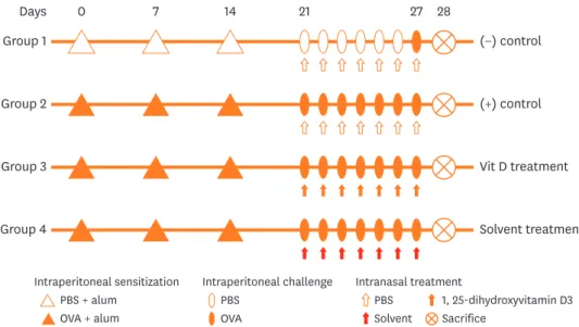
mL), anhydrous dibasic sodium phosphate (7.5 mg/mL) and monobasic sodium hydrogen phosphate monohydrate (1.84 mg/mL). Allergen sensitization, challenge for the development of the allergic rhinitis murine model, and treatment are summarized in Fig. 1. Briefly, the mice were sensitized by an intraperitoneal injection of 25 µg of ovalbumin (OVA; grade V;
Sigma-Aldrich, St. Louis, MO, USA) and 2 mg of aluminum hydroxide on days 0, 7, and 14.
The mice were then subjected to intranasal challenges with 100 µg of OVA on 7 consecutive days from days 21 to 27. The negative control mice were intraperitoneally injected and
intranasally challenged with phosphate-buffered saline (PBS), instead of OVA following the same schedule until day 27 at which mice were intranasally challenged with OVA. 1, 25-dihydroxyvitamin D3 (20 µL of Bonky® [Yuyu Pharma, Inc.] containing 0.02 µg per mouse) and solvent (20 µL per mouse) were administered by intranasal instillation on days 21 to 27 (3 hours before intranasal OVA challenge) to mice in groups 3 and 4, respectively.
Symptom scores
On day 27, after intranasal allergen provocation with 100 µg of OVA, the numbers of sneezing and nose rubbing bouts were counted for 15 minutes to evaluate early allergic responses by blinded observers.
Tissue preperation
All animal experiments were repeated twice. In both experiments, symptom scores were checked and the mice were then killed 24 hours after the last OVA challenge. Cervical lymph nodes (LNs) (primarily submandibular, 2-3 nodes from each side) were harvested from each mouse and were phyisically dissociated. In the first experiment, nasal tissues were obtained and further homogenized to measure cytokine expression. Serum samples from each mouse were also obtained for further analysis. Cells from the cervical LNs were analyzed by flow cytometry. In the second experiment, nasal tissuses from each mouse were fixed in formaldehyde solution for histological analysis. Cells from the cervical LNs were cultured to measure cytokine release (proliferation assay).
Reverse transcription polymerase chain reaction (RT-PCR) analysis of cytokines in the nasal tissue
After sacrifice, the head of each mouse was removed. After exposing the nasal cavity, the nasal mucosa in each mouse was carefully taken out by using a curette. Total RNA was isolated from the nasal mucosa using the TRIzol reagent (Invitrogen, Carlsbad, CA, USA).
Complementary DNA (cDNA) was synthesized using a cDNA synthesis kit (Gendepot, Katy, TX, USA). For the analysis of interleukin (IL)-4 (Mm 00445259_m1), IL-5 (Mm
Intraperitoneal sensitization PBS + alum
OVA + alum
Intraperitoneal challenge PBS
OVA
Intranasal treatment
PBS 1, 25-dihydroxyvitamin D3 Sacrifice
Solvent 0
Group 1 Days
(−) control
7 14 21 27 28
Group 2 (+) control
Group 3 Vit D treatment
Group 4 Solvent treatment
Fig. 1. Experimental protocol for sensitization with OVA and aluminum hydroxide gel and treatment with 1, 25-dihydroxyvitamin D3.
OVA, ovalbumin; PBS, phosphate-buffered saline.
00439646_m1), interferon (IFN)-γ (Mm 99999071_m1), IL-10 (Mm 00439616_m1), IL-13 (Mm 00434204_m1), and glyceraldehyde-3-phosphate dehydrogenase (GAPDH) (Mm 99999915_g1), pre-developed assay reagent kits of primers and probes were purchased from Applied Biosystems (Foster City, CA, USA). The amplification of IL-4, IL-5, IL-10, IL-13, IFN-γ and GAPDH cDNA was performed in MicroAmp optical 96-well reaction plates (Applied Biosystems). The reaction was performed using a QuantStudio™ 3 RT-PCR System (Applied Biosystems). The average transcript levels of genes are normalized to GAPDH expressed as 2−ΔCT values.
Histology of the nasal tissue
For the evaluation of nasal histology, the heads of mice in each group were fixed with 10%
formaldehyde solution. The nasal tissues were decalcified with ethylenediaminetetraacetic acid solution, embedded in paraffin, sectioned coronally into 4-µm slices and stained with hematoxylin and eosin (H&E) for the visualization of eosinophils. Under a light microscope (×400 magnification), infiltrating eosinophils were counted in 4 fields of the nasal septal mucosa by a single-blind observer. Eosinophils were morphologically defined by the presence of eosinophilic granules that were stained by H&E and the presence of a 2-lobed nucleus.8
Determination of serum levels of total and OVA-specific Igs
Serum samples from each mouse were obtained at the time of sacrifice. Serum levels of total IgE and OVA-specific IgE, IgG1 and IgG2a were measured by enzyme-linked immunosorbent assay (ELISA). For the analysis of total IgE, 96-well flat-bottom plates were coated with purified rat anti-mouse IgE (#553413; BD Bioscience, San Jose, CA, USA). A purified mouse IgE isotype (#557079; BD Bioscience) was used as a standard. Nonspecific antigen-antibody reactions were blocked with 3% bovine serum albumin. To detect total IgE, horseradish peroxidase (HRP)-conjugated anti-mouse Ig (#554002; BD Bioscience) was added to the wells. For the analysis of OVA-specific IgE, serum samples were added to OVA-coated 96-well flat-bottom plates. After washing, 100 µL of biotin-conjugated rat anti-mouse IgE mAb (#553414; BD Bioscience) was added, followed by the addition of streptavidin-HRP (#554066; BD Bioscience). For the analysis of OVA-specific IgG1 and IgG2a, serum samples were added to OVA-coated 96-well flat-bottom plates. After washing, biotinylated rat anti- mouse IgG1 and IgG2a (#553388 and #553441, respectively; BD Bioscience) were added to each well and incubated. After washing, streptavidin-HRP was added to each well. The reactions were developed using 3,3′,5,5′-tetramethylbenzidine (Moss Inc., Belfast, ME, USA) and terminated by adding 1 N HCl. The absorbance was measured in a microplate reader at 450 nm.
Flow cytometric analysis of cervical LNs
For the analysis, harvested cells from each mouse were distributed into 2 sets and were suspended in 50 µL of cold PBS containing 2% fetal calf serum. The first set of cells was stained with V450-conjugated anti-CD45 (BD Bioscience), BB515-conjugated anti-major histocompatibility complex II (MHCII) (BD Bioscience), phycoerythrin-conjugated anti- CD11c (BD Bioscience) and anaphase-promoting complex (APC)-conjugated anti-CD86 (BD Bioscience) for the detection of activated DCs. The other set of cells was stained with peridinin chlorophyll protein complex-conjugated anti-CD3 (BD Bioscience), fluorescein isothiocyanate-conjugated anti-CD4 (eBiosience, San Diego, CA, USA), APC-conjugated anti-CD25, and phycoerythrin-conjugated anti-Foxp3 for the detection of regulatory T cells.
The cells were analyzed by flow cytometry (LSR II; BD Bioscience). The CD11c+MHCIIhigh cells were considered as migratory DCs (Supplementary Fig. S1).9
Measurement of cytokines in the LN cell culture
Cervival LN single-cell suspensions from each mouse were plated in 24-well cell culture plates at a final concentration of 5 × 10⁶ cells/well using Roswell Park Memorial Institute 1,640 containing 10% fetal bovine serum supplemented with 100 U/mL penicillin and 100 µg/mL streptomycin (Gibco, Grand Island, NY, USA). The cells were incubated in a CO2
incubator at 37°C for 72 hours and stimulated with OVA for 72 hours. The culture supernatant was collected and stored at −70°C until cytokines were measured. Cytokine levels in the culture supernatant were assayed using a DuoSet ELISA kit (R&D Systems, Minneapolis, MN, USA) according to the manufacturer's protocol. After measuring the absorbance at 450 nm, the concentrations of IL-4, IL-5, IL-10 and IL-13 were determined by interpolation from the respective standard curves; all data are expressed in pg/mL.
Statistical analysis
The data are presented as mean ± standard error mean. The Mann-Whitney U test was used to compare results between the negative and positive control groups and between the treatment and positive control groups. P values of <0.05 were considered statistically significant.
Statistical analysis was performed using SPSS 22.0 software (IBM Co., Armonk, NY, USA).
RESULTS
Intranasal 1,25-dihydroxyvitamin D3 treatment alleviates allergic symptoms
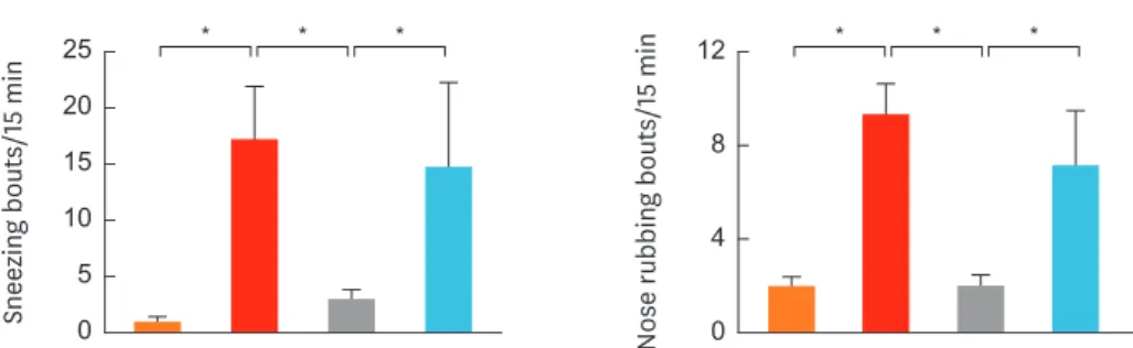
Fig. 2. shows symptom scores for each group after nasal challange with OVA. In the mice of group 2 (positive control), the mean numbers of sneezing and nose rubbing bouts were significantly higher than those of group 1 (negative control) (P < 0.001 and P = 0.001, respectively). The mice of group 3 (treatment group) showed significantly lower numbers of sneezing and nose rubbing bouts than those of groups 2 and 4 (P < 0.001 and P = 0.001, respectively). There was no signficiant difference in the mean numbers of sneezing of nose rubbing bouts between groups 2 and 4.1,25-dihydroxyvitamin D3 treament significantly decreases eosinophil infiltration in the nasal mucosa
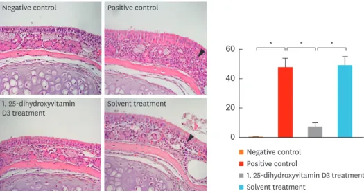
Fig. 3. shows eosinophil infiltration in the nasal mucosa for each group. Significantly higher number of eosinophils infiltrating the nasal mucosa per high-power field was observed in
Sneezing bouts/15 min
0 10 25 20
5 15
Nose rubbing bouts/15 min
0 4 12
8
Negative control Positive control 1, 25-dihydroxyvitamin D3 treatment Solvent treatment
* * * * * *
Fig. 2. Sneezing (left) and nose rubbing (right) bouts within 15 minutes. Intranasal treatment with 1, 25-dihydroxyvitamin D3 (group 3) suppressed allergic symptoms. Data are expressed as the mean ± standard error mean.
*P < 0.05.
group 2 than in group 1 (P = 0.004). The mean number of eosinophils infiltrating the nasal mucosa per high-power field was significantly lower in group 3 than in groups 2 (P = 0.002) and 4 (P = 0.004). However, no significant difference was observed between groups 2 and 4.
1,25-dihydroxyvitamin D3 treatment decreases Th2 cytokine expression in the nasal tissue
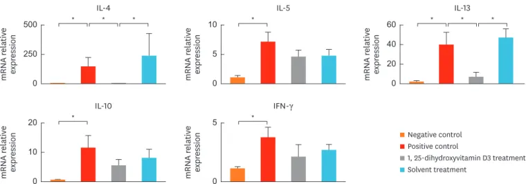
Fig. 4. shows cytokine expression in the nasal tissue. Compared to group 1 mice, significantly increased mRNA levels of IL-4 (P = 0.010), IL-5 (P = 0.010), IL-10 (P = 0.010), IL-13 (P = 0.010), and IFN- γ (P = 0.019) were observed in the nasal tissues of group 2 mice. The IL-4 and IL-13 mRNA levels were significantly decreased in group 3 compared to groups 2 (P = 0.010 and P = 0.038, respectively) and 4 (P = 0.029, both). The IL-5 expression in group 3 tended to follow the same pattern; however, the results were not significant, compared to groups 2 and 4. The mRNA levels of IFN-γ and IL-10 in group 3 did not significantly differ from those in groups 2 and 4. No significant difference was observed in the mRNA levels of IL-4, IL-5, IL-10, IL-13 or IFN-γ between groups 2 and 4.
1, 25-Dihydroxyvitamin D3 treatment down-regulates CD86 expression in cervical LNs
CD86 expression among CD11c+MHChigh cells from cervical LNs was examined. Group 2 showed significantly increased CD86 expression among CD11c+MHChigh cells compared to group 1 (P = 0.038). Although the number of CD86+ cells among CD11c+MHChigh cells was not significantly different among groups 2, 3 and 4 (data not shown), mean fluorescence intensity (MFI) showed that CD86 expression was significantly lower in group 3 than in groups 2 (P
= 0.041) and 4 (P = 0.010). The CD86 expression in group 3 did not significantly differ from that in group 1. Similarly, CD86 expression did not significantly differ between groups 2 and 4. The number of CD4+CD25+Foxp3+ cells tended to be higher in group 3 than in groups 2 and 4; however, the differences were not significant (Fig. 5). The MFI of Foxp3 expressed by CD4+CD25+ cells did not significantly differ from that expressed by most of the cells (≥90%) (data not shown).
0 20 60
40
Negative control Positive control
1, 25-dihydroxyvitamin D3 treatment Solvent treatment
* * *
Negative control Positive control
1, 25-dihydroxyvitamin
D3 treatment Solvent treatment
Fig. 3. The H&E staining (×400) (left). Arrow heads show eosinophilic granules with 2-lobed nuclei. Intranasal 1, 25-dihydroxyvitamin D3 treatment suppressed eosinophil infiltration in the nasal mucosa (right). Data indicate mean ± standard error mean.
H&E, hematoxylin and eosin.
*P < 0.05.
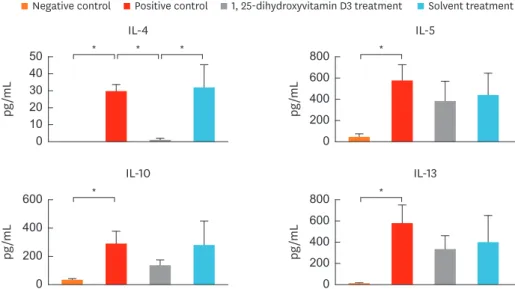
1,25-dihydroxyvitamin D3 treatment decreases IL-4 secretion in the lymphocyte culture from cervical LNs
To investigate whether the decreased CD86 expression in cervical LNs down-regulates T-cell activity, we measured the level of cytokines in the culture supernantants (Fig. 6). Levels of IL-4 (P = 0.016), IL-5 (P = 0.008), IL-10 (P = 0.008), and IL-13 (P = 0.008) were significanlty
mRNA relative expression mRNA relative expression mRNA relative expression
0 500
250
0 10
5
IL-4 IL-5 IL-13
0 60 40 20
* * * * * * *
Negative control Positive control
1, 25-dihydroxyvitamin D3 treatment Solvent treatment
* *
mRNA relative expression mRNA relative expression
0 20
10
0 5
IL-10 IFN-γ
Fig. 4. Decreased IL-4 and IL-13 mRNA levels in the nasal tissue in 1, 25-dihydroxyvitamin D3 treated mice (group 3). Data are expressed as the mean ± standard error mean.
IL, interleukin; IFN, interferon.
*P < 0.05.
MFI
0 100 150
50
A
%
0 20 30
10
B
%
CD86+ CD86+ CD25+Foxp3+Tregs
9 11 12
10
C
Negative control Positive control 1, 25-dihydroxyvitamin D3 treatment Solvent treatment
CD86 APC-A 102
500 400 300 200 100
0 103 104 105
D
P3 147
CD86 APC-A 102
500 400 300 200 100
0 103 104 105
P3 89
CD86 APC-A 102
500 400 300 200 100
0 103 104 105
P3 155
CD86 APC-A
Count
102 500 400 300 200 100
0 103 104 105
P3 52
Negative control Positive control 1, 25-dihydroxyvitamin
D3 treatment Solvent treatment
* * *
Fig. 5. (A) Decreased CD86 expression among DCs from mice treated with intranasal 1, 25-dihydroxyvitamin D3. (B, C) No significant difference in proportion (%) of CD86 + DCs or Tregs among the different groups. (D) Example histogram in each group representing expression of CD86 among DCs. Numerical values next to the histogram indicate MFI. Data are expressed as the mean ± standard error mean.
DC, dendritic cell; Treg, regulatory T cell; MFI, mean fluorescence intensity; APC, anaphase-promoting complex.
*P < 0.05.
higher in group 2 than in group 1. IL-4 level was significantly lower in group 3 than in groups 2 (P = 0.029) and 4 (P = 0.029). Levels of IL-5, IL-10, and IL-13 in group 3 showed a similar pattern; however, the differences were not significant when compared to groups 2 and 4.
Serum total IgE and OVA-specific Igs
Serum total IgE and OVA-specific IgE, IgG1, and IgG2a levels were significantly higher In group 2 than in group 1 (P = 0.010) (Fig. 7). However, no significant differences were observed among groups 2, 3 and 4.
pg/mL
0 30 50
20 40
10 pg/mL
IL-4 IL-5
0 400 800 600
200
Negative control Positive control 1, 25-dihydroxyvitamin D3 treatment Solvent treatment
* * *
pg/mL
0 400 600
200 pg/mL
IL-10 IL-13
0 400 800 600
200
*
*
*
Fig. 6. Cytokine levels in the lymphocyte culture from cervical LNs. Level of IL-4 was significantly decreased in mice treated with intranasal 1, 25-dihydroxyvitamin D3. Data are expressed as the mean ± standard error mean.
LN, lymph node; IL, interleukin.
*P < 0.05.
ng/mL
0 1,000 2,000
500 1,500
OD450
Total IgE OVA IgE
0 1 3 2
Negative control Positive control 1, 25-dihydroxyvitamin D3 treatment Solvent treatment
*
OD450
0 2 3
1 OD450
OVA IgG1 OVA IgG2a
0 1.0 1.5
0.5
*
*
*
Fig. 7. Levels of serum total IgE and OVA-specific IgE, IgG1, and IgG2a. Data are expressed as the mean ± standard error mean.
Ig, immunoglobulin; OVA, ovalbumin.
*P < 0.05.
DISCUSSION
In this study, we observed that intranasally instilled 1,25-dihydroxyvitamin D3 decreases symptom score, tissue eosinophil infiltration, and IL-4 and IL-13 expression in the nasal tissue. Moreover, 1,25-dihydroxyvitamin D3 treatment down-regulated CD 86 expression among CD11c+MHCIIhigh in cervical LNs and also decreased IL-4 production in the proliferation assay of cervical LN.
The initial production of IL-4 from naïve T cells by the T-cell receptor signaling pathway is important in eliciting the Th2 response because IL-4 is responsible for the polarization and maintenance of Th2 cells. Decreased IL-4 secretion in cervical LNs in the proliferation assay indicates the decreased activation of OVA-specific Th2 cells in cervical LNs.
These observations imply that 1,25-dihydroxyvitamin D3 exerts antiallergic effects, which may be partially due to decreased DC activation leading to decreased Th2 polarization in cervical LNs. Cervical LNs are the regional LNs of the upper airway tract, and nasal sensitization with an antigen activates DCs, which migrate to these regional LNs.10 Therefore, to identify activated DCs and their interaction with naïve T cells, we analyzed cytokine secretion in cervical LNs.
Naïve CD4 T cells are activated by several stimuli, including MHCII complexed with antigenic peptides and co-stimulatory factors expressed by DCs. The absence of these co-stimulatory signals makes T cells anergic.11 Among the co-stimulatory molecules, CD80 and CD86 expression by DCs is crucial for T cell activation. In the murine system, increased CD86 and decreased CD80 expressions indicate the maturation of bone marrow-derived DCs.12 The CD80 and CD86 levels are reported to be elevated in patients with asthma and allergic rhinitis,13,14 and an inhibition of their activities has been proved to be effective with less tissue eosinophil infiltration and Th2-mediated cytokine production.15,16
Densities of co-stimulatory molecules CD80 and CD86 on DCs are decreased in the presence of 1,25-dihydroxyvitamin D36,17; therefore, it holds potential as a therapeutic agent for diseases with an overactive immune system and inflammation.
In this study, no significant reduction in the expression of IL-5 had been observed in the nasal tissue. However, intranasal 1,25-dihydroxyvitamin D3 treatment significantly decreased eosinophil infiltration in the nasal mucosa. The IL-5 is a key regulator of eosinophil
proliferation in the bone marrow. It amplifies the tissue recruitment of eosinophils in response to locally elicited chemotactic signals. However, it does not play an obligatory role in the homing of eosinophils to local tissues and promoting peripheral eosinophilia in response to allergic stimulation.18 In this regard, reduced nasal tissue eosinophilia in 1,25-dihydroxyvitamin D3 treatment group may be associated with decreased IL-4 production.19 Moreover, 1,25-dihydroxyvitamin D3 is known to directly down-regulate and stabilize mast cells, which may further decrease the release of chemokines crucial for eosinophil recruitment.20,21 Additionally, it is reported to regulate the crosstalk between natural killer cells and eosinophils via IL-15/IL-8 axis.22
The OVA-specific IgG1 has been used as a Th2 marker, and it correlates with OVA-specific IgE levels.23 Howerver, in our study, the production of OVA-specific Igs, including IgE, IgG1 and IgG2a, were not affected by intranasal 1,25-dihydroxyvitamin D3 treatment. There had
been no treatment during sensitization period; therefore, intraperitoneal OVA and alum sensitization may have resulted in the production of a modest amount of OVA-specific Igs.
A previous study indicated that there is a strong association bewteen serum vitamin D level and allergen-specific IgE in children,24 while, these association had not been significant as in adults,3 suggesting an important role of vitamin D in sensizitation period. A study from a murine allergic asthma model had also shown that peri-natal vitamin D deficiency alone had immunomodulatory effects into Th2 skewing such that worse eosinophilic inflammation and airway remodelling with allergen exposure had been oberved in early life vitamin D deficienct mouse.25 Therefore, 1,25-dihydroxyvitamin D3 treatment during the sensitization period may play a preventive role and this should also be validated as well in the future studies.
There are several reasons for the intranasal instillation of 1,25-dihydroxyvitamin D3 in this study. First, little is known about intranasally applied vitamin D. To the best of our knowledge, this is the first study reporting the effect of topically applied vitamin D in allergic rhinitis. Secondly, when applied topically, its local concentration may increase in the nasal mucosa which may increase the efficacy. We have administrated 0.02 µg per mouse since we considered this amount as a maximal dosage (0.02 µg in 20 µL of undiluted solution) that lacks the possibility of aspiration into the lung. Our preliminary study had also shown that 0.02 µg resulted in a better antiallergic effect compared to lower doses (data not shown).
Moreover, intranasal administration facilitates the delivery of higher drug concentrations with reduced systemic adverse effects. Results of this study verify the antiallergic effect of intranasally instilled 1,25-dihydroxyvitamin D3, and suggest that it possesses therapeutic potential either alone or in combination with intranasal corticosteroids.26 It can be also applied in combination with allergen-specific immunotherapy.27
Systemically administered 1,25-dihydroxyvitamin D3 via intraperitoneal injection is also known to have a significant effect in alleviating allergic symptoms and some of Th2 cytokines such as IL-5, and IL-13 in allergic rhinitis murine model.28 Also, there have been studies that showed positive effect of systemically supplied vitamin D in preventing asthma exacerbations in humans.29 A systemic administration of 1,25-dihydroxyvitamin D3 systematically would be more clinically feasible considering the high prevalence of vitamin D deficiency.30
However, since the serum half-life of 1,25-dihydroxyvitamin D3 is short (5-8 hours) in adults,31 systemic injection of 1,25-dihydroxyvitamin D3 may not reach locally effective levels. Pharmacokinetics and local concentration at the target organ may differ according to the route of administration; therefore, further comparative studies regarding the dosage and route of administration of vitamin D are necessary to determine the optimal treatment regimen in animal studies. Lipophilic nature of 1,25-dihydroxyvitamin D3 and solvent composition of the drug may also affect pharmacokinetics. Therefore, such factors should be also taken into consideration when evaluating the efficacy.
Although the the mechanism by which 1,25-dihydroxyvitamin D3 affects the tolererogenic phenotype of DCs remains unclear, it is known to affect oxidative phosphoryation. Toll-like receptor activation induces metabolic transition in DCs from oxidative phosphorylation to aerobic glycolysis, which is essential for DC activation.32 1,25-dihydroxyvitamin D3 induces the transcriptional activation of oxidative phosphorylation.33,34 By sustaining oxidative phosphorylation in quiescent cells as a mode of glucose breakdown and inhibiting DC activation, 1,25-dihydroxyvitamin D3 may support immune quiescence and tolerance.35
There are several limitations in our study. First, the direct inhibitory effect of
1,25-dihydroxyvitamin D3-treated DCs on naïve T cells was not investigated. The antiallergic effect of 1,25-dihydroxyvitamin D3 may also have resulted from its effects on other types of inflammatory cells, such as mast cells, lymphocytes and macrophages because all these cells are known to express vitamin D receptor. Secondly, efficacy of 1,25-dihydroxyvitamin D3 treament during the sensitization period (to mimic prevention effect) had not been measured. Thrirdly, as for treament perspective, in order to be utilized as a potential therapeutic agent, an optimal treament regimen should have been given. Therefore, futher studies are necessary.
To summarize, intranasally administered 1,25-dihydroxyvitamin D3 alleviates allergic rhinitis symptoms in mice, and this is associated with downregulation of CD 86 expression of DC and decreased IL-4 production by T cells. Thus, intranasal 1,25-dihydroxyvitamin D3 may be a therapeutic option for treat allergic rhinitis.
SUPPLEMENTARY MATERIAL
Supplementary Fig. S1.
(A) CD11c+MHCIIhigh cells were considered as migratory DCs. Among them, CD86+ cells were considered as activated DCs. (B) CD4+, CD25+, and Foxp3+ cells were considered as Tregs.
Click here to view
REFERENCES
1. Lambrecht BN, Hammad H. Biology of lung dendritic cells at the origin of asthma. Immunity 2009;31:412-24.
PUBMED | CROSSREF
2. Aryan Z, Rezaei N, Camargo CA Jr. Vitamin D status, aeroallergen sensitization, and allergic rhinitis: a systematic review and meta-analysis. Int Rev Immunol 2017;36:41-53.
PUBMED | CROSSREF
3. Cheng HM, Kim S, Park GH, Chang SE, Bang S, Won CH, et al. Low vitamin D levels are associated with atopic dermatitis, but not allergic rhinitis, asthma, or IgE sensitization, in the adult Korean population. J Allergy Clin Immunol 2014;133:1048-55.
PUBMED | CROSSREF
4. Tian HQ, Cheng L. The role of vitamin D in allergic rhinitis. Asia Pac Allergy 2017;7:65-73.
PUBMED | CROSSREF
5. Bartels LE, Hvas CL, Agnholt J, Dahlerup JF, Agger R. Human dendritic cell antigen presentation and chemotaxis are inhibited by intrinsic 25-hydroxy vitamin D activation. Int Immunopharmacol 2010;10:922-8.
PUBMED | CROSSREF
6. Ferreira GB, van Etten E, Verstuyf A, Waer M, Overbergh L, Gysemans C, et al. 1,25-Dihydroxyvitamin D3 alters murine dendritic cell behaviour in vitro and in vivo. Diabetes Metab Res Rev 2011;27:933-41.
7. Hubo M, Trinschek B, Kryczanowsky F, Tuettenberg A, Steinbrink K, Jonuleit H. Costimulatory molecules on immunogenic versus tolerogenic human dendritic cells. Front Immunol 2013;4:82.
PUBMED | CROSSREF
8. Percy DH, Barthold SW. Pathology of laboratory rodents and rabbits. 3rd ed. Ames, IA: Blackwell Publishing Professional; 2007.
9. Shklovskaya E, Roediger B, Fazekas de St Groth B. Epidermal and dermal dendritic cells display differential activation and migratory behavior while sharing the ability to stimulate CD4+ T cell proliferation in vivo. J Immunol 2008;181:418-30.
PUBMED | CROSSREF
10. Kleinjan A, Lambrecht BN. Dendritic cells in rhinitis. Handb Exp Pharmacol 2009:115-36.
PUBMED | CROSSREF
11. Corthay A. A three-cell model for activation of naïve T helper cells. Scand J Immunol 2006;64:93-6.
PUBMED | CROSSREF
12. Inaba K, Inaba M, Romani N, Aya H, Deguchi M, Ikehara S, et al. Generation of large numbers of dendritic cells from mouse bone marrow cultures supplemented with granulocyte/macrophage colony- stimulating factor. J Exp Med 1992;176:1693-702.
PUBMED | CROSSREF
13. Hattori H, Okano M, Yoshino T, Akagi T, Nakayama E, Saito C, et al. Expression of costimulatory CD80/
CD86-CD28/CD152 molecules in nasal mucosa of patients with perennial allergic rhinitis. Clin Exp Allergy 2001;31:1242-9.
PUBMED | CROSSREF
14. Wong CK, Lun SW, Ko FW, Ip WK, Hui DS, Lam CW. Increased expression of plasma and cell surface co- stimulatory molecules CTLA-4, CD28 and CD86 in adult patients with allergic asthma. Clin Exp Immunol 2005;141:122-9.
PUBMED | CROSSREF
15. Chen ZR, Zhang GB, Wang YQ, Yan YD, Zhou WF, Zhu C, et al. Therapeutic effects of anti-B7-H3 antibody in an ovalbumin-induced mouse asthma model. Ann Allergy Asthma Immunol 2013;111:276-81.
PUBMED | CROSSREF
16. Li JG, Du YM, Yan ZD, Yan J, Zhuansun YX, Chen R, et al. CD80 and CD86 knockdown in dendritic cells regulates Th1/Th2 cytokine production in asthmatic mice. Exp Ther Med 2016;11:878-84.
PUBMED | CROSSREF
17. Pierrot-Deseilligny C, Souberbielle JC. Vitamin D and multiple sclerosis: an update. Mult Scler Relat Disord 2017;14:35-45.
PUBMED | CROSSREF
18. Foster PS, Mould AW, Yang M, Mackenzie J, Mattes J, Hogan SP, et al. Elemental signals regulating eosinophil accumulation in the lung. Immunol Rev 2001;179:173-81.
PUBMED | CROSSREF
19. KleinJan A, Willart M, van Rijt LS, Braunstahl GJ, Leman K, Jung S, et al. An essential role for dendritic cells in human and experimental allergic rhinitis. J Allergy Clin Immunol 2006;118:1117-25.
PUBMED | CROSSREF
20. Liu ZQ, Li XX, Qiu SQ, Yu Y, Li MG, Yang LT, et al. Vitamin D contributes to mast cell stabilization.
Allergy 2017;72:1184-92.
PUBMED | CROSSREF
21. Yip KH, Kolesnikoff N, Yu C, Hauschild N, Taing H, Biggs L, et al. Mechanisms of vitamin D3 metabolite repression of IgE-dependent mast cell activation. J Allergy Clin Immunol 2014;133:1356-64, 1364.e1-1364.e14.
PUBMED | CROSSREF
22. El-Shazly AE, Lefebvre PP. Modulation of NK cell autocrine-induced eosinophil chemotaxis by interleukin-15 and vitamin D(3): a possible NK-eosinophil crosstalk via IL-8 in the pathophysiology of allergic rhinitis. Mediators Inflamm 2011;2011:373589.
PUBMED | CROSSREF
23. Lewkowich IP, Rempel JD, HayGlass KT. Antigen-specific versus total immunoglobulin synthesis: total IgE and IgG1, but not IgG2a levels predict murine antigen-specific responses. Int Arch Allergy Immunol 2004;133:145-53.
PUBMED | CROSSREF
24. Chiu CY, Su KW, Tsai MH, Hua MC, Liao SL, Lai SH, et al. Longitudinal vitamin D deficiency is inversely related to mite sensitization in early childhood. Pediatr Allergy Immunol 2018;29:254-9.
PUBMED | CROSSREF
25. Vasiliou JE, Lui S, Walker SA, Chohan V, Xystrakis E, Bush A, et al. Vitamin D deficiency induces Th2 skewing and eosinophilia in neonatal allergic airways disease. Allergy 2014;69:1380-9.
PUBMED | CROSSREF
26. Litonjua AA. Vitamin D and corticosteroids in asthma: synergy, interaction and potential therapeutic effects. Expert Rev Respir Med 2013;7:101-4.
PUBMED | CROSSREF
27. Nelson HS. Allergen immunotherapy now and in the future. Allergy Asthma Proc 2016;37:268-72.
PUBMED | CROSSREF
28. Chen B, Qu S, Li M, Ye L, Zhang S, Qin T, et al. Effects of 1,25-dihydroxyvitamin D3 in an ovalbumin- induced allergic rhinitis model. Int Immunopharmacol 2017;47:182-9.
PUBMED | CROSSREF
29. Jolliffe DA, Greenberg L, Hooper RL, Griffiths CJ, Camargo CA Jr, Kerley CP, et al. Vitamin D supplementation to prevent asthma exacerbations: a systematic review and meta-analysis of individual participant data. Lancet Respir Med 2017;5:881-90.
PUBMED | CROSSREF
30. Park JH, Hong IY, Chung JW, Choi HS. Vitamin D status in South Korean population: seven-year trend from the KNHANES. Medicine (Baltimore) 2018;97:e11032.
PUBMED | CROSSREF
31. Wootton AM. Improving the measurement of 25-hydroxyvitamin D. Clin Biochem Rev 2005;26:33-6.
PUBMED
32. Everts B, Amiel E, Huang SC, Smith AM, Chang CH, Lam WY, et al. TLR-driven early glycolytic reprogramming via the kinases TBK1-IKKɛ supports the anabolic demands of dendritic cell activation.
Nat Immunol 2014;15:323-32.
PUBMED | CROSSREF
33. Ferreira GB, Overbergh L, Verstuyf A, Mathieu C. 1α,25-dihydroxyvitamin D3 and its analogs as modulators of human dendritic cells: a comparison dose-titration study. J Steroid Biochem Mol Biol 2013;136:160-5.
PUBMED | CROSSREF
34. Ferreira GB, van Etten E, Lage K, Hansen DA, Moreau Y, Workman CT, et al. Proteome analysis demonstrates profound alterations in human dendritic cell nature by TX527, an analogue of vitamin D.
Proteomics 2009;9:3752-64.
PUBMED | CROSSREF
35. Bscheider M, Butcher EC. Vitamin D immunoregulation through dendritic cells. Immunology 2016;148:227-36.
PUBMED | CROSSREF