Purpose: Live Mycobacterium bovis Bacille Calmette-Guérin (BCG) has a suppressive effect on asthma, but its use in clinical practice may be limit- ed due to adverse reactions. To develop a product that is effective for suppressing asthma with minimal adverse reactions, we investigated whether the heat-killed body or culture supernatants of mycobacteria could also prevent asthma development. Methods: Female BALB/c mice were treat- ed with live BCG, the heat-killed body, or culture supernatants of BCG or Mycobacterium tuberculosis intraperitoneally, while sensitizing and provok- ing with ovalbumin. Then they underwent a methacholine bronchoprovocation test, and the peribronchial inflammatory cell numbers and cytokine levels in splenocyte culture supernatants were assessed. Results: The airway sensitivity to methacholine decreased significantly after treatment with not only live BCG (30.8 versus 10.0 mg/mL, P<0.001) but also with the culture supernatant (BCG, 23.0 mg/mL, P<0.05; M. tuberculosis, 20.5 mg/mL, P<0.05). In contrast, heat-killed mycobacteria did not effectively decrease airway sensitivity. The peribronchial eosinophil counts and the goblet cell proportions in total epithelial cells decreased significantly in most of the groups. The interferon-g/interleukin-5 ratios increased signifi- cantly in most of the treatment groups except for the heat-killed groups, and were significantly related to airway sensitivity (r=0.312, P<0.01) and peribronchial eosinophil counts (r=-0.416, P<0.001). Interleukin-17A level was inversely related to airway sensitivity (r=-0.212, P<0.05) and was sig- nificantly lower in the live BCG group than in the control (137±20 versus 308±57 pg/mL, P<0.05). Conclusions: BCG and mycobacteria culture su- pernatants may effectively prevent the development of asthma associated with altered Th1/Th2 cytokines and interleukin-17A levels.
Key Words: Asthma; BCG vaccine; interferons; interleukins; mycobacterium
INTRODUCTION
The so-called “hygiene/old friends hypothesis” states that an increase in the prevalence of asthma/allergy is related to dimin- ished exposure to infections and certain relatively harmless en- vironmental organisms or components that the immune sys- tem has learned to tolerate.1 In this context, our group2 and oth- er investigators3-7 have shown that Mycobacterium bovis Bacille Calmette-Guérin (BCG) and other mycobacterial infections suppress airway hyperresponsiveness and eosinophilic inflam- mation, likely through a T-helper 1 (Th1) or regulatory T cell (Treg) response. We also demonstrated that BCG vaccination has therapeutic effects on asthma in adult patients.8 However, Shirtcliffe et al.9 reported that repeated heat-killed BCG vacci- nations had no beneficial effects, and they halted the trial early due to severe local reactions in asthma patients. Although the mycobacterial strains,10 route of delivery,11 and other factors might have affected the efficacy of the vaccinations, it is neces- sary to develop an effective anti-asthmatic agent devoid of ad- verse reactions.
Preventive effects of mycobacteria and their culture supernatants against asthma development in BALB/c mice
Eui-Ryoung Han,
1Inseon S. Choi,
1* Sun-Ho Eom,
2Hwa-Jung Kim
21Department of Allergy, Chonnam National University Medical School, Gwangju, Korea
2Department of Microbiology, College of Medicine, Chungnam National University, Daejeon, Korea
Mycobacterial components rather than whole mycobacteria may be more promising to avoid untoward vaccination effects.
The recognition of mycobacterial components by Toll-like re- ceptor (TLR) 2 associated with TLR1/TLR6 and TLR4 on den- dritic cells has been implicated in mycobacteria-induced Th1/
Treg cell responses.12 In fact, the mycobacterial cell wall lipogly- cans lipoarabinomannan (LAM) and phosphatidylinositol mannan suppress airway eosinophilia in association with in- creased interleukin (IL)-10 secretion by T cells.13 In addition, mannose-capped LAM from BCG induced Th1 cells,14 and that from M. tuberculosis induced Treg cells from human peripheral blood mononuclear cells.15 Synthetic lipopeptides that stimu- late TLR2 decrease airway eosinophilia and IgE levels but in- Allergy Asthma Immunol Res. 2010 January;2(1):34-40.
doi: 10.4168/aair.2010.2.1.34 pISSN 2092-7355 • eISSN 2092-7363
This is an Open Access article distributed under the terms of the Creative Commons Attribution Non-Commercial License (http://creativecommons.org/licenses/by-nc/3.0/) which permits unrestricted non-commercial use, distribution, and reproduction in any medium, provided the original work is properly cited.
Correspondence to: Inseon S. Choi, MD, Department of Allergy, Chonnam National University Medical School, 8 Hak-dong, Dong-gu, Gwangju 501-757, Korea.
Tel: +82-62-220-6571; Fax: +82-62-225-8578; E-mail: [email protected] Received: July 29, 2009; Accepted: December 10, 2009
•There are no financial or other issues that might lead to conflict of interest.
crease IL-10 and interferon (IFN)-g production by T cells.16 My- cobacterial chaperonin 60.1 also suppresses airway eosinophil- ia and methacholine airway hypersensitivity.17 However, only a few studies using mycobacterial components in vaccinations have investigated their effects on asthma, and no study has compared the efficacy of various mycobacterial strains. There- fore, we investigated whether whole heat-killed mycobacteria and their products in the culture supernatants, from both M.
tuberculosis and BCG, are effective for preventing the develop- ment of asthma.
MATERIALS AND METHODS Experimental animals
Specific pathogen-free, 6-week-old female BALB/c mice (Dae- han BioLink, Eumsung, Korea) were fed standard mouse chow and given water ad libitum in an animal care room at Chonnam National University Medical School, Korea. Animal care and treatment followed the guidelines of the Chonnam National University Research Institute of Medical Sciences.
Study design
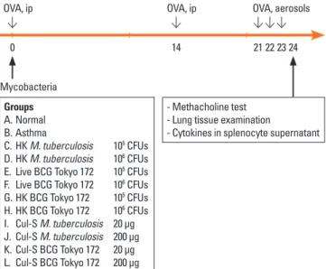
The mice were divided into 12 groups (n=10 per group): one normal control group, one asthma control group, four M. tuber- culosis groups (heat-killed or culture supernatants), and six BCG groups (live, heat-killed, or culture supernatants; Fig. 1).
On the first day of the study, the mice in the treatment groups were given 1×10⁵ or 1×10⁶ colony-forming units (CFUs) of my- cobacteria, or 20 or 200 μg of culture supernatant intraperitone- ally. Then, the mice were sensitized and provoked using ovalbu- min, underwent a methacholine bronchial challenge test, and were sacrificed so that the inflammatory cell numbers in the
peribronchial tissue and the cytokine levels in the supernatants of concanavalin A-stimulated splenocytes could be quantified.
Preparation of mycobacteria and culture supernatants
Pathogenic M. tuberculosis H37Rv (ATCC 27294) and M. bovis BCG Tokyo 172 strain (obtained from the Korean National Tu- berculosis Association, Seoul) were cultured as surface pellicles on Sauton’s medium at 37°C. After 6 weeks, the bacilli were re- moved by filtration through filter paper. The culture superna- tants were sequentially sterilized using a membrane filter (1.2- and 0.2-μm pore size) and concentrated by ultrafiltration (Ami- con Centriprep-10, Millipore, Bedford, MA, USA). The concen- tration of culture filtrate antigen was determined using a bicin- choninic acid protein assay kit (Pierce Biotechnology, Rock- ford, IL, USA).
To prepare the live bacterial stock, both mycobacteria were grown in roller bottles containing Middlebrook 7H9 broth (Dif- co, Detroit, MI, USA) supplemented with 0.05% Tween 80 and 10% oleic acid-albumin-dextrose-catalase (Becton Dickinson, Cockeysville, MD, USA) at 37°C until the optical density at 600 nm reached 0.8. The cells were washed three times with phos- phate-buffered saline (PBS) and centrifuged at 150×g for 5 min- utes to remove any clumps. Aliquots of the upper bacterial sus- pension were kept at -80°C until use. The thawed bacterial ali- quots used for infection were dispersed using a bath sonicator.
The CFUs in the dispersed supernatants were determined by preparing serial dilutions in 7H9 broth and plating them onto Middlebrook 7H10 agar (Difco). To protect the laboratory work- ers from mycobacterial infection, the M. tuberculosis inocu- lums were prepared like those that were heated for 15 minutes.
The culture supernatants, which contain most of the secreted antigens, were concentrated 10-fold for administration.
Ovalbumin sensitization and provocation
The animals were sensitized using two intraperitoneal injec- tions of 200 μL PBS containing 20 μg ovalbumin (Grade V; Sig- ma-Aldrich, St. Louis, MO, USA) and 2.25 mg aluminum hy- droxide (Imject Alum; Pierce) 2 weeks apart. One week after the second sensitization injection, the mice were provoked with 1% ovalbumin aerosol using an ultrasonic nebulizer (Ultra- Neb; DeVilbiss, Somerset, PA, USA) in an animal body plethys- mograph (OCP3000; All Medicus, Ahnyang, Korea) for 30 min- utes per day for three successive days.
Lung function tests
Airway responsiveness to methacholine (Sigma) was mea- sured using a OCP3000 body plethysmograph, as described previously.10 Briefly, 24 hours after the final ovalbumin inhala- tion challenge, the animals inhaled PBS aerosol and then in- haled progressively doubled concentrations of methacholine, starting from 3.125 mg/mL and increasing to 50 mg/mL. Aero- sols were generated using an Ultra-Neb nebulizer, and 3 mL of Groups
A. Normal B. Asthma
C. HK M. tuberculosis 105 CFUs D. HK M. tuberculosis 106 CFUs E. Live BCG Tokyo 172 105 CFUs F. Live BCG Tokyo 172 106 CFUs G. HK BCG Tokyo 172 105 CFUs H. HK BCG Tokyo 172 106 CFUs I. Cul-S M. tuberculosis 20 µg J. Cul-S M. tuberculosis 200 µg K. Cul-S BCG Tokyo 172 20 µg L. Cul-S BCG Tokyo 172 200 µg
- Methacholine test - Lung tissue examination
- Cytokines in splenocyte supernatant
0 14 21 22 23 24
OVA, ip OVA, ip OVA, aerosols
Mycobacteria
Fig. 1. The time course of the experiment. OVA, ovalbumin; IP, intraperitoneal;
M. tuberculosis, Mycobacterium tuberculosis; BCG, bacille Calmette-Guérin;
HK, heat-killed; Cul-S, culture-supernatant.
solution was aerosolized for 3 minutes. Enhanced pause (Penh) was used to represent airway resistance, as described previous- ly.10 The methacholine concentration required for a 200% in- crease in Penh from the post-saline value (PC200) was obtained as an index of airway sensitivity.
Histological analysis
The left lung was fixed in 4% formalin and embedded in par- affin for histopathological analysis. The embedded tissue was sectioned every 4 µm and stained with hematoxylin-eosin and periodic acid-Schiff. Small airways (circumference <500 μm) were selected,18 and the numbers of eosinophils, lymphocytes, and neutrophils within the peribronchial area from the base- ment membrane to a depth of 100 μm were determined and expressed as the number of cells/mm2 using a Nikon micro- scope with a computerized image analyzer program (AnalySIS® Pro; Soft Imaging System GmBH, Münster, Germany).19 Goblet cell hyperplasia was evaluated according to a 5-point scoring system (0-4), defined as follows: 0, no goblet cells; 1, <25%; 2, 25-50%; 3, 50-75%; and 4, ≥75%.20
Culture supernatant cytokine assays
Splenocyte culture and cytokine measurements were per- formed as described previously.10 Briefly, splenocytes were cul- tured in RPMI 1640 (BioWhittaker, Walkersville, NY, USA) sup- plemented with 10% fetal bovine serum (Gibco BRL, Grand Is- land, NY, USA) and a 1% penicillin-streptomycin-amphotericin B mixture (BioWhittaker) and were stimulated with 2.5 μg/mL concanavalin A (Sigma) for 48 hours. The IFN-g, IL-5, and IL- 17A concentrations were determined in the supernatant of stimulated splenocytes using commercial enzyme-linked im- munosorbent assay (ELISA) kits (BioLegend, Inc., San Diego, CA, USA). The sensitivities of the assays for IFN-g, IL-5, and IL- 17A were 4, 8, and 7.8 pg/mL, respectively.
Statistical analysis
The PC200 was Log10 transformed before analysis, and all sta- tistical results are expressed as the mean±SEM. The Kruskal- Wallis test and Mann-Whitney U-test were used to determine the significant differences between groups. Associations be- tween variables were examined using Spearman’s rank correla-
700 600 500 400 300 200 100 0
% for baseline Penh
normal control asthma control Cul-S BCG 20 µg Cul-S BCG 200 µg
baseline
Conc of MCh (mg/mL)
3.125 6.25 12.5 25 50
**
**†
700 600 500 400 300 200 100 0
% for baseline Penh
normal control asthma control Cul-S TB 20 µg Cul-S TB 200 µg
baseline
Conc of MCh (mg/mL)
3.125 6.25 12.5 25 50
**†
**†
700 600 500 400 300 200 100 0
% for baseline Penh
normal control asthma control Live BCG 105 CFUS Live BCG 106 CFUS
baseline
Conc of MCh (mg/mL)
3.125 6.25 12.5 25 50
**††
**††
**††
**
Fig. 2. Dose response curves for methacholine (MCh) challenges. n=10 per group. Penh, enhanced pause; BCG, bacille Calmette-Guérin; TB, Mycobacterium tuberculosis; Cul-S, culture-supernatant. *P<0.05 and **P<0.01 for low dose treatments, and †P<0.05 and ††P<0.01 for high dose treatments compared with the asthma control.
tion coefficient. A P value <0.05 was considered statistically sig- nificant.
RESULTS
Airway responsiveness
The percent increases in Penh relative to baseline after metha- choline inhalation were significantly higher in the asthma group than the normal control group at each concentration of
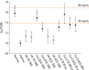
methacholine (P<0.01; Fig. 2). Compared to the asthma group, the Penh values were significantly lower in response to 3.125, 6.25, 12.5, and 25 mg/mL of methacholine in mice treated with 1×10⁵ CFUs of live BCG. Similarly, 1×10⁶ CFUs of live BCG sig- nificantly decreased the values obtained after 3.125, 6.25, and 12.5 mg/mL methacholine treatment. Moreover, 20 μg BCG cul- ture supernatant significantly decreased the Penh values at 3.125 and 12.5 mg/mL methacholine treatment while 200 μg BCG culture supernatant decreased the value at 12.5 mg/mL metha- choline treatment. Both 20 and 200 μg M. tuberculosis culture supernatant also significantly decreased the Penh values at 3.125 and 12.5 mg/mL methacholine treatment. In addition, metha- choline-PC200 was significantly higher in groups treated with live BCG (geometric mean, 30.8 versus 10.0 mg/mL, P<0.001) and groups treated with culture supernatant (BCG, 23.0 mg/
mL, P<0.05; M. tuberculosis, 20.5 mg/mL, P<0.05) than in the asthma control group (Fig. 3). However, the heat-killed BCG and M. tuberculosis mycobacteria did not significantly suppress airway responsiveness.
Inflammatory cells in peribronchial tissue and epithelial goblet cells
The asthma control group had significantly more peribron- chial eosinophils than the normal control group (Fig. 4). All treatments except 1×10⁶ CFUs of heat-killed M. tuberculosis and 1×10⁵ CFUs of heat-killed BCG significantly suppressed peri- bronchial eosinophilia. The peribronchial neutrophil counts were also significantly higher in the asthma control group than in the normal control group (251±64 versus 57±19/mm2, P<
0.01), and were lower in all of the treatment groups but only sig- nificantly lower in those treated with 200 μg BCG (85±22/mm2, 1.8
1.6
1.4
1.2
1.0
0.8
log PC200
Norm al Asthma
HK TB 10(5)
HK TB 10(6)
Live B CG 10
(5) Live B
CG 10 (6) HK BC
G 10(5) HK BC
G 10(6) Cul-S
TB 20 µg Cul-S
TB 200 µg Cul-S
BCG 20 µg Cul-S
BCG 2 00 µg 25 mg/mL 50 mg/mL
***
***
** * *
*
**
Fig. 3. Comparisons of the airway responsiveness to methacholine (PC200) be- tween the asthma control group and the other groups. n=10 per group. TB, My- cobacterium tuberculosis; BCG, bacille Calmette-Guérin; HK, heat-killed; Cul-S, culture-supernatant. *P<0.05, **P<0.01, and ***P<0.001 compared with the asthma control.
80
60
40
20
lstoell ciaelthpil eta tols Prelt cleob gofn tioorop 0
Norm al Asthma
HK TB 10(5)
HK TB 10(6)
Live B CG 10
(5) Live B
CG 10 (6) HK BC
G 10(5) HK BC
G 10(6) Cul-S
TB 20 µg Cul-S
TB 200 µg Cul-S
BCG 20 µg Cul-S
BCG 2 00 µg
***
*** *** *** ***
*** ***
***
** **
* 350
300 250 200 150 100 50 0
Peribronchial eosinophil counts /mm2
Norm al Asthma
HK TB 10(5)
HK TB 10(6)
Live B CG 10
(5) Live B
CG 10 (6) HK BC
G 10(5) HK BC
G 10(6) Cul-S
TB 20 µg Cul-S
TB 200 µg Cul-S
BCG 20 µg Cul-S
BCG 2 00 µg
*** ***
** **
** **
**
** **
Fig. 4. Comparisons of the peribronchial eosinophil numbers and goblet cell hyperplasia in the bronchial epithelium. n=10 per group. TB, Mycobacterium tuberculo- sis; BCG, bacille Calmette-Guérin; HK, heat-killed; Cul-S, culture-supernatant. *P<0.05, **P<0.01, and ***P<0.001 compared with the asthma control.
P<0.05) or M. tuberculosis (93±12/mm2, P<0.05) culture super- natant. Furthermore, the proportion of goblet cells in the epi- thelial cells was significantly lower in all of the treated groups than in the asthma control group (Fig. 4).
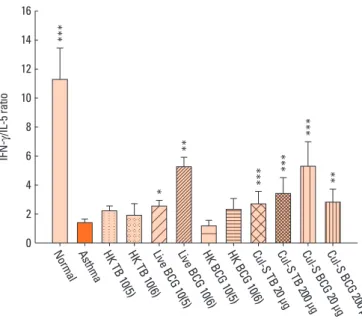
Cytokine concentrations in the cultured splenocyte supernatants
In the supernatants of concanavalin A-stimulated spleno- cytes, the IL-5 level was significantly higher, and the IFN-g con- centration and the IFN-g/IL-5 ratio were significantly lower, in the asthma control group than in the normal control group (Fig.
5). Compared to the asthma group, the IFN-g/IL-5 ratio was sig- nificantly higher in all treated groups except those treated with heat-killed mycobacteria. The IL-17A level was significantly higher in the asthma control group than in the normal control group. Only the treatment with 1×10⁵ CFUs of live BCG signifi- cantly decreased IL-17A levels (137.1±20.5 versus 308.2±57.4 pg/mL, P<0.05). The IFN-g/IL-5 ratio was significantly associat- ed with airway sensitivity (P<0.01), the number of peribronchi- al eosinophils (P<0.001), and goblet cell hyperplasia (P<0.001;
Table 1). However, IL-17A level had a weak inverse relationship only with airway sensitivity (P<0.05).
DISCUSSION
Treatment of mice with live BCG significantly suppressed air- way responsiveness, eosinophilia, and goblet cell hyperplasia.
These results are consistent with those of previous studies show-
ing that BCG2-4,6,10,11,21 and M. vaccae6,18 have suppressive effects on asthma.
Furthermore, we found that the BCG and M. tuberculosis cul- ture supernatants also effectively suppressed asthmatic reac- tions, although the effectiveness for suppressing airway respon- siveness was somewhat lower in the culture supernatant groups than in the live BCG group. Means et al.22 demonstrated that M.
tuberculosis culture media induce TLR2-dependent cellular ac- tivation, and suggested that TLR2 ligand was the non-protein- aceous heat-stable factor. Because TLR2 stimulation suppresses airway eosinophilia,16 a soluble factor released by cultured my- cobacteria might have acted as a TLR2 ligand in the present study.
We also found that vaccination with heat-killed mycobacteria was ineffective for airway responsiveness even though it signifi- cantly suppressed airway eosinophilia and goblet cell hyper- plasia. Major et al.23 also showed that the efficacy of heat-killed BCG against the development of airway eosinophilia was lower than that of live BCG. The lower effectiveness of the culture su- pernatants and heat-killed preparations used in this study may be explained at least in part as follows: a) live BCG may not only release an effective factor after inoculation, but it also has cell wall components such as LAM and phosphatidylinositol that increase IL-10 secretion;13 b) the effects are dose-dependent3 and live BCG must work longer in vivo due to its proliferation potential; c) some components that actively suppress allergic responses are heat-labile. Means et al.22 showed that a heat- sensitive cell-associated mycobacterial factor distinct from LAM mediated TLR4-dependent activation. However, Zuany-Amor- im et al.7 demonstrated that treating mice with heat-killed M.
vaccae induces allergen-specific Treg cells and effectively sup- presses methacholine airway responsiveness and airway eosin- ophilia. Therefore, if these less effective preparations are used instead of live mycobacteria as an asthma vaccine to avoid ad- verse effects, there may be a need to increase the vaccine dose or manipulate the vaccine content to increase their efficacy.
IFN-g production induced by M. tuberculosis is 4-fold greater than that induced by BCG,24 so we can speculate that M. tuber- Table 1. Relationship between levels of cytokines in splenocyte supernatants and markers of asthmatic airway reactions in mice
IL-5 IFN-g IFN-g/IL-5 IL-17A
PC200 (mg/mL) -0.303** 0.149 0.312** -0.212*
Peribronchial Eos (/mm2) 0.416*** -0.237** -0.416*** 0.051 Peribronchial Neut (/mm2) 0.152 -0.207* -0.214* 0.069 Goblet cell grade† 0.449*** -0.321*** -0.473*** 0.074 IL, interleukin; IFN, interferon; PC200, the concentration of methacholine re- quired for a 200% increase of lung function measurement from baseline; Eos, eosinophils; Neut, neutrophils. †, the proportion of the goblet cells in the epithe- lial cells; 0, no goblet cells; 1, <25%; 2, 25-50%; 3, 50-75%; 4, ≥75%. *P<0.05,
**P<0.01 and ***P<0.001.
***
***
***
***
**
**
* 16
14 12 10 8 6 4 2 0
IFN
-g/IL-5 ratio
Norm al Asthma
HK TB 10(5)
HK TB 10(6)
Live B CG 10
(5) Live B
CG 10 (6) HK BC
G 10(5) HK BC
G 10(6) Cul-S
TB 20 µg Cul-S
TB 200 µg Cul-S
BCG 20 µg Cul-S
BCG 2 00 µg Fig. 5. Comparisons of the interferon (IFN)-g/interleukin (IL)-5 ratios in the su- pernatants of concanavalin A-stimulated splenocytes. n=10 per group. TB, My- cobacterium tuberculosis; BCG, bacille Calmette-Guérin; HK, heat-killed; Cul-S, culture-supernatant. *P<0.05, **P<0.01, and ***P<0.05 compared with the asthma control.
culosis culture supernatants or heat-killed preparations would be more effective for suppressing asthmatic reactions than those of BCG. However, this was not the case in the present study, and there were no differences between them. A large ep- idemiological study by von Mutius et al.25 showed that an in- crease in the tuberculosis notification rates of 25 per 100,000 was associated with a decrease in the prevalence of asthma by 4.7%, while another large epidemiological study by Grüber et al.26 reported that BCG vaccination had only a weak protective effect against asthma. Thus, the difference in IFN-g levels be- tween BCG and M. tuberculosis and its effect on asthma may be related to any difference in a heat-sensitive cell-associated my- cobacterial factor. Further studies are needed to investigate this possibility.
The effects of the mycobacteria treatments, including the my- cobacteria culture supernatants, on asthmatic reactions were associated with increases in the IFN-g/IL-5 ratio. These results are consistent with those of previous studies showing that my- cobacterial infections2-6,10,11,21 or administering mycobacterial components17 shift the Th1/Th2 balance toward Th1. IFN-g seems to play a key role in the mycobacterial effects on Th2 cy- tokines and airway eosinophilia, because these effects are strongly impaired in IFN-g-deficient mice.23 In addition, provo- cation via allergen inhalation induces IL-17 mRNA expression and neutrophilic influx in the airways of mice.27 Recently, McKinley et al.28 showed that transferring Th17 cells that pro- duce IL-17 into mice results in neutrophilic airway inflamma- tion and methacholine airway hyperresponsiveness. Therefore, the inverse relationship between PC200 and IL-17A in the pres- ent study may be attributable to neutrophils despite the lack of a significant relationship between IL-17A and neutrophils.
However, IL-17 decreases airway eosinophilia27 and may re- duce matrix deposition by inducing metalloproteinases.29 The complex action of IL-17 on asthma needs further investigation.
Because the airway cytokines and the ovalbumin-specific cy- tokines in the splenocyte culture supernatants were almost un- detectable in a preliminary study, we did not check them in the present study. Future studies should consider airway cytokine profiles, especially for allergen-specific cytokines, using other methods such as Western blotting. Furthermore, lung function measurements should be validated using more rigorous meth- ods, even though several previous studies used Penh values as an airway hyperresponsiveness measure.
Taken together, our results show that both live BCG and my- cobacteria culture supernatants are effective for suppressing the development of asthmatic reactions. Therefore, the use of soluble culture supernatant components is promising for the development of asthma treatments without the adverse effects associated with whole live BCG. Heat-killed preparations were also effective for suppressing airway eosinophilia and remodel- ing. However, the loss of an effect on airway hyperresponsive- ness suggests that further studies of mycobacterial cell wall
components are needed to develop more effective vaccines to treat asthma. Finally, the effects of mycobacteria and their com- ponents were associated with altered levels of Th1/Th2 cyto- kines and interleukin-17A.
ACKNOWLEDGMENTS
We are indebted to Ms. Young-Ah Koh for her technical sup- port. This study was supported by a grant (CRI09066-1) from the Chonnam National University Hospital Research Institute of Clinical Medicine. The authors had full access to all of the data in this study, and the authors take complete responsibility for the integrity of the data and the accuracy of the data analysis.
REFERENCES
1. Rook GAW, Hamelmann EH, Brunet LR. Mycobacteria and aller- gies. Immunobiology 2007;212:461-73.
2. Koh YI, Choi IS, Kim WY. BCG infection in allergen-presensitized rats suppresses Th2 immune response and prevents the develop- ment of allergic asthmatic reaction. J Clin Immunol 2001;21:51-9.
3. Erb KJ, Holloway JW, Sobeck A, Moll H, Le Gros G. Infection of mice with Mycobacterium bovis-Bacillus Calmette-Guérin (BCG) sup- presses allergen-induced airway eosinophilia. J Exp Med 1998;187:
561-9.
4. Herz U, Gerhold K, Grüber C, Braun A, Wahn U, Renz H, Paul K.
BCG infection suppresses allergic sensitization and development of increased airway reactivity in an animal model. J Allergy Clin Immunol 1998;102:867-74.
5. Wang CC, Rook GA. Inhibition of an established allergic response to ovalbumin in BALB/c mice by killed Mycobacterium vaccae. Im- munology 1998;93:307-13.
6. Hopfenspirger MT, Agrawal DK. Airway hyperresponsiveness, late allergic response, and eosinophilia are reversed with mycobacteri- al antigens in ovalbumin-presensitized mice. J Immunol 2002;168:
2516-22.
7. Zuany-Amorim C, Sawicka E, Manlius C, Le Moine A, Brunet LR, Kemeny DM, Bowen G, Rook G, Walker C. Suppression of airway eosinophilia by killed Mycobacterium vaccae-induced allergen- specific regulatory T-cells. Nat Med 2002;8:625-9.
8. Choi IS, Koh YI. Therapeutic effects of BCG vaccination in adult asthmatic patients: a randomized, controlled trial. Ann Allergy Asthma Immunol 2002;88:584-91.
9. Shirtcliffe PM, Easthope SE, Weatherall M, Beasley R. Effect of re- peated intradermal injections of heat-inactivated Mycobacterium bovis bacillus Calmette-Guérin in adult asthma. Clin Exp Allergy 2004;34:207-12.
10. Choi IS, Lin XH, Koh YA, Koh YI, Lee HC. Strain-dependent sup- pressive effects of BCG vaccination on asthmatic reactions in BALB/c mice. Ann Allergy Asthma Immunol 2005;95:571-8.
11. Choi IS, Lin XH, Koh YA, Cui Y. Inoculation route-dependent and allergen-specific suppressive effects of bacille Calmette-Guérin vaccination on asthmatic reactions in BALB/c mice. Lung 2007;
185:179-86.
12. Barlan I, Bahceciler NN, Akdis M, Akdis CA. Bacillus Calmette- Guérin, Mycobacterium bovis, as an immunomodulator in atopic diseases. Immunol Allergy Clin North Am 2006;26:365-77.
13. Sayers I, Severn W, Scanga CB, Hudson J, Le Gros G, Harper JL.
Suppression of allergic airway disease using mycobacterial lipogly- cans. J Allergy Clin Immunol 2004;114:302-9.
14. Ito T, Hasegawa A, Hosokawa H, Yamashita M, Motohashi S, Naka T, Okamoto Y, Fujita Y, Ishii Y, Taniguchi M, Yano I, Nakayama T.
Human Th1 differentiation induced by lipoarabinomannan/lipo- mannan from Mycobacterium bovis BCG Tokyo-172. Int Immunol 2008;20:849-60.
15. Garg A, Barnes PF, Roy S, Quiroga MF, Wu S, Garcia VE, Krutzik SR, Weis SE, Vankayalapati R. Mannose-capped lipoarabinomannan- and prostaglandin E2-dependent expansion of regulatory T cells in human Mycobacterium tuberculosis infection. Eur J Immunol 2008;
38:459-69.
16. Akdis CA, Kussebi F, Pulendran B, Akdis M, Lauener RP, Schmidt- Weber CB, Klunker S, Isitmangil G, Hansjee N, Wynn TA, Dillon S, Erb P, Baschang G, Blaser K, Alkan SS. Inhibition of T helper 2-type responses, IgE production and eosinophilia by synthetic lipopep- tides. Eur J Immunol 2003;33:2717-26.
17. Riffo-Vasquez Y, Spina D, Page C, Tormay P, Singh M, Henderson B, Coates A. Effect of Mycobacterium tuberculosis chaperonins on bronchial eosinophilia and hyper-responsiveness in a murine model of allergic inflammation. Clin Exp Allergy 2004;34:712-9.
18. Ozdemir C, Akkoc T, Bahceciler NN, Kucukercan D, Barlan IB, Ba- saran MM. Impact of Mycobacterium vaccae immunization on lung histopathology in a murine model of chronic asthma. Clin Exp Allergy 2003;33:266-70.
19. Maestrelli P, Saetta M, Di Stefano A, Calcagni PG, Turato G, Rugg- ieri MP, Roggeri A, Mapp CE, Fabbri LM. Comparison of leukocyte counts in sputum, bronchial biopsies, and bronchoalveolar lavage.
Am J Respir Crit Care Med 1995;152:1926-31.
20. Tanaka H, Masuda T, Tokuoka S, Komai M, Nagao K, Takahashi Y, Nagai H. The effect of allergen-induced airway inflammation on airway remodeling in a murine model of allergic asthma. Inflamm Res 2001;50:616-24.
21. Yang X, Wang S, Fan Y, Zhu L. Systemic mycobacterial infection in- hibits antigen-specific immunoglobulin E production, bronchial mucus production and eosinophilic inflammation induced by al- lergen. Immunology 1999;98:329-37.
22. Means TK, Wang S, Lien E, Yoshimura A, Golenbock DT, Fenton MJ. Human toll-like receptors mediate cellular activation by Myco- bacterium tuberculosis. J Immunol 1999;163:3920-7.
23. Major T Jr, Wohlleben G, Reibetanz B, Erb KJ. Application of heat killed Mycobacterium bovis-BCG into the lung inhibits the devel- opment of allergen-induced Th2 responses. Vaccine 2002;20:1532- 40.
24. Chernousova LN, Smirnova TG, Afanasieva EG, Karpov VL, Timofeev AV. Ex vivo production of interferon-g, tumor necrosis factor-a, and interleukin-6 by mouse macrophages during infec- tion with M. bovis and M. tuberculosis H37Rv. Bull Exp Biol Med 2007;144:709-12.
25. von Mutius E, Pearce N, Beasley R, Cheng S, von Ehrenstein O, Bjorksten B, Weiland S. International patterns of tuberculosis and the prevalence of symptoms of asthma, rhinitis, and eczema. Tho- rax 2000;55:449-53.
26. Grüber C, Meinlschmidt G, Bergmann R, Wahn U, Stark K. Is early BCG vaccination associated with less atopic disease? An epidemi- ological study in German preschool children with different ethnic backgrounds. Pediatr Allergy Immunol 2002;13:177-81.
27. Hellings PW, Kasran A, Liu Z, Vandekerckhove P, Wuyts A, Over- bergh L, Mathieu C, Ceuppens JL. Interleukin-17 orchestrates the granulocyte influx into airways after allergen inhalation in a mouse model of allergic asthma. Am J Respir Cell Mol Biol 2003;28:42-50.
28. McKinley L, Alcorn JF, Peterson A, Dupont RB, Kapadia S, Logar A, Henry A, Irvin CG, Piganelli JD, Ray A, Kolls JK. TH17 cells mediate steroid-resistant airway inflammation and airway hyperrespon- siveness in mice. J Immunol 2008;181:4089-97.
29. Schmidt-Weber CB, Akdis M, Akdis CA. TH17 cells in the big pic- ture of immunology. J Allergy Clin Immunol 2007;120:247-54.