Role of Transarterial Embolization
in the Management of Urologic Diseases
비뇨기계 질환에서의 색전술의 역할
Kichang Han, MD*
Department of Radiology, Severance Hospital, Research Institute of Radiological Science, Yonsei University College of Medicine, Seoul, Korea
Transarterial embolization has been widely utilized in the management of urologic diseases.
Acute hemorrhage can be caused by tumor, trauma, and percutaneous procedures and is the most common indication for embolization. In the past, surgical intervention was the standard treatment for urological bleeding. However, it entailed high morbidity and mortality. The de- mand for less-invasive treatments led to the use of transarterial embolization as an alternative to surgery. Embolization is a safe and effective treatment option for hemorrhage in various uro- logical organs. Additionally, embolization has been employed to prevent or treat hemorrhage associated with angiomyolipoma or to treat high-flow priapism. Embolization of the target ves- sel in a superselective manner and use of appropriate embolic materials are key to a safe and effective procedure.
Index terms Hemorrhage; Embolization, Therapeutic; Urologic Neoplasm
서론
최근 인터벤션 시술이 비약적으로 발전하면서 경동맥 색전술은 다양한 비뇨기계 질환에 서 사용되어 오고 있다. 가장 대표적으로 외상, 암, 수술, 시술 등에 의해 발생할 수 있는 출혈 과 혈관근육지방종, 지속발기증과 같은 질환에서도 유용성이 입증되었다. 특히, 출혈의 경 우 불안정한 생체징후를 동반할 경우 즉시 교정을 필요로 하는 응급질환으로 분류될 수 있 다. 급성 동맥성 출혈의 경우 보존적 치료로는 대개 효과가 제한적이며, 수술적 치료는 합병 증을 초래할 가능성이 높아 색전술이 유용할 수 있다. 본 고찰에서는 장기별로 비뇨기과 계 통의 질환에서 쓰이고 있는 색전술의 역할에 대해 살펴보고자 한다.
Received May 1, 2019 Revised June 21, 2019 Accepted June 24, 2019
*Corresponding author Kichang Han, MD Department of Radiology, Severance Hospital, Research Institute of Radiological Science, Yonsei University College of Medicine,
50-1 Yonsei-ro, Seodaemun-gu, Seoul 03722, Korea.
Tel 82-2-2228-7400 Fax 82-2-393-3035
E-mail wowsaycheese@hanmail.
net
This is an Open Access article distributed under the terms of the Creative Commons Attribu- tion Non-Commercial License (https://creativecommons.org/
licenses/by-nc/4.0) which permits unrestricted non-commercial use, distribution, and reproduc- tion in any medium, provided the original work is properly cited.
ORCID iD Kichang Han https://
orcid.org/0000-0002-9701-9757
신장
부분 신절제술(Partial Nephrectomy) 후 발생한 출혈에 대한 색전술
부분 신절제술은 신종괴 치료성적 면에서 근치적 신절제술에 필적하는 치료법으로 자리 잡았다 (1-3). 최근 수술법의 발전에 따라 부분 신절제술 시행 시 개복하기보다는 복강경이나 로봇을 이용 하는 경우가 빠르게 늘고 있으며, 개복술과 대등한 치료 성적을 보이고 있다. 하지만, 어떠한 신절 제 술식을 사용하든 가성동맥류, 혈관외 유출, 동정맥루 등과 같은 혈관 관련 합병증은 꾸준히 발 생하고 있으며, 이러한 출혈은 때때로 혈역학적 쇼크를 초래하기도 한다. 신제술 후 혈관계 합병 증은 대개 수일 후에 발생하는 것으로 알려져 있으나, 때로는 수개월 후에 가성동맥류가 진단되는 경우도 있어 적절한 추적관찰이 필요하다(4).
색전술의 효과
술식별 혈관계 합병증을 비교한 메타분석에서는 개복술에서 1%, 최소침습적 술식에서 1.96%
로 유의한 차이를 보였다(5). 다기관 후향적 분석에서는 최소 침습적 수술 후에 약 2%의 환자에서 출혈이 발생했다(6). 최소 침습적 수술법 중에서는 복강경에 비해 로봇을 이용한 수술에 합병증이 적게 보고되고 있다. Shin 등(7)은 부분 신제술을 시행 받은 환자 1187명을 분석하였는데 약 3%의 환자에서 출혈 관련 합병증으로 색전술을 시행 받았다(Fig. 1). 술식에 따른 분석에서는 복강경 (5.9%)을 사용한 경우가 개복술(1.8%)이나 로봇(2.4%)을 사용하여 수술을 시행한 경우에 비해 유 의하게 출혈 관련 합병증을 유발하는 것으로 나타났다. 이는 로봇을 이용하는 경우 3차원 확대 시 야를 제공하며, 좀 더 정교하고 미세한 조작이 가능하기 때문인 것으로 생각된다(8, 9).
경동맥 색전술은 최소 침습적인 시술로 부분 신절제술 후에 발생하는 출혈 관련 합병증 치료에 사용되어 있다(Table 1). 색전술의 기술적 성공률은 100%, 임상적 성공률은 94.4~100%로 보고되 었다(7, 10). 초선택적 색전술이 전제된 경우 색전술 전후에 신기능은 유의한 차이를 보이지 않는 것으로 나타났다(7, 11-13). 색전 물질로는 단독 혹은 병합 요법으로 색전술이 이루어졌는데, 출혈 의 원인이 되는 혈관이 재개통되는 경우가 드물게 보고되었다(14). 특히, 응고장애 있는 환자에서 코일만을 단독으로 사용할 경우 재개통의 가능성이 있는데 이는 코일의 파이버(fiber)가 응고장애 로 인해 효과적인 혈전 형성으로 이어지지 못하기 때문이다. 최근 들어, 다양한 출혈 부위에 N-bu- tyl cyanoacrylate (이하 NBCA) glue가 쓰이고 있는데 응고인자와 상관없이 색전효과를 발휘하므 로 신절제술 후에 발생한 환자에서도 매우 유용한 게 쓰일 수 있다(15-17).
혈관근육지방종에 대한 색전술
혈관근육지방종(angiomyolipoma; 이하 AML)은 진단명에서 드러나듯이 혈관, 지방, 평활근으
로 이루어진 양성 종괴이다(18-20). AML은 비록 양성 종양에 속하지만 서서히 성장하는 것으로
알려져 있으며, 이로 인해 주변 조직 및 신장에 종괴 효과(mass effect)를 초래할 수 있다(21). 종양
이 성장하면서 내부에 혈관조직에 변형이 초래되면서 후복강으로 출혈을 유발할 수 있다(22). 특
히, 종양 내부의 혈관이 조직학적으로 내측 탄력막(elastic lamina)이 없으면서, 평활근층이 비정
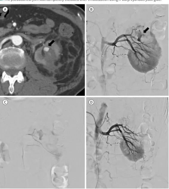
Fig. 1. Patient who developed hematuria after partial nephrectomy for renal cell carcinoma.
A. CT reveals a pseudoaneurysm (arrow) arising from the renal artery.
B, C. Renal angiography demonstrates a pseudoaneurysm (arrow), which is superselected using a micro- catheter.
D. The pseudoaneurysm was completely excluded after embolization using n-butyl cyanoacrylate glue.
Table 1. Outcomes of Transarterial Embolization for Hemorrhage after Partial Nephrectomy
Author Type of Surgery Number of Patient Hemorrhage Onset Technical Success
Heye et al. (56) OPN 251 7 8.9 (2–22)* 100
Netsch et al. (57) OPN 289 4 12.5 (6–25)† 100
Shapiro et al. (9) LPN 259 6 12.6 (5–23)* 100
Hyams et al. (6) LPN/RALPN 998 20 14.5 (3–24)* 100
Shin et al. (7) OPN/RALPN/LPN 1187 36 5 (0–89)† 94.4
*Presented as mean and range.
†Presented as median and range.
LPN = laparoscopic partial nephrectomy, OPN = open partial nephrectomy, RALPN = robot-assisted laparoscopic partial nephrectomy A
C
B
D
상적으로 형성되어 있어 가성동맥류나 파열에 취약한 것으로 알려져 있다(23). AML 환자의 약 20%는 결정성 경화증에 동반하여 발생하고, 결정성 경화증 환자에서 일생 동안 약 반수에서 AML 이 발생하는 것으로 알려져 있다(24).
과거에는 영상의학적으로 악성 종과의 감별이 쉽지 않아 산발성 AML (sporadic AML)은 대부 분 외과적으로 절제를 하였다(22). 하지만, MRI를 이용한 영상의학적 진단기술이 빠르게 발전하 여 현재는 AML의 진단 정확도가 크게 향상되어 수술보다는 보전적 치료를 시행하는 것으로 바뀌 게 되었다(25). 최소침습적 치료의 대표적인 방법으로 경동맥 색전술이 널리 받아들여지고 있다 (26). 경동맥 색전술은 수술적 절제와 비교하여 종양의 영양 동맥만을 초선택하여 시술하므로 잔 여 신기능을 최대한 보존할 수 있으며, 시술 후 회복이 빠르다는 장점이 있다. 특히, AML이 양측 성으로 발현할 수 있는 결절성 경화증 환자에게 있어 신기능 보존은 더욱 중요할 수 있다.
색전술 치료의 적응증과 치료 효과
현재까지 활동성 후복강 출혈 혹은 증상(통증, 혈뇨)을 동반한 AML이 색전술의 적응증으로 널 리 받아들여지고 있다(27). 종양의 크기가 4 cm 이상일 경우 활동성 출혈로 이어질 가능성이 높다 고 보고된 이후에 이를 기준으로 하기도 하였으나 후속 연구에서 크기 기준을 6~8 cm으로 늘리는 것을 제안하기도 하였다(28, 29). AML 환자에서 초선택적 경동맥 색전술은 90%를 상회하는 좋은 기술적 성공률을 보였으며, 종양 크기의 감소는 내부에 혈관과 근육층을 많이 포함할수록 병변의 크기가 많이 줄어드는 것으로 보고되었다(Fig. 2) (30, 31). AML에 대한 색전술에는 다양한 색전 물질이 단독 혹은 병용하여 사용되고 있다. 색전 물질에 대한 메타연구에서 367명 포함되었으며, 172명(46.8%)의 환자에서 2가지 이상의 색전 물질이 병용되어 사용되었으며, 코일만 단독으로 사 용한 환자가 23명(46.8%)이었다(27). 에탄올만을 사용한 환자는 153명(41.7%)이었으며, 코일만 단독으로 사용한 환자가 23명(6.2%)이었다. 에탄올 단독 요법을 시행한 연구의 경우 색전술의 기 술적 성공률이 95.5% 였으며, 색전술 후 평균 크기 감소는 2.3 cm였다. 2개 이상의 색전 물질을 사 용한 경우 기술적 성공률 95% 색전술 후 종괴의 평균 크기 감소는 4.6 cm이었다. 따라서, AML의 색전술 치료에 있어 어느 색전 물질이 다른 색전 물질보다 치료 효과가 우수하다고 특정하기는 어 려우며, 시술자에게 익숙한 색전 물질을 적절히 사용하여 초선택적으로 시술하는 것이 중요하다 고 할 수 있다.
경피적 시술과 관련된 신장 손상(Iatrogenic Renal Injury)에 대한 색전술
임상현장에서 신장을 대상으로 조직검사, 신루술, 신석절제술 등 다양한 경피적 시술이 이루어 지며, 이로 인해 합병증이 발생하기도 한다. 시술과 관련된 출혈은 1~7%의 환자에서 발생하며, 대 개 자연적으로 치유되나 때로는 심한 혈뇨나 불안정한 생체징후로 인해 색전술과 같은 적극적인 치료법이 필요할 수 있다(32).
색전술의 효과
의인성 신장 혈관 손상에 대한 색전술의 기술적 그리고 임상적 성공률은 각각 87~100%, 57~
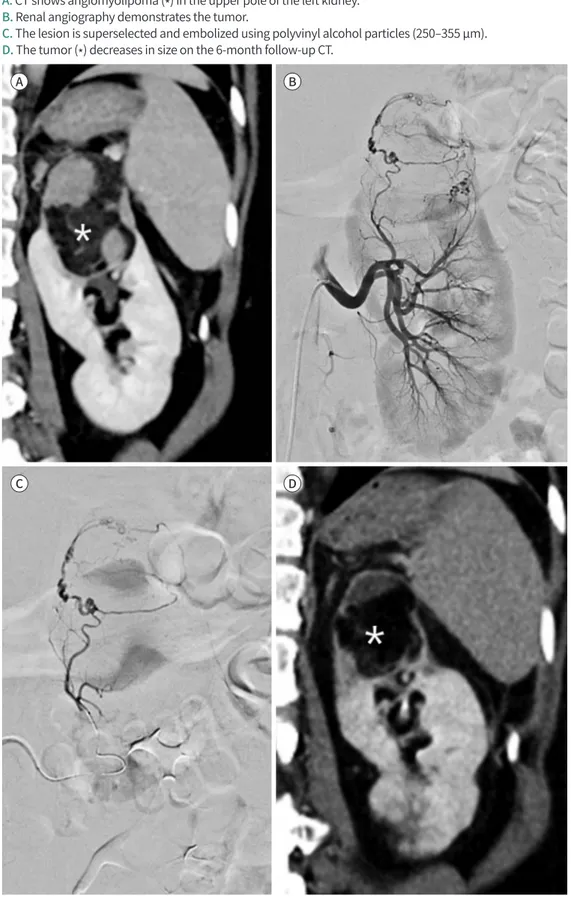
Fig. 2. Angiomyolipoma patient who complained of flank pain.
A. CT shows angiomyolipoma (*) in the upper pole of the left kidney.
B. Renal angiography demonstrates the tumor.
C. The lesion is superselected and embolized using polyvinyl alcohol particles (250–355 µm).
D. The tumor (*) decreases in size on the 6-month follow-up CT.
A
C
B
D
100%로 보고되었다(Table 2) (33-39). Choi 등(38)은 경피적 신장 시술 후에 혈관조영술을 시행한 79명의 환자를 후향적으로 분석하였다(Fig. 3). 81%의 환자에서 혈관조영술에서 출혈이 확인되어 단독 혹은 2개 이상의 색전 물질을 이용해서 색전술을 시행 받았으며, 출혈이 확인되지 않은 환자 15명 중 6명이 경험적 색전술을 시행 받았다. 기술적 성공률은 100%였으며, 임상적 성공률은 85.7%였으며, 경험적 색전술을 시행 받은 6명의 환자는 모두 임상적으로 호전되었다. 드물게 출 혈이 신장 동맥이 아닌 신장 피막 동맥, 요추 동맥, 표재성 장골동맥 등에서도 출혈이 발생한 경우 가 있어 주의해야 한다. Park 등(40)은 간이식, 신장 조직검사, 신루술 등을 시행 받은 환자에서 생 긴 신장피막동맥(renal capsular artery)으로 부터의 출혈에 대한 색전술의 효과를 분석하였다(40).
기술적 성공률은 90.9%, 임상적 성공률은 80%로 보고하였는데, 신장 또는 신장 주변의 경피적 시 술, 간 하부 공간(subhepatic space) 혹은 신장주변(perirenal space)에 혈종이나 출혈이 있는 경 우 신장피막동맥을 포함해서 혈관조영술을 시행하는 것이 필요하다.
신장에 대한 경피적 시술로 인한 출혈은 신장 동맥을 비롯한 다양한 혈관에서 발생할 수 있으므 로 이에 대한 이해를 바탕으로 출혈 혈관에 대한 초선택적인 색전술을 시행하는 것이 중요하겠다.
전립선과 방광
방광이나 전립선에서도 심한 출혈이 유발될 수 있으며, 대개 혈뇨로 나타나게 된다. 심한 혈뇨 의 원인으로는 방광암, 방사선 방광염, 사이클로스포린(cyclosporine) 유발성 방광염, 전립선의 경요도 치료, 전립선암 등이 있다. 이러한 환자들에서 혈뇨가 보존적 치료로 호전되지 않는 경우 가 많다(41). 수술적 지혈은 위험성이 높고 접근도 어려워 흔하게 시행되기 어려운 가운데 경동맥 색전술로 방광 동맥이나 전립선 동맥을 색전 하는 것이 좋은 치료법으로 대두되고 있다.
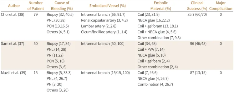
Table 2. Outcomes of Transarterial Embolization for Vascular Injury Associated with Percutaneous Procedures Author Number
of Patient
Cause of
Bleeding (%) Embolized Vessel (%) Embolic
Material (%)
Clinical Success (%)
Major Complication Choi et al. (38) 79 Biopsy (32, 40.5)
PNL (30,38) PCN (13,16.5) Others (4, 5.1)
Intrarenal branch (66, 91.7) Renal capsular artery (3, 4.2) Lumbar artery (2, 2.8) Cicumflex iliac artery (1, 1.4)
Coil (23, 31.9) NBCA glue (16,22.2) Coil + gelforam (13, 18.1) Coil + NBCA glue (4, 5.6) Other combination (7, 9.8)
85.7 (60/70) 0
Sam et al. (37) 50 Biopsy (17, 34) PNL (14, 28) PN (11,22) PCN (5, 10) Others (3, 6)
Intrarenal branch (50, 100) Coil (34, 68) Coil + PVA (7, 14) NBCA glue (5, 10) Coil + gelfoam (2, 4) Other combination (2, 4)
96 (46/48) 0
Mavili et al. (39) 15 Biopsy (5, 33.3) PNL (4, 26.7) PN (3, 20) Others (3, 20)
Intrarenal branch (15/15, 100) Coil (7, 46.6) NBCA glue (4, 26.7) Combination (4, 26.7)
87 (13/15) 0
NBCA = N-butyl cyanoacrylate, PCN = percutanous nephrostomy, PN = partial nephrectomy, PNL = pecutaneous nephrolithotomy, PVA = poly- vinyl alcohol
색전술의 효과와 합병증
방광이나 전립선에서 발생한 출혈로 인한 시행된 색전술의 효과에 대한 보고는 많지 않지만, 초 선택적 색전술의 기술적 성공률이 92.6~100%로 알려져 있다(Fig. 4) (42-44). 골반강 내 동맥은 작 은 분지들 사이에 많은 연결성을 보이므로 가급적 초선택적 색전술이 중요하다. 초선택 색전술을 시행하지 않은 경우 허혈성 합병증이 68.5%의 많은 환자에서 발생했다는 보고가 있는 반면, 초선 택적 시술 시 10% 미만의 낮은 합병증을 보고하였다(42-45). 다만, 하부요도에 출혈이 대개 고령 의 환자에서 발생하여 접근 동맥에 협착이 있거나 구불구불한 경우가 많아 내장골동맥의 접근이
Fig. 3. A 49-year-old male who developed hemorrhage after percutaneous renal biopsy.A. CT reveals a pseudoaneurysm and hematoma in the kidney.
B. Renal angiography shows a pseudoaneurysm (arrow).
C. The affected branch is embolized using n-butyl cyanoacrylate glue and vascular plug.
D. Follow-up CT shows mild atrophy of the renal parenchyma, supplied by the embolized vessel.
CT = computed tomography A
C
B
D
어렵거나 표적 동맥의 초선택이 어려운 경우가 있어, C-arm CT를 이용한 혈관 평가 및 시술자의 숙련도 매우 중요하다.
보존적 치료로 조절되지 않는 방광이나 전립선에서 발생한 출혈에 대해 색전술을 시도해 볼 수 있 으며, 이로 인해 합병증의 위험성이 높은 수술적 치료의 필요성을 낮출 수 있다. 하지만, 다른 장기와 비교해서 색전술의 효과와 안정성에 대한 근거가 적은 편으로 이에 대한 추가 연구가 필요하다.
음경(Penis)
지속발기증(Priapism)
지속발기증(arterial, high-flow priapism)은 성적 자극이나 욕구와는 상관없이 지속적으로 음 경이 발기되어 있는 질환이다(46, 47). 병태생리에 따라 고혈류(high-flow) 혹은 저혈류성(low- flow) 지속발기증으로 분류된다. 고혈류성 지속발기증은 음경이나 회음부의 둔상으로 인해 해면 체 동맥(cavernosal artery)가 손상되어 동맥과 열공(arterio-lacular) 간에 누공(fistula)이 형성되 어 발생한다(48).
경동맥 색전술 치료
경동맥 색전술은 동맥 손상이 원인이 되는 고혈류성 지속발기증 치료에 사용되어 왔다(49). 경 동맥 색전술은 1977년 Wear 등(50)에 의해 처음 시도된 이후로 초선택적 색전술의 도입으로 고혈 류성 지속발기증 치료에 널리 쓰이고 있는데, 이를 위해서는 내장골동맥에서 기원하는 음경 동맥 (penile artery)의 혈류를 다양한 색전 물질을 이용해 차단한다(Fig. 5). 색전 물질로는 자가 혈전 (autologous blood clot), NBCA glue, polyvinyl alcohol (PVA), 코일 등 다양하게 사용되고 있다.
2003년 미국 비뇨기과학회(American Urological Association) 가이드라인에 따르면 지속발기증 치료에 있어 일시적 색전 물질의 사용을 권장했다(51). 그 이유는 시술 성공률(74% vs. 78%)은 비
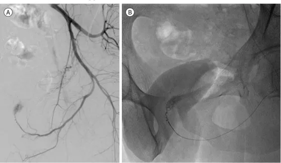
Fig. 4. Bladder cancer patient who complained of hematuria.A. CT shows a tumor on the left side of the bladder.
B. The mass is opacified on left internal iliac angiography.
C. The feeding artery is superselected and embolized using Gelfoam.
A B C
슷하면서도 색전술 후 발기 부전 발생률(5% vs. 39%)이 영구 색전 물질을 사용했을 경우에 높았 기 때문이다. 일부 연구자들은 색전술 후에 혈관 재개통 및 발기능 보존을 위해 일시적인 색전 물 질의 사용을 주장하였으나, 다른 연구자들은 영구 색전 물질을 사용해서도 발기능을 보전하면서 색전술이 가능하다고 보고하였다(52, 53).
Kim 등(54)은 다기관 후향적 연구에서 고혈류성 지속발기증으로 초선택적 색전술을 시행 받은 27명의 환자를 대상으로 색전술의 효용성과 안정성에 대해 분석하였다. 24명(89%)의 환자에서 1 회의 색전술로 지속발기증이 효과적으로 치료되었으며, 2명(7%)의 환자는 반복 시술이 필요하였 으며 나머지 2명(7%)은 정맥폐쇄성 병변을 동반하여 단락(shunt) 수술을 추가로 받아야 했다. 평 상시 정상적인 발기능을 가졌던 23명의 환자 중 18명(78%) 이 색전술 후에도 정상적인 발기능을 유지할 수 있었다. 색전 물질로 사용된 젤폼과 자가혈전은 반복 시술이나 시술 후 발기능과 유의한 연관 관계를 가지지는 않았다( p=0.537). Kojima 등(55)은 96예의 고혈류성 지속발기증을 분석하였 는데, 일시적 색전 물질을 사용했던 33명의 환자 중 28명에서 증상 호전을 경험했으며, 32명에서 발기능을 보존할 수 있었다. 영구 색전 물질을 사용한 경우 63명 모두에서 증상 호전 및 61명에서 발기능을 보존할 수 있었다고 보고하였다. 임상 증상 호전에서는 영구 색전 물질을 사용한 경우에 서 유의하게 효과가 좋았으나, 발기능 보존 측면에서는 두 군 간에 유의한 차이를 보이지 않았다.
고혈류성 지속발기증 치료에 있어 색전술은 뛰어난 치료 효과를 보였으며, 시술 후 발기능 보존 에도 탁월한 효과를 보여왔다. 시술자에서 익숙한 색전 물질을 이용해서 초선택적으로 시술하는 것이 치료 효과를 극대화하는데 가장 중요한 것으로 여겨지며, 향후 좀 더 많은 환자를 대상으로 보다 안전하고 효과적으로 시술할 수 있는 색전 물질에 대한 연구가 더 필요하다.
Fig. 5. A 34-year-old male who developed priapism.
A. Penile angiography shows contrast extravasation.
B. Embolization is performed using gelfoam.
A B
결론
본 종설에서 살펴본 것과 같이 출혈을 비롯하여 다양한 비뇨기 계통의 질환에 대해 색전술은 유 용하게 사용되고 있다. 안전하고 효과적인 색전술을 위해서는 가급적 출혈이 있는 혈관을 초선택 해야 하며 혈역동학과 해부학을 고려하여 적절한 색전 물질을 사용하는 것이 필수적이다.
Conflicts of Interest
The author has no potential conflicts of interest to disclose.
REFERENCES
1. MacLennan S, Imamura M, Lapitan MC, Omar MI, Lam TB, Hilvano-Cabungcal AM, et al. Systematic review of perioperative and quality-of-life outcomes following surgical management of localised renal cancer. Eur Urol 2012;62:1097-1117
2. Thompson RH, Siddiqui S, Lohse CM, Leibovich BC, Russo P, Blute ML. Partial versus radical nephrectomy for 4 to 7 cm renal cortical tumors. J Urol 2009;182:2601-2606
3. Pahernik S, Roos F, Hampel C, Gillitzer R, Melchior SW, Thüroff JW. Nephron sparing surgery for renal cell carcinoma with normal contralateral kidney: 25 years of experience. J Urol 2006;175:2027-2031
4. Albani JM, Novick AC. Renal artery pseudoaneurysm after partial nephrectomy: three case reports and a lit- erature review. Urology 2003;62:227-231
5. Jain S, Nyirenda T, Yates J, Munver R. Incidence of renal artery pseudoaneurysm following open and mini- mally invasive partial nephrectomy: a systematic review and comparative analysis. J Urol 2013;189:1643- 1648
6. Hyams ES, Pierorazio P, Proteek O, Sukumar S, Wagner AA, Mechaber JL, et al. Iatrogenic vascular lesions af- ter minimally invasive partial nephrectomy: a multi-institutional study of clinical and renal functional out- comes. Urology 2011;78:820-826
7. Shin J, Han K, Kwon JH, Kim GM, Kim D, Han SC, et al. Clinical results of transarterial embolization to control postoperative vascular complications after partial nephrectomy. J Urol 2019;201:702-708
8. Leslie S, Goh AC, Gill IS. Partial nephrectomy--contemporary indications, techniques and outcomes. Nat Rev Urol 2013;10:275-283
9. Shapiro EY, Hakimi AA, Hyams ES, Cynamon J, Stifelman M, Ghavamian R. Renal artery pseudoaneurysm following laparoscopic partial nephrectomy. Urology 2009;74:819-823
10. Jeon CH, Seong NJ, Yoon CJ, Byun SS, Lee SE. Clinical results of renal artery embolization to control post- operative hemorrhage after partial nephrectomy. Acta Radiol Open 2016;5:2058460116655833
11. Chen J, Yang M, Wu P, Li T, Ning X, Peng S, et al. Renal arterial pseudoaneurysm and renal arteriovenous fis- tula following partial nephrectomy. Urol Int 2018;100:368-374
12. Gahan JC, Gaitonde M, Wadskier L, Cadeddu JA, Trimmer C. Renal function outcomes following selective angioembolization for iatrogenic vascular lesions after partial nephrectomy. J Endourol 2013;27:1516-1519 13. Strobl FF, D’Anastasi M, Hinzpeter R, Franke PS, Trumm CG, Waggershauser T, et al. Renal pseudoaneurysms
and arteriovenous fistulas as a complication of nephron-sparing partial nephrectomy: technical and func- tional outcomes of patients treated with selective microcoil embolization during a ten-year period. Rofo 2016;188:188-194
14. Ghoneim TP, Thornton RH, Solomon SB, Adamy A, Favaretto RL, Russo P. Selective arterial embolization for pseudoaneurysms and arteriovenous fistula of renal artery branches following partial nephrectomy. J Urol 2011;185:2061-2065
15. Han K, Ahmed BM, Kim MD, Won JY, Lee DY, Kim GM, et al. Clinical outcome of transarterial embolization for postgastrectomy arterial bleeding. Gastric Cancer 2017;20:887-894
16. Huang YS, Chang CC, Liou JM, Jaw FS, Liu KL. Transcatheter arterial embolization with N-butyl cyanoacry- late for nonvariceal upper gastrointestinal bleeding in hemodynamically unstable patients: results and pre- dictors of clinical outcomes. J Vasc Interv Radiol 2014;25:1850-1857
17. Koo HJ, Shin JH, Kim HJ, Kim J, Yoon HK, Ko GY, et al. Clinical outcome of transcatheter arterial emboliza-
tion with N-butyl-2-cyanoacrylate for control of acute gastrointestinal tract bleeding. AJR Am J Roentgenol 2015;204:662-668
18. Koo KC, Kim WT, Ham WS, Lee JS, Ju HJ, Choi YD. Trends of presentation and clinical outcome of treated re- nal angiomyolipoma. Yonsei Med J 2010;51:728-734
19. Soulen MC, Faykus MH Jr, Shlansky-Goldberg RD, Wein AJ, Cope C. Elective embolization for prevention of hemorrhage from renal angiomyolipomas. J Vasc Interv Radiol 1994;5:587-591
20. Murray TE, Lee MJ. Are we overtreating renal angiomyolipoma: a review of the literature and assessment of contemporary management and follow-up strategies. Cardiovasc Intervent Radiol 2018;41:525-536 21. Mues AC, Palacios JM, Haramis G, Casazza C, Badani K, Gupta M, et al. Contemporary experience in the
management of angiomyolipoma. J Endourol 2010;24:1883-1886
22. Oesterling JE, Fishman EK, Goldman SM, Marshall FF. The management of renal angiomyolipoma. J Urol 1986;135:1121-1124
23. Lenton J, Kessel D, Watkinson AF. Embolization of renal angiomyolipoma: immediate complications and long-term outcomes. Clin Radiol 2008;63:864-870
24. Nelson CP, Sanda MG. Contemporary diagnosis and management of renal angiomyolipoma. J Urol 2002;
168:1315-1325
25. Lienert AR, Nicol D. Renal angiomyolipoma. BJU Int 2012;110 Suppl 4:25-27
26. Faddegon S, So A. Treatment of angiomyolipoma at a tertiary care centre: the decision between surgery and angioembolization. Can Urol Assoc J 2011;5:E138-141
27. Murray TE, Doyle F, Lee M. Transarterial embolization of angiomyolipoma: a systematic review. J Urol 2015;
194:635-639
28. Mourikis D, Chatziioannou A, Antoniou A, Kehagias D, Gikas D, Vlahos L. Selective arterial embolization in the management of symptomatic renal angiomyolipomas. Eur J Radiol 1999;32:153-159
29. Dickinson M, Ruckle H, Beaghler M, Hadley HR. Renal angiomyolipoma: optimal treatment based on size and symptoms. Clin Nephrol 1998;49:281-286
30. Hocquelet A, Cornelis F, Le Bras Y, Meyer M, Tricaud E, Lasserre AS, et al. Long-term results of preventive em- bolization of renal angiomyolipomas: evaluation of predictive factors of volume decrease. Eur Radiol 2014;
24:1785-1793
31. Ramon J, Rimon U, Garniek A, Golan G, Bensaid P, Kitrey ND, et al. Renal angiomyolipoma: long-term results following selective arterial embolization. Eur Urol 2009;55:1155-1161
32. Summerton DJ, Kitrey ND, Lumen N, Serafetinidis E, Djakovic N; European Association of Urology. EAU guidelines on iatrogenic trauma. Eur Urol 2012;62:628-639
33. Dinkel HP, Danuser H, Triller J. Blunt renal trauma: minimally invasive management with microcatheter em- bolization experience in nine patients. Radiology 2002;223:723-730
34. Dorffner R, Thurnher S, Prokesch R, Bankier A, Turetschek K, Schmidt A, et al. Embolization of iatrogenic vascular injuries of renal transplants: immediate and follow-up results. Cardiovasc Intervent Radiol 1998;21:
129-134
35. Perini S, Gordon RL, LaBerge JM, Kerlan RK Jr, Wilson MW, Feng S, et al. Transcatheter embolization of biop- sy-related vascular injury in the transplant kidney: immediate and long-term outcome. J Vasc Interv Radiol 1998;9:1011-1019
36. Poulakis V, Ferakis N, Becht E, Deliveliotis C, Duex M. Treatment of renal-vascular injury by transcatheter em- bolization: immediate and long-term effects on renal function. J Endourol 2006;20:405-409
37. Sam K, Gahide G, Soulez G, Giroux MF, Oliva VL, Perreault P, et al. Percutaneous embolization of iatrogenic arterial kidney injuries: safety, efficacy, and impact on blood pressure and renal function. J Vasc Interv Radiol 2011;22:1563-1568
38. Choi MJ, Kim PH, Shin JH, Kim JW, Gwon DI, Kim JH, et al. Angiographic management of percutaneous re- nal procedure-related bleeding: a single-center experience. Int J Urol 2019;26:406-412
39. Mavili E, Dönmez H, Ozcan N, Sipahiog˘lu M, Demirtas¸ A. Transarterial embolization for renal arterial bleed- ing. Diagn Interv Radiol 2009;15:143-147
40. Park HJ, Shin JH, Han KC, Yoon HK, Ko GY, Sung KB. Transcatheter arterial embolization of angiographically visible and occult renal capsular artery hemorrhage in 28 patients. J Vasc Interv Radiol 2016;27:973-980 41. Choong SK, Walkden M, Kirby R. The management of intractable haematuria. BJU Int 2000;86:951-959 42. Delgal A, Cercueil JP, Koutlidis N, Michel F, Kermarrec I, Mourey E, et al. Outcome of transcatheter arterial
embolization for bladder and prostate hemorrhage. J Urol 2010;183:1947-1953
43. Prasad V, Sacks BA, Kraus S, Clouse ME. Embolotherapy for lower urinary tract hemorrhage. J Vasc Interv Radiol 2009;20:965-970
44. Rastinehad AR, Caplin DM, Ost MC, VanderBrink BA, Lobko I, Badlani GH, et al. Selective arterial prostatic embolization (SAPE) for refractory hematuria of prostatic origin. Urology 2008;71:181-184
45. Pisco JM, Martins JM, Correia MG. Internal iliac artery: embolization to control hemorrhage from pelvic neo- plasms. Radiology 1989;172:337-339
46. Pautler SE, Brock GB. Priapism. From priapus to the present time. Urol Clin North Am 2001;28:391-403 47. Sánchez-López S, González-Gómez S, Di Lizio-Miele K, González-Gómez J. High-flow priapism treated with
superselective transcatheter embolization using polyvinyl alcohol particles. SAGE Open Med Case Rep 2017;
5:2050313X17693179
48. Burnett AL, Bivalacqua TJ. Priapism: new concepts in medical and surgical management. Urol Clin North Am 2011;38:185-194
49. Liu BX, Xin ZC, Zou YH, Tian L, Wu YG, Wu XJ, et al. High-flow priapism: superselective cavernous artery em- bolization with microcoils. Urology 2008;72:571-573; discussion 573-574
50. Wear JB Jr, Crummy AB, Munson BO. A new approach to the treatment of priapism. J Urol 1977;117:252-254 51. Montague DK, Jarow J, Broderick GA, Dmochowski RR, Heaton JP, Lue TF, et al. American Urological Associ-
ation guideline on the management of priapism. J Urol 2003;170:1318-1324
52. Bastuba MD, Saenz de Tejada I, Dinlenc CZ, Sarazen A, Krane RJ, Goldstein I. Arterial priapism: diagnosis, treatment and long-term followup. J Urol 1994;151:1231-1237
53. Park JK, Jeong YB, Han YM. Recanalization of embolized cavernosal artery: restoring potency in the patient with high flow priapism. J Urol 2001;165:2002-2003
54. Kim KR, Shin JH, Song HY, Ko GY, Yoon HK, Sung KB, et al. Treatment of high-flow priapism with superselec- tive transcatheter embolization in 27 patients: a multicenter study. J Vasc Interv Radiol 2007;18:1222-1226 55. Kojima H, Tanigawa N, Kariya S, Komemushi A, Shomura Y, Yanishi M, et al. High-flow priapism undergoing
arterial embolization: review of literature following American Urological Association guideline on the man- agement of priapism. Minim Invasive Ther Allied Technol 2009;18:1-5
56. Heye S, Maleux G, Van Poppel H, Oyen R, Wilms G. Hemorrhagic complications after nephron-sparing sur- gery: angiographic diagnosis and management by transcatheter embolization. AJR Am J Roentgenol 2005;
184:1661-1664
57. Netsch C, Brüning R, Bach T, Gross AJ. Management of renal artery pseudoaneurysm after partial nephrec- tomy. World J Urol 2010;28:519-524
비뇨기계 질환에서의 색전술의 역할
한 기 창*
비뇨기계 질환에서의 경동맥색전술은 널리 쓰이고 있다. 대표적으로 종양, 외상, 경피적 시술 등 여러 원인에 의해 출혈이 발생하는 경우 대개 보존적 치료로 조절이 가능하지만, 때로는 혈역학적 불안정이 초래될 중대한 출혈이 발생할 경우 경동맥 색전술이 유용하게 쓰일 수 있 다. 이는 수술적 치료법에 비해 합병증은 덜 유발하면서도 뛰어난 치료성적과 안정성을 보여 왔기 때문이다. 장기별로는 부분신제술 후에 발생한 출혈이나, 신종괴로 인한 출혈에 대한 고 식적 색전술을 비롯하여 외상으로 인해 발생한 출혈에 대해 색전술이 활발하게 이루어지고 있다. 혈관근육지방종의 파열 예방이나 파열 시 지혈 목적으로의 색전술이 사용될 수 있다.
전립선이나 방광의 경우 종양이나 외상으로 인해 발생한 출혈 및 음경에서 발생한 지속 발기 증에도 색전술의 효용성이 입증되었다. 다양한 비뇨기 질환에서의 색전술은 지혈을 위한 일 차적 치료로 고려될 수 있으며, 가장 적합한 색전물질을 이용해서 초선택적으로 시행하는 것 이 시술의 효용성 및 안전성을 높이는데 중요하다.
연세대학교 의과대학 세브란스병원 영상의학과