70 서 론
일조량, 계절, 시공간에따라환경수내의용존산소량은현 저히다른양상을나타낸다. 이러한환경적용존산소량(dis- solved oxygen, DO)의변동성은어류의성장, 생식발달과밀 접한영향을가진다(Wagner et al. 1986, Soitamo et al. 2001).
어류는저산소(hypoxia) 환경조건에서생존하기위하여호흡, 대사변화및산소전달을수행하는헤모글로빈(hemo-globin) 분자의변형 등다양한생리적, 생화학적적응과정을거치 게된다(Shoubridge and Hochachka, 1981; Weber, 1982). 포 유동물의경우, 체내 hypoxia에의해높은발현이유도되는 포도당수용체(glucose transporter), 당분해효소(glycolytic enzymes), 적혈구생성촉진인자(erythropoietin), 트렌스페린 (transferrin), 혈관내피성장인자(vascular endothelial growth factor, VEGF) 등 40가지이상의유전자가보고된바있다(Se-
menza 1999). 하지만, 포유동물과비교하여어류의관련유전 자의분리와발현조절, 기능에대한연구는상대적으로부족한 실정이다. 포유동물에서 hypoxia 의존적인유전자들의전사활 성은주로저산소증유발인자(hypoxia-inducible factor-1, HIF- 1)에의해조절된다. HIF-1은 HIF-1α과 HIF-1β로구성된이 형이량체(heterodimer)로서전사활성을조절하며, 특히 HIF-1
α는세포내 hypoxia에특이적으로반응하는전사조인자로
보고되고있다(Wang et al. 1995, Wang and Semenza 1995).
HIF-1α mRNA와단백질은 hypoxia 가유발될경우발현량이 현저히증가하게되며(Huang et al. 1996, Kallio et al. 1997), 반대로 정상적인혈중 산소조건에서 HIF-1α 단백질은 26S proteasome에의해신속히분해되어낮은수준으로회복되어 진다(Salceda and Caro 1997, Huang et al. 1998).
최근포유동물의 hypoxia에의해발현이유도되는단백질로
Article history;
Received 17 September 2012; Revised 6 December 2012; Accepted 12 February 2013
*Corresponding author: Tel: +82. 33. 640. 2348 Fax: +82. 33. 640. 2348 E-mail address: [email protected]
Kor J Fish Aquat Sci 46(1) 070-076, February 2013 http://dx.doi.org/10.5657/KFAS.2013.0070 pISSN:0374-8111, eISSN: 2281-8815
ⓒ The Korean Society of Fishereis and Aquatic Science. All rights reserved
Some fish live in aquatic environments with low or temporally changing O2 availability. Variation in dissolved oxy- gen (DO) levels requires behavioral, physiological, and biochemical adaptations to ensure the uptake of sufficient O2. Several species are relatively well adapted to tolerate low O2 partial pressures (hypoxia). The medaka (Oryzias dancena) is an important model organism for biomedical research that shows remarkable tolerance to hypoxia. We investigated the regulation and role of hypoxia-inducible factor-1 (HIF-1α) as a general hypoxia-response gene and stanniocalcin-2 (STC2), which is one of the genes regulated by HIF-1α in mammals under hypoxia. We subjected adult male medaka to the following three acute hypoxia regimes: 1, 24, and 72 h at DO = 1.8±0.5 ppm. The changes in STC2 and HIF-1α mRNA were monitored using quantitative real-time reverse-transcription PCR. We found strong upregulation of HIF-1α mRNA in the livers of fish exposed to hypoxia. Hypoxia rapidly upregulated STC- 2 mRNA expression in muscle, but not in the brain, gills, liver, or intestine. Therefore, unlike in mammals, hypoxia
might regulate O. dancena STC-2 expression in an HIF-1α-independent manner.
Key words : Oryzias dancera, Hypoxia, Hypoxia-inducible factor, Medaka, Real-time PCR, Stanniocalcin
저산소환경에 의한 송사리( Oryzias dancena)의 Stanniocalcin-2와 Hypoxia-Inducible Factor-1αmRNA 발현의 변화
강릉원주대학교 해양분자생명공학과
Ji hye Shin and Young Chang Sohn* 신지혜·손영창*
Changes in Stanniocalcin-2 and Hypoxia-Inducible Factor-1α mRNA Expression in Medaka Oryzias dancena Exposed to Acute Hypoxia
Department of Marine Molecular Biotechnology, Gangneung-Wonju National University, Gangneung 210-702, Korea
서스타니오칼신(stannio-caincin, STC1)이보고된바있다. STC1은경골어류에서칼슘의흡수를억제시키는항과잉칼슘 호르몬으로서그기능이최초로밝혀진호르몬이다(Butkus et al. 1987, Sundell et al. 1992). 포유류 STC1은항과잉칼슘작 용과더불어위암과난소암세포의증식, 전이및종양형성에 관여하여, 마우스난소의성스테로이드분비를조절한다(Luo et al. 2004, Liu et al. 2010, He et al. 2011). STC1 상동단백 질인 STC2는포유동물에서최초로분리되었으며, STC-1과 는비교적낮은상동성(~30%)을나타낸다(Chang and Red- del 1998, DiMattia et al. 1998, Ishibashi et al. 1998). 하지만, STC2는유방암, 신장, 췌장암의발달에따라특이적으로발현 이증가하고, 종양성장과전이를촉진한다는점에서 STC1과 유사한생리적기능을나타낸다(Bouras et al. 2002, Ieta et al.
2009, Meyer et al. 2009). 대부분의종양성장과전이는세포 내 hypoxia와소포체스트레스(endoplasmic reticulum stress) 와밀접한연관성을지니고있다. 이러한현상은암세포의생 존을위한신생혈관형성(angiogenesis)의조건에필수적이며,
이과정중 HIF-1에의해발현이중재되는다양한유전자들이
암세포발달에관여된다(Koumenis et al. 2002, Koumenis and Wouters 2006). STC-1은인두암세포주 CNE-2에서 STC-2는 난소암세포주 SKOV3에서세포의증식에따른 hypoxia에의 해발현이증가된다고보고된바있다(Yeung et al. 2005, Law and Wong 2010). STC-1과 STC-2 유전자의프로모터영역내 HIF-1α 단백질의결합영역(hypoxia response element, HRE) 이존재하며, HIF-1α에의해 STC-1과 STC-2 유전자의전사 활성이유도된다(Yeung et al. 2005, Law and Wong 2010).
어류의 hypoxia에의한 HIF α subunits (HIF-1α, -2α, -3α) 유전자, 단백질의발현유도와전사조절메커니즘은포유류의 경우와유사하게나타나며(Soitamo et al. 2001, Rahman and Thomas 2007, Chen et al. 2012), 그외 hypoxia 의존적발현 양상을보이는유전자들이동정되고있다(Ju et al. 2007, Waw- rowski et al. 2011). 어류의 STC2는복어(Takifugu rubripes), 제브라피쉬(Danio rerio), 넙치(Paralichthys olivaceus)에서
유전자가분리되고일부기능이연구되어있지만(Luo et al.
2005, Shin and Sohn 2009), hypoxia에의한어류내생리적 작용및 HIF-1와의연관성또한명확하지않다. 본연구는광 염성 Oryzias dancena의 hypoxia 반응유전자 HIF-1α mRNA 발현및낮은용존산소에따른 STC2 mRNA의발현변화를분 자수준에서밝히고자하였다. 향후, 환경중의용존산소부족 에따른항상성유지에필요한생리적조절기구를이해하는기 초연구가될것으로사료된다.
재료 및 방법 실험어와 실험조건
실험어 O. dancena는부경대학교해양바이오신소재학과남
윤권교수님으로부터제공받았다. 수컷O. dancena(약 6월령) 를대조군(control; 15마리) 및 hypoxia 실험군(hypoxia; 15마 리)으로나누어수용하였다. 각사육수조(35×19×22 cm)의 수온은 25℃ 내외로유지하였으며, 실험기간중사료는공급하 지않았다. 실험전 2일동안안정화를시킨후, hypoxia 실험 군의사육수는질소가스(100% N2)를주입하여낮은용존산소 조건을유도하였다. 각군의용존산소농도와수온은용존산소 측정기(Hanna Instruments, Smithfield, RI, USA)를이용하여 측정하였으며, 대조군과 hypoxia 실험군의 DO는각각 6.8±
0.5 ppm과 1.8±0.5 ppm으로유지시켜진행하였다. 실험개 시 1시간, 24시간, 72시간경과시점에서각그룹의실험어로부 터뇌, 아가미, 장, 간, 근육을적출하였다(각각 5마리). 조직적 출은실체현미경을이용하여수행하였으며, 각조직은액체질 소에급속동결하여 total RNA를추출하기전까지 -80℃에동 결보관하였다.
RNA 추출
Total RNA는 RNeasy Mini Kit (QIAGEN, Valencia, CA, USA)로추출하였으며, genomic DNA 제거및 cDNA 합성은 QuantiTect Reverse Transcription Kit (QIAGEN)를이용하여 수행하였다. 모든조직의 cDNA는최종농도 0.5 μg으로정량 된 total RNA로부터합성하였다. 각 cDNA는 qRT-PCR 분석 전까지 -20℃에동결보관하였다.
Quantitative real-time reverse-transcription PCR
HIF-1α, STC2 유전자발현분석은 quantitative real-time reverse-transcription PCR (qRT-PCR) 방법으로조사하였 다. 표적유전자 STC2는클로닝한 O. dancena STC2 partial cDNA (GenBank Accession No. JX680809) 염기서열을바탕 으로 oligo primers를제작하였으며(Table 1 and Fig. 1), HIF-1 α (GenBank Accession No. DQ317443)와 18S ribosomal RNA (18S rRNA, GenBank Accession No. HM347347)에대 한 oligo primers는 GenBank에등록된염기서열을바탕으로 Primer Express v3.0 software (Applied Biosystems, Foster
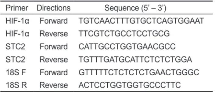
Table 1. Oligo primer sequences used in quantitative real-time reverse-transcription PCR (qRT-PCR)
Primer Directions Sequence (5’ – 3’)
HIF-1α Forward TGTCAACTTTGTGCTCAGTGGAAT HIF-1α Reverse TTCGTCTGCCTCCTGCG
STC2 Forward CATTGCCTGGTGAACGCC STC2 Reverse TGTTTGATGCATTCTCTCTGGA 18S F Forward GTTTTTCTCTCTCTGAACTGGGC 18S R Reverse ACTCCTGGTGGTGCCCTTC
City, CA, USA)를이용하여제작하였다(Table 1). 뇌, 아가미, 장, 간, 근육의 total RNA (0.5 μg)로부터합성된 cDNA를주 형으로 oligo primers (10 μM), SYBR premix Ex-Taq, ROX Referance Dye II와함께총량 20 μL로 qRT-PCR을수행하였 다. PCR은 ABI 7500 Fast Real-Time PCR Systems (Applied Biosystems)을이용하여 50℃에서 2분그리고 95℃에서 10분 반응후, two-step PCR 방법으로 95℃에서 15초, 60℃에서 1 분, 총 40 cycles을수행하였다. qRT-PCR 결과를분석하기전, 각시료의희석배수에따른증폭산물의임계값(cycle thresh-
old, Ct)을통한표준정량과해리곡선분석을통하여사용된
oligo primers의표적유전자에대한특이성을검증하였다. 증 폭된 HIF-1α와 STC2 유전자발현량은 ABI 7500 Sequence Detection Software V.1.3.1 (Applied Biosystems)를이용하여 분석하였으며, 18S rRNA 유전자발현량으로값을보정하였 다.
통계처리
각그룹내 15마리개체의표적유전자발현에대한평균간의
유의성검정은 SPSS V.18.0 통계패키지를이용하여분산분석 후, post hoc Tukey test와 t-test로분석하였다(P<0.05).
결 과 HIF-1α mRNA 발현변화
O. dancena 간의경우, hypoxia 실험군(hypoxia) 송사리의
HIF-1αmRNA 발현은 1시간시점에서일시적으로감소하는
경향을나타낸후, 72 시간시점에서발현이유의적으로증가
하였다(Fig. 2C). Hypoxia 실험군의아가미와장에서 HIF-1
α mRNA 발현이경시적으로증가하는경향을나타내었으나,
대조군(control)과의유의적인차이는없었으며(Fig. 2B and
2D), 뇌와근육의경우는두군간의유의적인차이가확인되지
않았다(Fig. 2A and 2E).
STC2 mRNA 발현변화
O. dancena 근육의경우, hypoxia 유도 1시간시점에서 STC2 mRNA 발현이유의적으로증가하였으며, 72시간시점 Fig. 1. Alignment of medaka STC2 partial sequence and other vertebrate STC2s. The STC2 sequences used for this alignment were ex- tracted from the NCBI GenBank databases. GenBank accession nos. are as follows: Medaka (JX680809; confidential until Oct 1, 2013 or published), Flounder (EU816770), Pufferfish (AY688945), Zebrafish (AY688947), Human (NM003714).
Fig. 2. Effects of hypoxia on HIF-1α mRNA levels. Expression levels of HIF-1α mRNA in medaka that was exposed to hypoxia at 1, 24 or 72 h after inducing hypoxia. The relative HIF-1α mRNA levels were normalized by 18S rRNA values, respectively.
Data were represented by the mean S.E.M of five independent samples (P<0.05). *, t-test; (A) Brain; (B) Gills; (C) Liver; (D) Intestine; (E) Muscle.
Fig. 3. Effects of hypoxia on STC2 mRNA levels. Expression levels of STC2 mRNA in medaka that was exposed to hypoxia at 1, 24 or 72 h after inducing hypoxia. The relative STC2 mRNA levels were normalized by 18S rRNA values, respectively. Data were represented by the mean S.E.M of five independent samples (P<0.05). *, t-test; (A) Brain; (B) Gills; (C) Liver; (D) Intestine;
(E) Muscle.
7 6 5 4 3 2 1 0
Control Hypoxia
1h 24h 72h a
a
a a
a a
HIF-1 α/18rRNAmRNAlevels
(A)
6 5 4 3 2 1 0
Control
Hypoxia b
a a
1h 24h 72h
HIF-1 α/18rRNAmRNAlevels
(B)
ab ab
ab
a
*
a a
a a
b
1h 24h 72h 2.5
2
1.5
1
0.5
0
HIF-1 α/18rRNAmRNAlevels
(C) Control
Hypoxia
a
a a a
a
a
1h 24h 72h 16
14 12 10 8 6 4 2 0
HIF-1 α/18rRNAmRNAlevels
(D) Control
Hypoxia
a
a
a
a
a
1h 24h 72h 14
12 10 8 6 4 2 0
HIF-1 α/18rRNAmRNAlevels
(E)
Control Hypoxia
a
12 10 8 6 4 2 0
Control Hypoxia
1h 24h 72h
a a
a ab
ab b
STC2/18SrRNAmRNAlevels
(A)
7 6 5 4 3 2 1 0
Control Hypoxia
1h 24h 72h a
a
a a
a b
STC2/18SrRNAmRNAlevels
(B)
*
*
7 6 5 4 3 2 1 0
Control Hypoxia
1h 24h 72h a
a
ab a
ab b
STC2/18SrRNAmRNAlevels
(D)
12 10 8 6 4 2 0
Control Hypoxia
1h 24h 72h a
C
ab bc
ab
STC2/18SrRNAmRNAlevels a (E)
8 7 6 5 4 3 2 1 0
Control Hypoxia
1h 24h 72h a
a
a
a
a a
STC2/18SrRNAmRNAlevels
(C)
에서대조군수준으로회복되었다(Fig. 3E). 반면, 뇌의 STC2
mRNA 발현은경시적으로증가하였고, 이는대조군과 hy-
poxia 실험군에서공통적으로나타났다(Fig. 3A). 대조군의아 가미, 간및장의 STC2 mRNA 발현도경시적증가패턴을나 타내었다(Fig. 3B-3D). 특히, 아가미조직의 STC2 mRNA는 hypoxia에의한유의적인발현변화가나타나지않았지만, 대
조군의증가된 STC2 mRNA 발현수준과비교하여상대적으
로낮은발현수준을나타내었다(Fig. 3D).
고 찰
최근송사리과에속하는일본송사리(O. javanicus)와인도송 사리(바다송사리, O. dancena 혹은 O. melastigma)는새로운 동물실험모델로주목을받고있는광염성어종이다 (Kang et al. 2008). 특히, O. dancena는 hypoxia 환경수(0.8-1.8 ppm) 에대한높은적응력과생존력을나타내고, 어류의 hypoxia 유 도실험에적합한동물모델로사용되고있다(Yu et al. 2006).
본연구는 qRT-PCR 분석을통하여 hypoxia 유발에따른 O.
dancena HIF-1α와 STC-2 mRNA의발현변화를조사하였다. 어류의 HIF-1α 유전자는뇌, 심장, 신장, 비장, 간, 생식소를포 함한다양한조직에서광범위하게발현되어지는데(Ju et al.
2007, Chen et al. 2012), 담수어 Chinese sucker (Beaufortia kweichowensis)의 HIF-α subunit mRNAs (HIF-1α, HIF-2α, HIF-3α)는특히간에서높게발현되며, 이중 HIF-1α과 HIF-3 α는 HIF-2α 보다상대적으로 높은 mRNA 발현수준을보 였으며(Chen et al. 2012). 또한, 농어(Dicentrarchus labrax)
의 HIF-1α mRNA는다른조직과비교하여간에서높은발
현수준이관찰되었다(Terova et al., 2008). 하지만조직특이 적인 HIF-1α의발현양상만으로는 hypoxia에대한 HIF-1α 의발현과조절을이해하는것으로는충분하지않다. 본연구 는 O. dancena의간 HIF-1α mRNA가 hypoxia에의존적으 로발현이증가되는경향을확인하였다(Fig. 2). 이결과는이 미보고된바있는 O. melastigma의경우와일치하며(Yu et al.
2006), hypoxia에대한어류의항상성에관여하는주된조직 이간으로추정된다. 어류의경우, hypoxia에의한다양한생 리적, 생화학적변화가수반되며, 특히대사율의저하(Chen et al. 2012), 산소에대한헤모글로빈의친화성증가(Jensen et al.
1993), 높은무기호흡율(Virani and Rees 2000)을나타낸다는 보고가있다. Hypoxia에의한 O. dancena 간 HIF-1α mRNA 의발현증가는체내항상성유지를위한대사작용의변화가 우선적으로일어난것이라사료된다.
일련의 O. dancena 유전자클로닝과정에서 STC2 프로모 터영역(-2397 bp) 내부에후보 HRE 영역(-RCGTG-)을확인 하였다(Data not shown). Hypoxia에의한간특이적인 HIF-1 α mRNA 발현과일치하여 STC2 전사가조절될것이라는가 설을바탕으로 hypoxia에의한 STC2 mRNA의발현을조
사하였다(Fig. 3). 아가미에서는경시적으로 발현이증가하
는대조군의 STC2 mRNA와대조적으로 hypoxia 실험군의
STC2 mRNA 발현은낮은수준으로유지되는경향을나타내
었다(Fig. 3B). 또한, 근육에서는 hypoxia 의존적으로 STC2
mRNA 발현이급증한후, 경시적으로발현이감소하여회복
되는경향을나타냈다(Fig. 3E).
본연구의 hypoxia에의한 O. dancena STC2 mRNA의발
현양상과조직반응은 HIF-1α와이질적인경향을나타내었으
며, 이는 STC2에대한전사가 HIF-1α 의존적으로조절될것 이라는본연구의가설과상응하지않는다. 어류 STC2는포유
동물과달리 HIF-1α와독립적인발현메커니즘에의해조절될
수있으며, hypoxia에대한차별적인생리작용을담당할것이 라사료된다. 프로모터내부에 HRE를가지고있는글로빈유 전자의경우, 일본송사리와제브라피쉬는 hypoxia에의해서 로다른발현양상을나타내는데, 이는같은유전자라도어류 의종에따라 hypoxia의반응역가나조절메카니즘이상이할 수있다는점을시사한다(Wawrowski et al. 2011). 이를명확
히하기위해서는 STC2 전사에대한상위조절자의연구와종
특이적인발현양상에대한조사가요구된다. 포유동물의경 우, STC1과 STC2는이온전이, 스테로이드호르몬합성, 미 토콘드리아의 ATP 합성을포함하여세포내작용과생리적기 능에대한연구결과가있다(Luo et al. 2004, Luo et al. 2005, Ellard et al. 2007, Yeung et al. 2012). STC2의과잉발현을유 도한형질전환 마우스는정상개체보다지연된성장율과 산 소소비량의급증, 섭이량의증가를나타낸다는보고가있으 며, 이는 STC2가호흡과대사조절에밀접하게관여됨을시사 한다(Gagliardi et al. 2005). 어류는환경수로부터유발되는 hypoxia에의해체내의다양한생리적변화가유도되는데, 특 히대사율은감소하고, 무기호흡률은증가시키면서체내항상 성을유지시킨다(Virani and Rees 2000, Chen et al. 2012). 환 경수의낮은 DO에의한 hypoxia는일종의스트레스로작용하 여어류내대사활동을급증시키게되는데, 이는 STC2에의하 여촉발되는효과이며점차체내항상성이유지되는과정에서
STC2 mRNA 발현의회복이근육에서나타난것이라사료된
다. 아가미는항상성과정에서호흡계를조절하게되는데, 무 기호흡으로의효율적인전환을유도하기위하여 STC2의발 현수준이 hypoxia 특이적으로낮게유지된것이라사료된다. 이러한 STC2의 hypoxia에의한발현특이성은실험조직들중 아가미와근육에서국한되어나타나게되는데이는두조직이 환경수의영향을직접적으로접하는 1차적인반응조직으로서 나타나는결과라고생각된다.
사 사
실험어를제공해주신부경대학교해양바이오신소재학과남 윤권교수님께심심한사의를표합니다. 본연구는국토해양부
의재원으로한국해양과학기술진흥원의지원 (20088033-1), 한국연구재단 (2012R1A1A2044506) 및강릉원주대학교교 내연구비(2011-0190)를지원받아수행되었음을밝히며사의 를표합니다.
참고문헌
Bouras T, Southey MC, Chang AC, Reddel RR, Willhite D, Glynne R, Henderson MA, Armes JE and Venter DJ. 2002.
Stanniocalcin 2 is an estrogen-responsive gene coexpressed with the estrogen receptor in human breast cancer. Cancer Res 62, 1289-1295.
Butkus A, Roche PJ, Fernley RT, Haralambidis J, Penschow JD, Ryan GB, Trahair JF, Tregear GW and Coghlan JP.
1987. Purification and cloning of a corpuscles of Stannius protein from Anguilla australis. Mol Cell Endocrinol 54, 123-133.
Chang AC and Reddel RR. 1998. Identification of a second stanniocalcin cDNA in mouse and human: stanniocalcin 2.
Mol Cell Endocrinol 141, 95-99.
Chen N, Chen LP, Zhang J, Chen C, Wei XL, Gul Y, Wang WM and Wang HL. 2012. Molecular characterization and expression analysis of three hypoxia-inducible factor alpha subunits, HIF-1alpha/2alpha/3alpha of the hypoxia- sensitive freshwater species, Chinese sucker. Gene 498, 81- 90. http://dx.doi.org/10.1016/j.gene.2011.12.058
DiMattia GE, Varghese R and Wagner GF. 1998. Molecular cloning and characterization of stanniocalcin-related pro- tein. Mol Cell Endocrinol 146, 137-140.
Ellard JP, McCudden CR, Tanega C, James KA, Ratkovic S, Staples JF and Wagner GF. 2007. The respiratory effects of stanniocalcin-1 (STC-1) on intact mitochondria and cells: STC-1 uncouples oxidative phosphorylation and its actions are modulated by nucleotide triphosphates. Mol Cell Endocrinol 264, 90-101. http://dx.doi.org/10.1016/
j.mce.2006.10.008
Gagliardi AD, Kuo EY, Raulic S, Wagner GF and DiMattia GE. 2005. Human stanniocalcin-2 exhibits potent growth- suppressive properties in transgenic mice independently of growth hormone and IGFs. Am J Physiol Endocrinol Metab 288, 92-105. http://dx.doi.org/10.1152/ajpendo.00268.2004 He LF, Wang TT, Gao QY, Zhao GF, Huang YH, Yu LK and
Hou YY. 2011. Stanniocalcin-1 promotes tumor angiogen- esis through up-regulation of VEGF in gastric cancer cells.
J Biomed Sci 18, 39. http://dx.doi.org/10.1186/1423-0127- 18-39
Huang LE, Arany Z, Livingston DM and Bunn HF. 1996. Ac- tivation of hypoxia-inducible transcription factor depends primarily upon redox-sensitive stabilization of its alpha subunit. J Biol Chem 271, 32253-32259.
Huang LE, Gu J, Schau M and Bunn HF. 1998. Regulation of hypoxia-inducible factor 1alpha is mediated by an O2-de-
pendent degradation domain via the ubiquitin-proteasome pathway. Proc Natl Acad Sci U S A 95, 7987-7992.
Ieta K, Tanaka F, Yokobori T, Kita Y, Haraguchi N, Mimori K, Kato H, Asao T, Inoue H, Kuwano H and Mori M. 2009.
Clinicopathological significance of stanniocalcin 2 gene expression in colorectal cancer. Int J Cancer 125, 926-931.
http://dx.doi.org/10.1002/ijc.24453
Ishibashi K, Miyamoto K, Taketani Y, Morita K, Takeda E, Sasaki S and Imai M. 1998. Molecular cloning of a second human stanniocalcin homologue (STC2). Biochem Bio- phys Res Commun 250, 252-258.
Jensen K, Freundlich M, Bunemann L, Therkelsen K, Hansen H and Cold GE. 1993. The effect of indomethacin upon cerebral blood flow in healthy volunteers. The influence of moderate hypoxia and hypercapnia. Acta Neurochir (Wien) 124, 114-119.
Ju Z, Wells MC, Heater SJ and Walter RB. 2007. Multiple tis- sue gene expression analyses in Japanese medaka (Oryzias latipes) exposed to hypoxia. Comp Biochem Physiol C Toxicol Pharmacol 145, 134-144. http://dx.doi.org/10.1016/
j.cbpc.2006.06.012
Kallio PJ, Pongratz I, Gradin K, McGuire J and Poellinger L.
1997. Activation of hypoxia-inducible factor 1alpha: post- transcriptional regulation and conformational change by recruitment of the Arnt transcription factor. Proc Natl Acad Sci U S A 94, 5667-5672.
Kang CK, Tsai SC, Lee TH and Hwang PP. 2008. Differen tial expression of branchial Na+/K(+)-ATPase of two medaka species, Oryzias latipes and Oryzias dancena, with different salinity tolerances acclimated to fresh water, brackish water and seawater. Comp Biochem Physiol A Mol Integr Physiol 151, 566-575. http://dx.doi.org/10.1016/j.cbpa.2008.07.020 Koumenis C, Naczki C, Koritzinsky M, Rastani S, Diehl A,
Sonenberg N, Koromilas A and Wouters BG. 2002. Regu- lation of protein synthesis by hypoxia via activation of the endoplasmic reticulum kinase PERK and phosphorylation of the translation initiation factor eIF2alpha. Mol Cell Biol 22, 7405-7416.
Koumenis C and Wouters BG. 2006. "Translating" tumor hy- poxia: unfolded protein response (UPR)-dependent and UPR-independent pathways. Mol Cancer Res 4, 423-436.
doi: 10.1158/1541-7786.MCR-06-0150
Law AY and Wong CK. 2010. Stanniocalcin-2 promotes epi- thelial-mesenchymal transition and invasiveness in hypoxic human ovarian cancer cells. Exp Cell Res 316, 3425-3434.
http://dx.doi.org/10.1016/j.yexcr.2010.06.026
Liu G, Yang G, Chang B, Mercado-Uribe I, Huang M, Zheng J, Bast RC, Lin SH and Liu J. 2010. Stanniocalcin 1 and ovar- ian tumorigenesis. J Natl Cancer Inst 102, 812-827. http://
dx.doi.org/10.1093/jnci/djq127
Luo CW, Kawamura K, Klein C and Hsueh AJ. 2004. Para- crine regulation of ovarian granulosa cell differentiation by
stanniocalcin (STC) 1: mediation through specific STC1 receptors. Molecular Endocrinology 18, 2085-2096.
Luo CW, Pisarska MD and Hsueh AJ. 2005. Identification of a stanniocalcin paralog, stanniocalcin-2, in fish and the para- crine actions of stanniocalcin-2 in the mammalian ovary.
Endocrinology 146, 469-476. http://dx.doi.org/10.1210/
en.2004-1197
Meyer HA, Tolle A, Jung M, Fritzsche FR, Haendler B, Kris- tiansen I, Gaspert A, Johannsen M, Jung K and Kristiansen G. 2009. Identification of stanniocalcin 2 as prognostic marker in renal cell carcinoma. Eur Urol 55, 669-678.
http://dx.doi.org/10.1016/j.eururo.2008.04.001
Rahman MS and Thomas P. 2007. Molecular cloning, charac- terization and expression of two hypoxia-inducible factor alpha subunits, HIF-1alpha and HIF-2alpha, in a hypoxia- tolerant marine teleost, Atlantic croaker (Micropogonias undulatus). Gene 396, 273-282. http://dx.doi.org/10.1016/
j.gene.2007.03.009
Salceda S and Caro J. 1997. Hypoxia-inducible factor 1alpha (HIF-1alpha) protein is rapidly degraded by the ubiquitin- proteasome system under normoxic conditions. Its stabi- lization by hypoxia depends on redox-induced changes. J Biol Chem 272, 22642-22647.
Semenza GL. 1999. Regulation of mammalian O2 homeostasis by hypoxia-inducible factor 1. Annu Rev Cell Dev Biol 15, 551-578.
Shin J and Sohn YC. 2009. cDNA cloning of Japanese flounder stanniocalcin 2 and its mRNA expression in a variety of tissues. Comp Biochem Physiol A Mol Integr Physiol 153, 24-29. http://dx.doi.org/ 10.1016/j.cbpa.2008.11.014 Soitamo AJ, Rabergh CM, Gassmann M, Sistonen L and Ni-
kinmaa M. 2001. Characterization of a hypoxia-inducible factor (HIF-1alpha) from rainbow trout. Accumulation of protein occurs at normal venous oxygen tension. J Biol Chem 276, 19699-19705.
Sundell K., Bjornsson BT, Itoh H and Kawauchi H. 1992.
Chum Salmon (Oncorhynchus-Keta) Stanniocalcin Inhibits Invitro Intestinal Calcium-Uptake in Atlantic Cod (Gadus- Morhua). Journal of Comparative Physiology B-Biochemi-
cal Systemic and Environmental Physiology 162, 489-495.
Virani NA and Rees BB. 2000. Oxygen consumption, blood lactate and inter-individual variation in the gulf killifish, Fundulus grandis, during hypoxia and recovery. Comp Bio- chem Physiol A Mol Integr Physiol 126:397-405.
Wagner GF, Hampong M, Park CM and Copp DH. 1986. Puri- fication, characterization, and bioassay of teleocalcin, a gly- coprotein from salmon corpuscles of Stannius. Gen Comp Endocrinol 63, 481-491.
Wang GL, Jiang BH, Rue EA and Semenza GL. 1995. Hypox- ia-inducible factor 1 is a basic-helix-loop-helix-PAS het- erodimer regulated by cellular O2 tension. Proc Natl Acad Sci U S A 92, 5510-5514.
Wang GL and Semenza GL. 1995. Purification and character- ization of hypoxia-inducible factor 1. J Biol Chem 270, 1230-1237.
Wawrowski A, Gerlach F, Hankeln T and Burmester T. 2011.
Changes of globin expression in the Japanese medaka (Oryzias latipes) in response to acute and chronic hypoxia.
J Comp Physiol B 181, 199-208. http://dx.doi.org/10.1007/
s00360-010-0518-2
Weber RE. 1982. Intraspecific adaptation of hemoglobin function in fish to oxygen availability. In: Exogenous and Endogenous Influences on Metabolic and Neural Control.
Addink ADF and Spronk N, eds. Pergamon Press, Oxford, U.K., 87-102.
Yeung BH, Law AY and Wong CK. 2012. Evolution and roles of stanniocalcin. Mol Cell Endocrinol 349, 272-280. http://
dx.doi.org/10.1016/j.mce.2011.11.007
Yeung HY, Lai KP, Chan HY, Mak NK, Wagner GF and Wong CK. 2005. Hypoxia-inducible factor-1-mediated activation of stanniocalcin-1 in human cancer cells. Endocrinology 146, 4951-4960. http://dx.doi.org/10.1210/en.2005-0365 Yu RM, Chen EX, Kong RY, Ng PK, Mok HO and Au DW.
2006. Hypoxia induces telomerase reverse transcriptase (TERT) gene expression in non-tumor fish tissues in vivo:
the marine medaka (Oryzias melastigma) model. BMC Mol Biol 7, 27. http://dx.doi.org/10.1186/1471-2199-7-27