ISSN 1225-6552, eISSN 2287-7630 http://dx.doi.org/10.7853/kjvs.2014.37.1.29
< Original Article >
Veterinary Service
Available online at http://kjves.org
*Corresponding author: Young-ho Kim, Tel. +82-63-850-6795, Fax. +82-63-850-7308, E-mail. [email protected] Ok-jin Kim, Tel. +82-63-850-6668, Fax. +82-63-850-7308, E-mail. [email protected]
A highly sensitive molecular diagnosis method for detecting Toxoplasma gondii tachyzoite: a PCR/dot blot hybridization
Sun-hwa Hong1, Yun-Seong Lee1, Young-ho Kim2*, Ok-jin Kim1*
1Center for Animal Resources Development, Wonkwang University, Iksan 570-749, Korea
2Department of Biochemistry, School of Medicine, Wonkwang University, Iksan 570-749, Korea (Received 13 August 2013; revised 30 December 2013; accepted 15 January 2014)
Abstract
This study aimed at finding a fast, sensitive, and efficient protocol for molecular identification of intra- cellular protozoa Toxoplasma (T.) gondii. For molecular detection of T. gondii, we developed a poly- merase chain reaction coupled with dot blot hybridization assay (PCR/DBH). For DBH analysis, the am- plified DNA of T. gondii tachyzoite was labeled by incorporation of digoxigenin. The DBH assay alone was capable of detecting down to 1×10⁴ pg of T. gondii genomic DNA. The PCR alone was capable of detecting down to 1×10³ pg of T. gondii genomic DNA, whereas the PCR/DBH assay was capable of detecting down to 1×10² pg of T. gondii genomic DNA, indicating that sensitivity of the PCR/DBH method was approximately 10 to 100 times higher than PCR or DBH alone. Our PCR/DBH assay will be useful for confirming the presence of T. gondii on the samples and differentiating T. gondii infection from other intracellular protozoa infections.
Key words : Toxoplasma gondii, Toxoplasmosis, Polymerase chain reaction, Dot blot hybridization, PCR/DBH
Toxoplasma (T.) gondii, an obligate intracellular api- complexan parasite protozoan, is widely distributed and can infect many species of warm-blooded animals; thus, it is considered a significant zoonotic pathogen (Weiss and Dubey, 2009). Toxoplasmosis is a parasitic disease of great importance for veterinary medicine, husbandry, and public health because it causes productive and eco- nomic losses and, further, damages to human health due to consumption of contaminated meat and milk (Jittapalapong et al., 2005). This disease often induces huge economic losses in raising livestock because it is a frequent cause of early embryonic death and resorption, fetal death and mummification, abortion, stillbirth, and neonatal death in livestock (Buxton et al., 2007; Dubey 2009).
T. gondii is one of the most prevalent zoonotic para- sites worldwide. While only felidae can act as definitive hosts and thus shed oocysts in their faeces, almost all warm-blooded animals can serve as intermediate hosts.
A tachyzoite stage of T. gondii appears on the primary infection, and, thereafter, the bradyzoite-containing tissue cysts occur primarily in brain or muscles (Paquet and Yudin, 2013; Tenter, 2009). Humans may acquire a T.
gondii infection via oral uptake of sporulated oocysts from the environment, consumption of raw or under- cooked meat containing tissue cysts, or transplacental transmission of the parasite from the non-immune moth- er to the foetus. Studies in Europe have shown that 35
∼58% of women at child-bearing age were seropositive for T. gondii (Tenter, 2009).
Experimental infections of food animals such as cat- tle, pigs, sheep and goats, have shown that these ani- mals are susceptible to T. gondii contamination by in-
take of oocysts or tissue cysts, and that following ex- perimental infection T. gondii can be isolated from their tissues, with the exception of beef (Zia-Ali et al., 2007).
T. gondii cysts in pork can persist for a long time, and has been considered an important source of infection for humans (Bayarri et al., 2012). Raw or undercooked lamb meat is considered a delicacy in certain countries such as France and is therefore considered an important source of infection in that country (Bayarri et al., 2012).
Birds can serve as a potential source of infection for humans. In chickens, T. gondii was found in skeletal muscles, heart, brain, ovary, oviduct, kidney, spleen, liv- er, lung, pancreas, gizzard, proventriculus, intestine and retina, and even in eggs (Kaneto et al., 1997).
Polymerase chain reaction (PCR) provides a powerful technique of identifying T. gondii and studying homol- ogy between their nucleic acids. However, PCR has a limitation of their susceptibility to contamination or to enzymatic inhibitors (Switaj et al., 2005). In order to avoid problems related to nucleic acid amplification, ef- forts have been made to obtain specific hybridization as- says like as dot blot hybridization (DBH) and in situ hybridization (McNicol and Farquharson, 1997). DBH is a simple and specific method for detection of pathogens and has been reported as a method with higher specific- ity and lower sensitivity as compare as PCR assay (Duggan et al, 1994; Xia et al, 1995).
This study aimed to find a fast, sensitive and efficient protocol for molecular identification of intracellular pro- tozoa T. gondii. For reliable and specific detection of T.
gondii, we developed a PCR coupled with dot blot hy- bridization assay (PCR/DBH).
Tachyzoites of T. gondii were obtained from peri- toneal washings in mice inoculated with the QHO strain provided by Professor H. Park at Wonkwang University in Korea. DNAs were extracted from the tachyzoites us- ing an AccuPrep Genomic DNA extraction kit (Bioneer Co., Korea) according to the manufacturer's instructions.
The DNA was eluted in Tris-EDTA buffer (pH 8.0), and an aliquot was used for the PCR amplification. All DNA samples were stored at −20oC until the PCR as- says were performed. The template DNA (50 ng) and 20 pmol of each primer were added to a PCR mixture tube (AccuPower PCR PreMix; Bioneer Co., Korea)
containing 2.5 U of Taq DNA polymerase, 250 μM each deoxynucleoside triphosphate, 10 mM Tris-HCl (pH 8.3), 40 mM KCl, 1.5 mM MgCl2, and the gel loading dye. The volume was adjusted with distilled wa- ter to 20 μl. Detection of T. gondii DNA was based on amplification of the first internal transcribed spacer (ITS-1) of ribosomal DNA by one pair of primers; for- ward primer, 5′-AGTTTAGGAAGCAATCTGAAAGCA- CATC-3′, and reverse primer, 5′-GATTTGCATTCAAG- AAGCGTGATAGTAT-3′ as described previously (Xie et al., 2005). The target size of PCR amplification was 529 base pairs. PCR using the ITS-1 primer pairs could be used to detect both bradyzoites and tachyzoites of T.
gondii in the previous our study (data not shown). The reaction mixture was subjected to denaturation at 94oC for 5 min followed by 35 cycles of 94oC for 30 s, 55oC for 30 s, and 72oC for 30 s and a final extension step of 72oC for 7 min as described previously (Xie et al., 2005). Reactions were conducted using My Genie 32 Thermal Block PCR (Bioneer Co., Korea).
For DBH analysis, T. gondii-specific DNA probes were prepared by digoxigenin (DIG)-labeling after am- plification of the genomic DNA by PCR as described previously (Kim, 2003). To prepare T. gondii-specific DNA probes, the PCR products amplified with ITS-1 primers were purified using Wizard PCR preps (Promega, Medison, WI, USA) and then labeled by ran- dom priming with DIG-dUTP (Roche Applied Science, Mannheim, Germany) according to the manufacturer's instructions. Dot blotting was achieved by direct appli- cation on a positively charged nylon membrane (Roche Applied Science, Mannheim, Germany).
The sensitivity of PCR assay with T. gondii-specific ITS-1 primers was evaluated. Purified T. gondii DNA samples ranging from 105 to 1 pg were used for the pri- mary target amplification. For PCR/DBH analysis, the PCR products after primary amplification were dotted on the nylon membrane. The membrane was immersed in 0.4 M NaOH for 5 min and then in neutralizing buf- fer for 5 min. After rinsing in 2 x saline-sodium citrate buffer (SSC), cross-linking between the applied DNA and the membranes was done using UV cross-linker (Stratagene, La Jolla, CA, USA). Hybridization solutions contained 5 x SSC, 2% buffered blocking solution
Fig. 1. Dot blot hybridization with purified genomic DNA of T.
gondii. A DIG-labeled probe derived from the genomic DNA of T.
gondii was used for detection. 1×10⁵ pg (lane 1), 1×10⁴ pg (lane 2), 1×10³ pg (lane 3), 1×10² pg (lane 4), 10 pg (lane 5), and 1 pg (lane 6) of sample DNA.
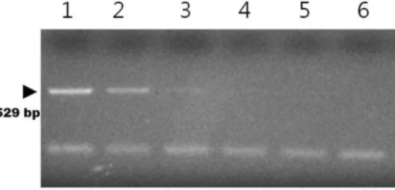
Fig. 2. PCR assay with T. gondii-specific ITS-1 primers. 1×10⁵ pg (lane 1), 1×10⁴ pg (lane 2), 1×10³ pg (lane 3), 1×10² pg (lane 4), 10 pg (lane 5), and 1 pg (lane 6) of template DNA.
Fig. 3.PCR/dot blot hybridization with PCR-amplified DNA of T.
gondii. A DIG-labeled probe derived from the genomic DNA of T.
gondii was used for detection. 1×10⁵ pg (lane 1), 1×10⁴ pg (lane 2), 1×10³ pg (lane 3), 1×10² pg (lane 4), 10 pg (lane 5), and 1 pg (lane 6) of PCR-amplified genomic DNA of T. gondii.
Fig. 4.Specificity of PCR/dot blot hybridization. The T. gondii probe was not reacted with other pathogens, E. tenella and E. maxima.
However, PCR/DBH using T. gondii template DNA resulted in strong positive signal.
(Roche Applied Science, Mannheim, Germany), 0.1%
N-lauroylsarcosine, and 0.02% sodium dodecyl sulfate.
DIG-labeled probe was denatured by boiling for 10 min and chilled in ice, and then added into hybridization solution at 0.1 g/mL. After pre-hybridization at 50oC for 1 h, the membrane was hybridized at 50oC for 3 h and then washed with 1 x SSC at 60oC for 10 min. For de- tection of hybridization, the membrane was incubated with anti-DIG conjugated with alkaline phosphatase (Roche Applied Science, Mannheim, Germany) and then colorized with nitroblue tetrazolium (NBT) and 5-bro- mocresyl-3-indolyl-phosphate (BCIP) (Roche Applied Science, Mannheim, Germany). The development of a dark purple positive reaction was allowed to proceed for 10∼30 min in the dark. The specificity of PCR/DBH was evaluated by using the template DNA samples like as Eimeria tenella and Eimeria maxima, which DNAs were provided by Professor W. Min at Gyeongsang National University in Korea.
In our results, the DBH assay alone was capable of detecting down to 1×10⁴ pg of T. gondii genomic DNA (Fig. 1). The PCR alone was capable of detecting down to 1×10³ pg of T. gondii genomic DNA (Fig. 2).
However, the PCR/DBH assay was capable of detecting down to 1×10² pg of T. gondii genomic DNA, indicat-
ing that sensitivity of the PCR/DBH method was ap- proximately 100 times higher than the DBH method alone (Fig. 3). The specificity of PCR/DBH was con- firmed by the study using other intracellular protozoa DNAs with high homology in their sequences. No pos- itive signals were observed in the template DNA sam- ples of Eimeria(E.) tenella and E. maxima in PCR/DBH assay. However, PCR/DBH using T. gondii template DNA resulted in strong positive signal (Fig. 4).
Toxoplasmosis transmission by unpasteurized or in- adequately processed milk or fresh cheese, important food sources in rural areas, can be a significant means of contamination by this agent (Hiramoto et al., 2001).
In this study, we used a non-radioactive probe for DBH, which makes these techniques more attractive for diag- nostic laboratories because the troublesome problems re- lated to the short half-life of radioactive compounds, their disposal, and personnel safety can be avoided (Gauthier and Blais, 2003; Mansfield et al., 1995).
PCR with specific primers is considered sensitive as- say for detecting T. gondii DNA from biological sam- ples directly, especially if nested PCR is used (Su et al., 2002). However, PCR assays are subject to a high risk of contamination through DNA carry-over and may re- sult frequently in false positive reactions (Borst et al., 2004; Maurer 2011; Szöllsi et al., 2008). To get around
these problems, PCR/DBH assay may be an alternative choice for sensitive and specific detection of T. gondii, in which PCR sensitivity and specificity is increased by hybridization methods of the replicated DNA with spe- cific labeled probe.
In this study, we used non-radioactive labels for DBH probe and it has made these techniques more attractive for diagnostic laboratories, because those avoid problems relative to the short life of radioactive compounds, their disposal, and personnel safety (Burns et al, 1987;
Syrjanen et al, 1988). Described DNA probe labeling was used in this study and the method was shown to be rapid, sensitive and specific, making it suitable for the detection of primary amplified T. gondii species DNA products, which was allowed the increased sensitivity and specificity and T. gondii species DNA densitometry quantification. Complete time including PCR procedure and DBH detection is 8 hours. The PCR/DBH, which was established in this study, is much more sensitive and specific compared with one step PCR assay and DBH detection alone. Our PCR/DBH assay will be use- ful for confirming the presence of T. gondii on the sam- ples like as meat.
In conclusion, the PCR/DBH assay is a more sensi- tive and specific method than PCR or DBH alone and will be diagnostically useful for detecting intracellular protozoa T. gondii.
ACKNOWLEDGMENTS
This study was supported by the research fund of Wonkwang University in 2012.
REFERENCES
Bayarri S, Gracia MJ, Lázaro R, Pérez-Arquillué C, Herrera A.
2012. Toxoplasma gondii in Meat and Food Safety Implications - A Review. pp.229-254. In: Lorenzo- Morales J(Ed.). Zoonosis. 1st ed. InTech, Rijeka, Croatia.
Borst A, Box AT, Fluit AC. 2004. False-positive results and con- tamination in nucleic acid amplification assays: sugges- tions for a prevent and destroy strategy. Eur J Clin Microbiol Infect Dis 23: 289-299.
Burns J, Graham AK, Frank C, Fleming KA, Evans MF, McGee JO. 1987. Detection of low copy human papilloma virus DNA and mRNA in routine paraffin sections by non iso- topic in situ hybridization. J Clin Pathol 40: 858-864.
Buxton D, Maley SW, Wright SE, Rodger S, Bartley P, Innes EA. 2007. Toxoplasma gondii and ovine toxoplasmosis:
new aspects of an old story. Vet Parasitol 149: 25-28.
Dubey JP. 2009. Toxoplasmosis in sheep - the last 20 years. Vet Parasitol 163: 1-14.
Duggan MA, Inoue M, McGregor SE, Stuart GC, Morris S, Chang-Poon V, Schepansky A, Honore L. 1994. A paired comparison of dot blot hybridization and PCR amplification for HPV testing of cervical scrapes in- terpreted as CIN 1. Eur J Gynaecol Oncol 15: 178-187.
Gauthier M, Blais BW. 2003. Comparison of different approaches for the incorporation of non-radioactive labels into poly- merase chain reaction products. Biotechnol Lett 25:
1369-1374.
Hiramoto RM, Mayrbaurl-Borges M, Galisteo Jr. AJ, Meireles LR, Macre MS, Andrade Jr. HF. 2001. Infectivity of cysts of the ME-49 Toxoplasma gondii strain in bovine milk and homemade cheese. Rev Saude Publica 35: 113- 118.
Jittapalapong S, Sangvaranond A, Pinyopanuwat N, Chimnoi W, Khachaeram W, Koizumi S, Maruyama S. 2005.
Seroprevalence of Toxoplasma gondii infection in do- mestic goats in Satun Province, Thailand. Vet Parasitol 127: 17-22.
Kaneto CN, Costa AJ, Paulillo AC, Moraes FR, Murakami TO, Meireles MV. 1997. Experimental toxoplasmosis in broiler chicks. Vet Parasitol 69: 203-210.
Kim O. 2003. Development of in situ nest PCR and Comparison of five molecular biological diagnostic methods for the detection of intracellular viral DNAs in paraffin sections.
J Vet Med Sci 65: 231-235.
Mansfield ES, Worley JM, McKenzie SE, Surrey S, Rappaport E, Fortina P. 1995. Nucleic acid detection using non-radio- active labeling methods. Mol Cell Probes 9: 145-156.
Maurer JJ. 2011. Rapid detection and limitations of molecular techniques. Annu Rev Food Sci Technol 2: 259-279.
McNicol AM, Farquharson MA. 1997. In situ hybridization and its diagnostic applications in pathology. J Pathol 182:
250-261.
Paquet C, Yudin MH. 2013. Toxoplasmosis in pregnancy: pre- vention, screening, and treatment. J Obstet Gynaecol Can 35: 78-79.
Su C, Howe DK, Dubey JP, Ajioka JW, Sibley LD. 2002.
Identification of quantitative trait loci controlling acute virulence in Toxoplasma gondii. Proc Natl Acad Sci USA 99: 10753-10758.
Switaj K, Master A, Skrzypczak M, Zaborowski P. 2005. Recent trends in molecular diagnostics for Toxoplasma gondii infections. Clin Microbiol Infect 11: 170-176.
Syrjanen S, Partanen P, Mantvjarvi R, Syrjanen K. 1988.
Sensitivity of the in situ hybridization techniques using
biotin and 35S labeled human papillomavirus(HPV) DNA probes. J Virol Methods 19: 225-238.
Szöllsi E, Hellgren O, Hasselquist D. 2008. A cautionary note on the use of nested PCR for parasite screening--an exam- ple from avian blood parasites. J Parasitol 94: 562-564.
Tenter AM. 2009. Toxoplasma gondii in animals used for human consumption. Mem Inst Oswaldo Cruz 104: 364-369.
Weiss LM, Dubey JP. 2009. Toxoplasmosis: A history of clinical observations. Int J Parasitol 39: 895-901.
Xia JQ, Yason CV, Kibenge FS. 1995. Comparison of dot blot hybridization, polymerase chain reaction, and virus iso-
lation for detection of bovine herpesvirus-1 (BHV-1) in artificially infected bovine semen. Can J Vet Res 59:
102-109.
Xie DH, Zhu XQ, Cui HL, Qiu CJ, Fan WH, Liao SQ, Zhai ML, Lin RQ, Weng YB. 2005. Development of a PCR assay for diagnosing swine toxoplasmosis. Chin J Vet Sci Technol 35: 289-293.
Zia-Ali N, Fazaeli A, Khoramizadeh M, Ajzenberg D, Darde M, Keshavarz-Valian H. 2007. Isolation and molecular char- acterization of Toxoplasma gondii strains from different hosts in Iran. Parasitol Res 101: 111-115.