pISSN 1738-3544 eISSN 2288-1662
Development of Real-time PCR Assays for Detection of Dirofilaria immitis from Infected Dog Blood
In Young Oh
1, Kyung Tae Kim
2, Jin Hyun Jun
1,3, Jae-Ho Shin
1,3, and Ho Joong Sung
1,31Department of Biomedical Laboratory Sciences, College of Health Science, Eulji University, Seongnam 13135, Korea
2ALPHAGENE Co., Ltd., Singu University, Business Incubation Center, Seongnam 13174, Korea
3BK21 Plus Program, Department of Senior Healthcare, Graduate School, Eulji University, Daejeon 34824, Korea
심장사상충에 감염된 개의 혈액에서 심장사상충 유전자를 검출할 수 있는 실시간 중합효소연쇄반응 기법 개발
오인영
1, 김경태
2, 전진현
1,3, 신재호
1,3, 성호중
1,31을지대학교 임상병리학과, 2(주)알파젠, 3을지대학교 대학원 시니어헬스케어학과
Dirofilaria immitis is a filarial nematode parasite that causes cardiopulmonary dirofilariasis in dogs. The purpose of this study was the development of real-time PCR assays for efficient detection of D. immitis. The D. immitis-specific primers confirmed in our previous study and a newly designed TaqMan probe were used for quantitative diagnostics. First, SYBR Green real-time PCR was performed using the specific primers and serially diluted genomic DNA or plasmid DNA, and melting curve analyses were performed after amplification. The melting curve showed one specific peak in each of the genomic and plasmid DNA reactions, suggesting that the primers specifically amplify the D. immitis cytochrome c oxidase subunit I gene. Comparison of SYBR Green and TaqMan real-time PCR using serially diluted plasmid DNA showed higher efficiency and specificity with TaqMan real-time PCR. The real-time PCR assays developed in this study will provide improved diagnostic methods to overcome the limitations of conventional diagnostic tools and facilitate more rapid and accurate diagnoses.
Keywords: Dirofilaria immitis, Molecular diagnosis, SYBR Green real-time PCR, TaqMan real-time PCR, PCR efficiency
Corresponding author: Ho Joong Sung Department of Biomedical Laboratory Sciences, College of Health Science, Eulji University, 553 Sanseong-daero, Soojeong-gu, Seongnam 13135, Korea Tel: 82-31-740-7306
Fax: 82-31-740-7425 E-mail: [email protected]
This is an Open Access article distributed under the terms of the Creative Commons Attribution Non-Commercial License (http://creativecommons.org/licenses/by-nc/4.0) which permits unrestricted non-commercial use, distribution, and reproduction in any medium, provided the original work is properly cited.
Copyright © 2016 The Korean Society for Clinical Laboratory Science. All rights reserved.
Received: April 21, 2016 Revised: May 11, 2016 Accepted: May 13, 2016
Introduction
Dirofilaria immitis is the causative agent of cardio- pulmonary dirofilariasis. Mosquitoes are the main vectors for accidental infection with D. immitis . D. immitis larvae develop in the mosquito at 18∼34
oC and are thereafter transmitted to the new final hosts such as canines, felines, various wild mammals, and some human populations in
tropical, subtropical, and some temperate regions in the world. With progressing globalization and climate change, the incidence of infection is increasing [1-9].
Diagnosis is recommended before administration of anti-parasite medication to avoid complications from drug abuse. The most common diagnostic tool for detection of D.
immitis is microscopic examination of microfilariae from
blood samples. However, this approach has limited
× × ×
× ×
sensitivity, and considerable expertise is required to distinguish among filarial parasite species because of their similar morphologies [8,10]. Other methods leveraging antigen detection are limited by their propensity to produce false-negative results during the first 5∼8 months of infection because they only detect antigens released from the adult female worm’s reproductive tract, leading to errors due to low worm counts, immature infections, and all-male infections [11,12]. To overcome these limitations, methods for PCR-based molecular detection are under development.
5.8S, 16S, or 18S ribosomal RNA (rRNA) genes have been used as target genes to detect filarial nematodes using end-point PCR-based detection from dog bloods or heads of mosquitoes [1,2,6,7].
This study aimed to develop quantitative PCR methods for detecting D. immitis . In our previous study, we designed primers targeting a D. immitis -specific gene region and developed an end-point PCR method. The specific gene region focused on was cytochrome c oxidase subunit I ( COI ) gene. COI is one of mitochondrial DNA (mtDNA) genes which are maternally inherited and generally do not undergo recombination. Amino acid sequences of COI gene have changed more slowly than any other mtDNA genes.
Therefore, COI gene is so-called “barcode” for identifying of species diversity [13-17].
The specificity and sensitivity of the developed assay were very high in our previous study. However, end-point PCR only allows determination of whether a sample is infected.
For more precise therapeutic monitoring, therefore, additional development of a method for quantitative analysis is needed.
Materials and Methods
1. Primers and probes
We employed the primers designed in our previous study for SYBR Green and TaqMan real-time PCR assays to detect a 150-bp fragment of D. immitis cytochrome c oxidase subunit I ( COI ) (forward: ATT GGG TGC CCC TGA AAT GG; reverse:
CCC TCT ACA CTC AAA GGA GGA). For TaqMan real-time PCR, a probe was designed within the fragment and labeled
with 6-carboxy-fluorescein (FAM, excitation wavelength 494 nm, emission wavelength 521 nm) at the 5'-end and ZEN
TM-Iowa Black
ⓇFQ quencher at the 3'-end. All primers and probes used in this study were synthesized by Integrated DNA Technologies (Coralville, IA, USA).
2. Extraction of genomic DNA
D. immitis -infected blood samples isolated from random infected source dogs were gifted from Seoul National University. The peripheral blood samples were from 51 blood donation dogs, Beagle species, and were collected in July 2015 in the Republic of Korea. Genomic DNA (gDNA) from blood samples collected in EDTA (ethylenediaminetetraacetic acid) tubes was extracted using the QIAamp DNA Mini Kit (Qiagen, Hilden, Germany) according to the manufacturer’s protocol. The concentration of gDNA was determined with a NanoDrop spectrophotometer (ThermoFisher Scientific, Sunnyvale, CA, USA), and twofold serial dilutions were prepared for analysis.
3. Preparation of plasmid DNA
PCR products amplified using the primers were purified and inserted into pLUG-Prime
ⓇTA-cloning vector (iNtRON Biotechnology, Republic of Korea), and the plasmid DNA (pDNA) was cloned. The DNA copy number was estimated from the molecular weight of the D. immitis pDNA:
DNAs were 10-fold serially diluted for analysis.
4. Real-time PCR
All amplification reactions were performed on a StepOnePlus
TMinstrument (Applied Biosystems, Foster City, CA, USA) in 20 L total volume. All analyses were performed in triplicate. Three samples without DNA template were routinely included as a no-template control. The PCR efficiency was calculated from the dilution factor and the slope of the trend line as follows:
PCR efficiency =-1+ dilution factor
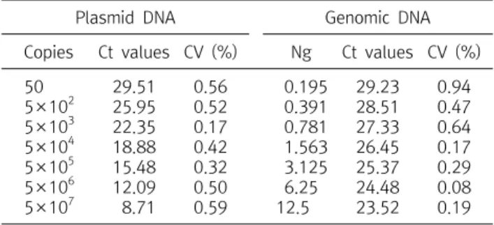
(-1/slope)Table 1. Ct values for SYBR Green real-time PCR using serially diluted plasmid DNA and genomic DNA
Plasmid DNA Genomic DNA
Copies Ct values CV (%) Ng Ct values CV (%) 50
5×10² 5×10³ 5×10⁴ 5×10⁵ 5×10⁶ 5×10⁷
29.51 25.95 22.35 18.88 15.48 12.09 8.71
0.56 0.52 0.17 0.42 0.32 0.50 0.59
0.195 0.391 0.781 1.563 3.125 6.25 12.5
29.23 28.51 27.33 26.45 25.37 24.48 23.52
0.94 0.47 0.64 0.17 0.29 0.08 0.19 Abbreviation: CV, coefficient of variation.
1) SYBR green real-time PCR
SYBR Green real-time PCR was performed with D. immitis COI primers. The PCR mixture was prepared with 1× SYBR Green Master Mix (Applied Biosystems, Foster City, CA, USA), 500 nM primers, and serially diluted pDNA or gDNA. The PCR protocol included an initial denaturation step at 95
oC for 10 min followed by 40 cycles of denaturation at 95
oC for 15 s and annealing and extension at 60
oC for 1 min. The cycle threshold (Ct) was determined automatically. Data collection was performed during each annealing step. Following amplification, melting curve analysis was performed at 95
oC for 15 s and 60
oC for 1 min, and the temperature was increased from 60 to 95
oC with 0.3
oC increments to obtain product-specific melting temperatures.
2) TaqMan real-time PCR
TaqMan real-time PCR was performed with D. immitis COI primers and TaqMan probe (5'-/56-FAM/TGC TTT ATC /ZEN/TTT TTG GAT TAC TTT TGT TGC GTT GTT GAT GG/3IABkFQ/-3'). The PCR mixture was prepared with 1×
TaqMan Master Mix (Applied Biosystems, Foster City, CA, USA), 500 nM primers, 250 nM probe, and serially diluted pDNA or gDNA. The PCR protocol included a UNG (uracil-N-glycosylase) incubation step at 50
oC for 2 min, initial denaturation at 95
oC for 10 min, and 40 cycles of denaturation at 95
oC for 15 s and annealing and extension at 60
oC for 1 min.
Results
In our previous study, we designed primers targeting a D.
immitis -specific region of COI based on multiple sequence alignment of seven filarial COI genes. Primer specificities were evaluated by end-point PCR experiments and sequencing.
1. Specificity and sensitivity of the D. immitis SYBR green real-time PCR assay
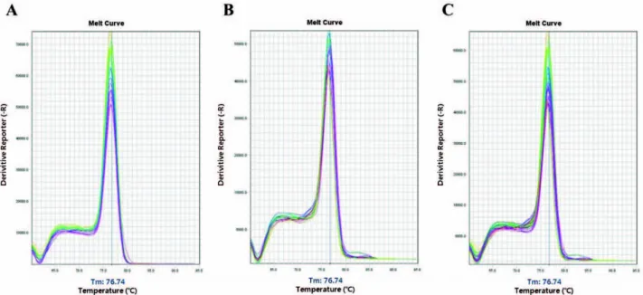
In this study, to confirm primer specificity, we performed melting curve analyses after amplification in the SYBR Green real-time PCR assays. DNA samples were serially diluted as
follows: pDNA was 10-fold serially diluted from 5×10
7copies to 50 copies, and gDNA was twofold serially diluted from 12.5 ng to 0.195 ng. The Ct values for three replicates of serially diluted D. immitis COI pDNA and gDNA are listed in Table 1, and the melting curves are presented in Fig. 1. Ct values were analyzed by linear regression analysis and showed good linearity (pDNA: R
2value, 0.9998; slope, -3.464; PCR efficiency, 94% and gDNA: R
2value, 0.9984;
slope, -0.9684; PCR efficiency, 104%). The gDNA melting curves matched those of pDNA with one specific peak at a melting temperature of 76.74
oC. These results suggested that the primers could amplify D. immitis COI specifically and efficiently in SYBR Green real-time PCR.
2. Comparison of SYBR green and TaqMan real-time PCR To improve specificity, TaqMan real-time PCR was developed using the same primers and a TaqMan probe. D.
immitis COI pDNA was 10-fold serially diluted from 5×10
7copies to 5×10
2copies the template for SYBR Green and TaqMan real-time PCR assays. Ct values were analyzed by linear regression analysis, and the results showed good linearity (SYBR Green real-time PCR: R
2value, 0.9998; slope, -3.4213; PCR efficiency, 96% and TaqMan real-time PCR: R
2value, 0.9999; slope, -3.4624; PCR efficiency, 94%) (Fig. 2).
3. Limit of detection
To determine the detection limit, SYBR Green and TaqMan
real-time PCR assays were performed with serially diluted
pDNA, from 5×10
8to 50 and 25 copies. The minimum
detection level was less than 25 copies in both assays. The Ct
Fig. 1. Melting curves for products of SYBR Green real-time PCR. The templates were (A) 10-fold serially diluted plasmid DNA (5×10⁷ to 50 copies) and (B) twofold serially diluted genomic DNA (12.5 ng to 0.195 ng). (C) Overlay of curves from (A) and (B). The curves show one specific peak at 76.74oC.
Fig. 2. Comparison of SYBR Green and TaqMan real-time PCR assays. The graph showed the results of linear regression analysis using Ct values of SYBR Green and TaqMan real-time PCR using serially diluted pDNA, respectively. Blue circles represent the Ct values of the SYBR Green real-time PCR assay, and the blue line is the trend line for those values. Red crosses show the Ct values of the TaqMan real-time PCR assay, and the red line is the trend line for those values.
Table 2. Ct values and detection limits of TaqMan and SYBR Green qPCR assays
pDNA (copies)
SYBR Green real-time PCR
TaqMan real-time PCR Ct values CV (%) Ct values CV (%) 25
50 5×10² 5×10³ 5×10⁴ 5×10⁵ 5×10⁶ 5×10⁷
31.7 30.66 27.09 23.5 20.02 16.66 13.3
9.94
1.19 0.61 0.55 0.15 0.55 0.24 0.35 0
32.33 31.04 27.16 23.87 20.24 16.77 13.32 9.94
1.71 0.88 0.26 0.02 0.14 0.03 0.23 0.32 Abbreviation: CV, coefficient of variation; pDNA, plasmid DNA.
values are listed in Table 2.
Discussion
Development of molecular diagnostic tools is essential for improving the clinical benefit of pathogen detection. In
particular, real-time PCR assays produce quantitative results with improved specificity, sensitivity, and reproducibility and reduced risk of carry-over contamination. Furthermore, real-time PCR requires less time and labor than conventional PCR. Therefore, real-time PCR assays are promising tools for detecting pathogen gDNA from biological fluids such as blood, urine, and sputum [18-23].
Researchers developed methods to differentiate filarial
nematodes using high resolution melt (HRM) real-time qPCR
which were performed PCR amplification and then melting
curve analysis which showed different melting peaks among
filarial nematodes. However, those developed methods could
be used for only detection and differentiation [10,24].
In this study, the SYBR Green real-time PCR assay using primers targeting D. immitis COI showed high PCR efficiency, specificity, and sensitivity. The Ct values of gDNA (12.5 ng to 0.195 ng) were similar to those of pDNA (5×10
3to 50 copies), and the melting curves for products from both templates had one specific peak. This assay provides a diagnostic tool that could be used to detect D. immitis levels quantitatively in real-time. Furthermore, a TaqMan real-time PCR assay was developed using the specific primers and a TaqMan probe designed to specifically detect the sequence of D. immitis COI between the forward and reverse primers. The results from TaqMan real-time PCR showed efficiency similar to SYBR Green real-time PCR with high specificity and sensitivity. The detection limit was 25 copies for both assays. Thus, the TaqMan real-time PCR assay exhibited greater specificity in less time than the SYBR Green real-time PCR assay.
The assays developed herein will allow D. immitis -specific real-time monitoring from the early stage of infection before administration of medication. Additionally, further development of these real-time PCR-based assays will facilitate efficient therapeutic control of D. immitis infection.
요 약
선형 사상충의 일종인 심장사상충은 개의 심폐 사상충증을 유발 한다. 이에 본 연구의 목적은 심장사상충을 효과적으로 검출할 수 있는 실시간 중합효소연쇄반응 기법을 개발함에 있다. 연구에 있어 서 사용된 프라이머 및 프로브는 선행연구에서 제작된 심장사상충 특이 프라이머 및 새롭게 제작된 TaqMan 프로브를 이용하였다. 선 행연구에서 제작된 프라이머 및 농도별로 희석된 게놈유전자와 플 라스미드유전자가 SYBR Green 실시간 중합효소연쇄반응 수행에 이용되었으며, 중합효소연쇄반응 과정 중 증폭 이후의 녹는 곡선의 결과를 분석하였다. 분석결과 사용된 프라이머는 각각 게놈유전자 및 플라스미드 유전자에서 특이 녹는 곡선을 나타냄에 따라 심장사 상충 특이 사이토크롬 C 산화효소 유전자만을 증폭하고 있음을 확 인 할 수 있었다. 새롭게 제작된 TaqMan 프로브는 SYBR Green 실 시간 중합효소연쇄반응과의 결과를 농도별로 희석된 플라스미드 유전자를 이용하여 비교 분석하였고, 분석결과 TaqMan 프로브를 이용한 실시간 중합효소연쇄반응이 검출효율 및 특이도에 있어서 우수함을 확인할 수 있었다. 본 연구를 통하여 개발한 실시간 중합
효소연쇄반응은 기존의 전통적인 진단기법의 한계를 극복할 수 있 는 신속하고 정확한 향상된 진단기법을 제시한다.
Acknowledgements: We thank Prof. Byeong Chun Lee, Seoul National University, Republic of Korea for providing dog blood samples.
Funding: Small and Medium Business Administration, Republic of Korea, Grant number: C0300247.
Conflict of interest: None
References
1. Watts KJ, Courteny CH, Reddy GR. Development of a PCR- and probe-based test for the sensitive and specific detection of the dog heartworm,
Dirofilaria immitis
, in its mosquito inter- mediate host. Mol Cell Probes. 1999;13:425-430.2. Kronefeld M, Kampen H, Sassnau R, Werner D. Molecular de- tection of
Dirofilaria immitis, Dirofilaria repens and Setaria tundra
in mosquitoes from Germany. Parasit Vectors.2014;7:30.
3. Khedri J, Radfar MH, Borji H, Azizzadeh M, Akhtardanesh B.
Canine heartworm in southeastern of Iran with review of dis- ease distribution. Iran J Parasitol. 2014;9:560-567.
4. Fu Y, Lan J, Wu X, Yang D, Zhang Z, Nie H, et al. Expression of translationally controlled tumor protein (TCTP) gene of
Dirofilaria immitis
guided by transcriptomic screening. Korean J Parasitol. 2014;52:21-26.5. Rossi MI, Aguiar-Alves F, Santos S, Paiva J, Bendas A, Fernandes O, et al. Detection of Wolbachia DNA in blood from dogs in- fected with
Dirofilaria immitis
. Exp Parasitol. 2010;126:270-272.
6. Nuchprayoon S, Junpee A, Poovorawan Y, Scott AL. Detection and differentiation of filarial parasites by universal primers and polymerase chain reaction-restriction fragment length poly- morphism analysis. Am J Trop Med Hyg. 2005;73:895-900.
7. Masetti A, Rivasi F, Bellini R. Mosquito-based survey for the de- tection of flaviviruses and filarial nematodes in
Aedes albo- pictus
and other anthropophilic mosquitoes collected in north- ern Italy. New Microbiol. 2008;31:457-465.8. Mazzariol S, Cassini R, Voltan L, Aresu L, Frangipane di Regalbono A. Heartworm (
Dirofilaria immitis
) infection in a leopard (Panthera pardus pardus
) housed in a zoological park in north-eastern Italy. Parasit Vectors. 2010;3:25.9. Furtado AP, Do Carmo ES, Giese EG, Vallinoto AC, Lanfredi RM, Santos JN. Detection of dog filariasis in Marajo island, Brazil by classical and molecular methods. Parasitol Res. 2009;105:
1509-1515.
10. Rojas A, Rojas D, Montenegro VM, Baneth G. Detection of
Dirofilaria immitis
and other arthropod-borne filarioids by an HRM real-time qPCR, blood-concentrating techniques and a serological assay in dogs from Costa Rica. Parasit Vectors.2015;8:170.
11. Schnyder M, Deplazes P. Cross-reactions of sera from dogs in- fected with
Angiostrongylus vasorum
in commercially availableDirofilaria immitis
test kits. Parasit Vectors. 2012;5:258.12. Oi M, Sato Y, Nakagaki K, Nogami S. Detection of
Dirofilaria immitis
DNA in host serum by nested PCR. Parasitol Res.2015;114:3645-3648.
13. Hwang IK, Lee HY, Kim MH, Jo HS, Choi DH, Kang PW, et al.
Development of real-time PCR assay for genetic identification of the mottled skate,
Beringraja pulchra
. Forensic Sci Int.2015;255:80-84.
14. Carracedo A, Bär W, Lincoln P, Mayr W, Morling N, Olaisen B, et al. DNA commission of the international society for forensic ge- netics: guidelines for mitochondrial DNA typing. Forensic Sci Int. 2000;110(2):79-85.
15. Dawnay N, Ogden R, McEwing R, Carvalho GR, Thorpe RS.
Validation of the barcoding gene COI for use in forensic genetic species identification. Forensic Sci Int. 2007;173(1):1-6.
16. Hebert PD, Ratnasingham S, deWaard JR. Barcoding animal life:
cytochrome c oxidase subunit 1 divergences among closely re- lated species. Proc Biol Sci. 2003;270(Suppl 1):S96-99.
17. Hebert PD, Cywinska A, Ball SL, deWaard JR. Biological identi- fications through DNA barcodes. Proc Biol Sci. 2003;270(1512):
313-321.
18. De Gregorio E, Roscetto E, Iula VD, Martinucci M, Zarrilli R, Di Nocera PP, et al. Development of a real-time PCR assay for the
rapid detection of
Acinetobacter baumannii
from whole blood samples. New Microbiol. 2015;38:251-257.19. Liu T, Yang B, Song X, Wang X, Yuan Y, Liu L, et al. Detection and quantification of hepatopancreatic parvovirus in penaeid shrimp by real-time PCR assay. J Invertebr Pathol. 2013;114:
309-312.
20. Liu CM, Kachur S, Dwan MG, Abraham AG, Aziz M, Hsueh PR, et al. FungiQuant: a broad-coverage fungal quantitative re- al-time PCR assay. BMC Microbiol. 2012;12:255.
21. Li K, Gao H, Gao L, Qi X, Qin L, Gao Y, et al. Development of TaqMan real-time PCR assay for detection and quantitation of reticuloendotheliosis virus. J Virol Methods. 2012;179:402-408.
22. Qu S, Shi Q, Zhou L, Guo Z, Zhou D, Zhai J, et al. Ambient stable quantitative PCR reagents for the detection of
Yersinia pestis
. PLoS Negl Trop Dis. 2010;4:e629.23. Cho JA, Kim DW, Jeong SD, Cheon JS, Na GA, Kim HR, et al.
Utility of real time RT-PCR for the quantitative detection of minimal residual disease in hematological malignancy. Korean J Clin Lab Sci. 2009;41:11-23.
24. Latrofa MS, Montarsi F, Ciocchetta S, Annoscia G, Gantas-Torres F, Ravaqnan S, et al. Molecular xenomonitoring of