© Copyright
Keimyung University School of Medicine 2015
A 74-year-old woman who had paroxysmal atrial fibrillation without mitral stenosis was hospitalized for syncope and right-sided weakness.
Echocardiography revealed a large free-floating thrombus in the left atrium, sometimes prolapsing partially into the left ventricle in diastole. Because of her poor neurological status, she was managed with anticoagulation. On the 12th day, the thrombus had disappeared on the follow-up echocardiography, and aortoiliac embolization was later detected on computed tomography. Unfortunately, she developed various complications of stroke and limb infarction, and died after 4 months of hospital care. In addition to this case report, we reviewed a total 70 cases of left atrial free-floating thrombus. Atrial fibrillation and mitral pathology were two major causative factors. All the cases, except 1, were confirmed on echocardiography. The most common presentation that led to echocardiography was systemic embolization, followed by heart failure. Others were acute hemodynamic decompensation from mitral obstruction, chest pain, palpitation, and bacteremia. Cardiac thrombectomy was the preferred treatment modality with favorable outcomes.
Key Words: Echocardiography, Free floating, Left atrium, Thrombus
Introduction
Left atrial free-floating thrombus (LAFFT) is rare type of left atrial thrombus that causes stroke, limb infarction, dyspnea, syncope, or sudden death. The thrombus is thought to begin as a small mural thrombus attached to the atrial wall or left atrial appendage [1]. The thrombus gradually grows to form a projecting mass that is connected by a pedicle
Received: September 18, 2015 Revised: October 5, 2015 Accepted: October 19, 2015
Corresponding Author: Wook Hyun Cho, M.D., Department of Internal Medicine,
Sam Yook Medical Center,
82 Mangwoo-ro, Dongdemoon-gu, Seoul 02500, Korea
Tel: +82-2-2210-3507 E-mail: [email protected]
• The authors report no conflict of interest in this work.
Department of Internal Medicine, Sam Yook Medical Center, Seoul, Korea
Dong Keun Kim, M.D., Jung Seok Kim, M.D., Ha Ram Yi, M.D., In Zoo Choi, M.D., Hyo Seung Ahn, M.D., Wook Hyun Cho, M.D.
A Case of Left Atrial Free-Floating Thrombus
to the atrial wall. As the bulbous end of the thrombus enlarges, the pedicle becomes longer and thinner and finally vanishes. After the thrombus becomes free, it moves around in the left atrium, is polished by the atrial structure, and acquires a smooth, ball-like shape.
Wood in 1814 first used the term ball thrombus in a description of the autopsy findings of a 15-year-old girl who had rheumatic mitral stenosis and syncope [2]. In the pre-echographic era, the diagnosis depended on the presence of a ball-shaped thrombus during autopsy. In the echographic era, researchers described not only various echographic findings, especially free-floating movement of the thrombus, but also treatment modalities and outcomes.
Wrisley et al. [2] reviewed 11 reported cases of the echographic era (from 1976 to 1990) and literature reported in the pre-echographic era. Since echocardiography became widely available, numerous cases have been reported. In this study, we report our experience of a case of LAFFT and review other cases reported in the literature.
Case Report
A 74-year-old woman presented to our hospital with syncope and weakness of the right limbs. Her medical history revealed type 2 diabetes and hypertension. She showed confused mentality and neurologic examination confirmed right-sided weakness. Her blood pressure was 110/80 mmHg, heart rate was regular at 80 beats/min, and respiratory rate was 20 breaths/min. Chest radiography revealed cardiomegaly, but no pulmonary congestion. Her laboratory finding was non-specific. Initial electrocardiography revealed sinus rhythm, but paroxysmal atrial fibrillation was detected during the admission period. Non-contrast brain computed tomography (CT) did not reveal any
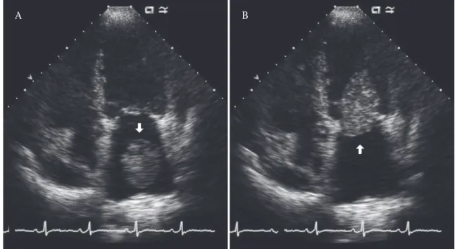
brain hemorrhage. Magnetic resonance imaging of the brain revealed multiple foci with T2-weighted hyperintensity in the left basal ganglion and both the occipital and parietal lobes, which were consistent with thromboembolic stroke. Transthoracic echocardiography revealed a 5.7-×3.1-cm ovoid floating mass in the enlarged left atrium (Fig. 1A).
The mass rotated on its own axis and sometimes prolapsed partially into the left ventricle in diastole (Fig. 1B). The left ventricle showed apical ballooning. Mild mitral regurgitation without mitral stenosis was observed. Cardiac surgery was not performed because of her progressive neurological deterioration, and anticoagulation with heparin and warfarin was started. On the follow-up echocardio- graphy performed on the 12th admission day, the mass in the left atrium was observed to have disappeared completely. On the 26th admission day, gangrene developed in the right toes, and CT angiography revealed a saddle embolus occluding the aortoiliac arteries and infarction of both the kidneys (Fig. 2A&B). Her later course was complicated by infective sequelae and she finally died after 4 months of hospital care.
Discussion
We reviewed 70 cases of LAFFT, including 69 cases from 65 papers written in English since 1990 and the present case. As of April 2015, a search of PubMed by using the keyword “left atrial free floating thrombus” returned 122 articles. Of these articles, 17 were discarded because they were published before 1990. Of the remaining 105 articles, 4 were unavailable in full text, 1 reported a case that was already reported, and 1 was a duplicate. Nine articles dealt with either right-sided thrombus or thrombus with a patent foramen ovale, 1 article dealt with aortic free-floating thrombus,
and 1 reported on a cardiac angiosarcoma, not a thrombus. Two articles described mural thrombi, which were not identified in spite of preoperative
evaluation and became free-floating thrombi during open-heart procedure. Twenty-one cases were written in languages other than English. Finally, after Fig. 1. (A) Transthoracic echocardiogram shows a large free-floating thrombus (arrow) in the left atrium.
(B) Transthoracic echocardiogram shows a thrombus (arrow) transiently impacted on the mitral orifice.
A B
Fig. 2. (A) Abdominal computed tomographic image shows the upper tip of a large embolus (arrow) in the abdominal aorta and infarctions of both the kidneys. (B) Abdominal computed tomographic image shows a large embolus (arrow) in the aortoiliac bifurcation.
A B
the process of elimination, we collected 65 full-text articles that reported cases of left atrial free-floating thrombus.
In the opinion of Hewitt, the diagnosis of free- floating ball thrombus should be based on two criteria, namely the thrombus must be larger than the mitral orifice and it should have a smooth surface without attachment to the atrial wall [3].
However, strict discrimination between a true free- floating ball thrombus and other large spherical thrombi with a pedicle in the left atrium is just for academic purpose and may be dangerous if applied in clinical practice [1]. In the 7 cases we reviewed, the thrombus had a pedicle that was demonstrated on echocardiography or in cardiac surgery. But, it was highly mobile and large enough to be confined in LA, so we included these cases in the analysis.
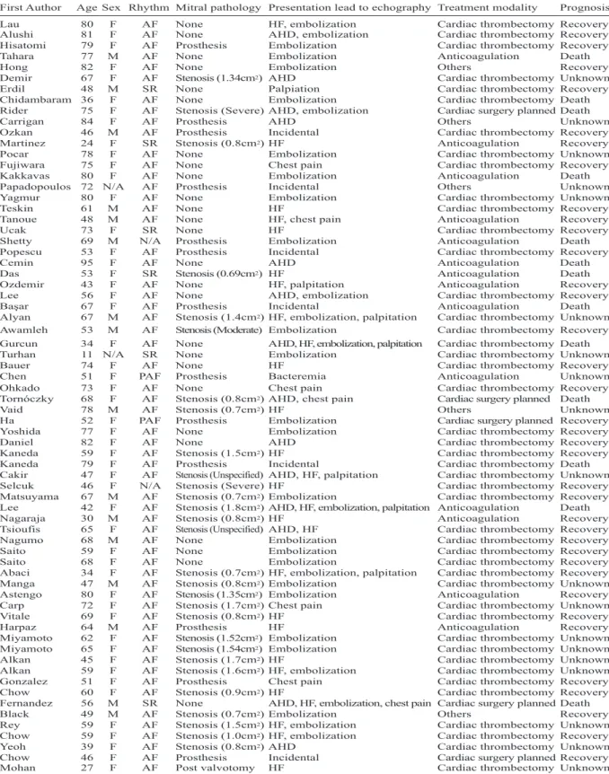
Table 1 summarizes the demographic characteristics, rhythm and mitral pathology (stenosis or prosthesis), clinical presentation, treatment modality, and outcomes of the 69 cases published since 1990.
Rhythm and Mitral Pathology
According to Virchow’s triad, thrombosis is caused by blood stasis, hypercoagulability, and endothelial injury. Mitral stenosis and atrial fibrillation are the two most well-known factors that trigger blood stasis and induce LAFFT. Large LA diameter and absence of significant mitral regurgitation were associated with blood stasis and thrombus formation in Beppu’s study [4]. The presence of mitral prosthesis with a large LA diameter was also associated with blood stasis [5]. In 1982, Schechter [6] reported that in their autopsy cases of LA ball thrombus, only 11.3% (19/168) were without mitral valve disease. In our review, 38.5%
cases (27/70) of LAFFT developed without mitral pathology (Table 2). The difference in the frequency of mitral pathology between the previous report and our review may be a consequence of the
increased prevalence of atrial fibrillation and/or other structural cardiac conditions. Other possible explanations were early detection of LAFFT before fatality or selection bias. A large consecutive study by Agmon et al. [7] about echocardiographic characteristics of patients with LA thrombus and sinus rhythm reported that 6.1% (23/380) of thrombus cases presented with sinus rhythm, which was not much different from the 8.8% that we derived from our own review (6/68, 2 unknown cases of rhythms excluded). Our review showed that 4 of 6 patients with LAFFT and sinus rhythm had no mitral pathology. For the 4 cases of sinus rhythm without mitral pathology, the suggested possible etiologies of thrombus formation were malignancy, restrictive cardiomyo-pathy, use of cyclosporine after heart transplantation, and hypercoagulable state after off-pump coronary artery bypass surgery [8-11]. Other conditions associated with thrombus formation in the reviewed cases were dilated cardiomyopathy, hypertrophic cardiomyopathy, low cardiac output, pregnancy, infertility treatment, and lobectomy of the lung due to squamous cell carcinoma [12-16].
Clinical Presentation
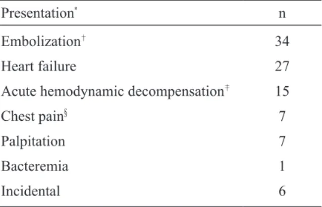
All the cases, except 1, were confirmed by using transthoracic echocardiography (TTE) and/or transesophageal echocardiography (TEE). Thus, we identified which presentations led patients to undergo echocardiography. The leading cause was evaluation for systemic embolization (Table 3). The second most common cause was evaluation for heart failure. Other causes were evaluation for acute hemodynamic decompensation, chest pain (including 3 cases of myocardial infarction), palpitation, and bacteremia. Some cases were detected incidentally during preoperative evaluation, routine follow-up after mitral valve surgery, and evaluation for an unidentified
Table 1. Summary of 69 reported cases with left atrial free-floating thrombus
AF: atrial fibrillation, HF: heart failure, N/A: not available, PAF: paroxysmal atrial fibrillation, SR: sinus rhythm, AHD:
acute hemodynamic decompensation.
First Author Age Sex Rhythm Mitral pathology Presentation lead to echography Treatment modality Prognosis
Lau 80 F AF None HF, embolization Cardiac thrombectomy Recovery
Alushi 81 F AF None AHD, embolization Cardiac thrombectomy Recovery Hisatomi 79 F AF Prosthesis Embolization Cardiac thrombectomy Recovery
Tahara 77 M AF None Embolization Anticoagulation Death
Hong 82 F AF None Embolization Others Recovery
Demir 67 F AF Stenosis (1.34cm2) AHD Cardiac thrombectomy Unknown
Erdil 48 M SR None Palpiation Cardiac thrombectomy Recovery
Chidambaram 36 F AF None Embolization Cardiac thrombectomy Death
Rider 75 F AF Stenosis (Severe) AHD, embolization Cardiac surgery planned Death
Carrigan 84 F AF Prosthesis AHD Others Unknown
Ozkan 46 M AF Prosthesis Incidental Cardiac thrombectomy Recovery
Martinez 24 F SR Stenosis (0.8cm2) HF Anticoagulation Recovery
Pocar 78 F AF None Embolization Cardiac thrombectomy Unknown
Fujiwara 75 F AF None Chest pain Cardiac thrombectomy Recovery
Kakkavas 80 F AF None Embolization Anticoagulation Death
Papadopoulos 72 N/A AF Prosthesis Incidental Others Unknown
Yagmur 80 F AF None Embolization Cardiac thrombectomy Unknown
Teskin 61 M AF None HF Cardiac thrombectomy Recovery
Tanoue 48 M AF None HF, chest pain Anticoagulation Recovery
Ucak 73 F SR None HF Cardiac thrombectomy Recovery
Shetty 69 M N/A Prosthesis Embolization Anticoagulation Death
Popescu 53 F AF Prosthesis Incidental Cardiac thrombectomy Recovery
Cemin 95 F AF None AHD Anticoagulation Death
Das 53 F SR Stenosis (0.69cm2) HF Anticoagulation Death
Ozdemir 43 F AF None HF, palpitation Anticoagulation Recovery
Lee 56 F AF None AHD, embolization Cardiac thrombectomy Recovery
Başar 67 F AF Prosthesis Incidental Anticoagulation Death
Alyan 67 M AF Stenosis (1.4cm2) HF, embolization, palpitation Cardiac thrombectomy Unknown Awamleh 53 M AF Stenosis (Moderate) Embolization Cardiac thrombectomy Recovery Gurcun 34 F AF None AHD, HF, embolization, palpitation Cardiac thrombectomy Death
Turhan 11 N/A SR None Embolization Cardiac thrombectomy Unknown
Bauer 74 F AF None HF Cardiac thrombectomy Recovery
Chen 51 F PAF Prosthesis Bacteremia Anticoagulation Unknown
Ohkado 73 F AF None Chest pain Cardiac thrombectomy Recovery
Tornóczky 68 F AF Stenosis (0.8cm2) AHD, chest pain Cardiac surgery planned Death
Vaid 78 M AF Stenosis (0.7cm2) HF Others Unknown
Ha 52 F PAF Prosthesis Embolization Cardiac surgery planned Recovery
Yoshida 77 F AF None Embolization Cardiac thrombectomy Recovery
Daniel 82 F AF None AHD Cardiac thrombectomy Recovery
Kaneda 59 F AF Stenosis (1.5cm2) HF Cardiac thrombectomy Recovery
Kaneda 79 F AF Prosthesis Incidental Cardiac thrombectomy Death
Cakir 47 F AF Stenosis (Unspecified) AHD, HF, palpitation Cardiac thrombectomy Unknown
Selcuk 46 F N/A Stenosis (Severe) HF Cardiac thrombectomy Recovery
Matsuyama 67 M AF Stenosis (0.7cm2) Embolization Cardiac thrombectomy Recovery Lee 42 F AF Stenosis (1.8cm2) AHD, HF, embolization, palpitation Anticoagulation Death
Nagaraja 30 M AF Stenosis (0.8cm2) HF Anticoagulation Recovery
Tsioufis 65 F AF Stenosis (Unspecified) AHD, HF Cardiac thrombectomy Recovery
Nagumo 68 M AF None Embolization Cardiac thrombectomy Recovery
Saito 59 F AF None Embolization Cardiac thrombectomy Recovery
Saito 68 F AF None Embolization Cardiac thrombectomy Recovery
Abaci 34 F AF Stenosis (0.7cm2) HF, embolization, palpitation Cardiac thrombectomy Recovery Manga 47 M AF Stenosis (0.8cm2) Embolization Cardiac thrombectomy Unknown Astengo 80 F AF Stenosis (1.35cm2) Embolization Anticoagulation Recovery Carp 72 F AF Stenosis (1.7cm2) Chest pain Cardiac thrombectomy Unknown Vitale 69 F AF Stenosis (0.8cm2) HF Cardiac thrombectomy Recovery
Harpaz 64 M AF Prosthesis HF Anticoagulation Recovery
Miyamoto 62 F AF Stenosis (1.52cm2) Embolization Cardiac thrombectomy Unknown Miyamoto 65 F AF Stenosis (1.54cm2) Embolization Cardiac thrombectomy Unknown
Alkan 45 F AF Stenosis (1.7cm2) HF Cardiac thrombectomy Unknown
Alkan 59 F AF Stenosis (1.6cm2) HF, embolization Cardiac thrombectomy Unknown Gonzalez 51 F AF Prosthesis Chest pain Cardiac thrombectomy Recovery
Chow 60 F AF Stenosis (0.9cm2) HF Cardiac thrombectomy Recovery
Fernandez 56 M SR None AHD, HF, embolization, chest pain Cardiac surgery planned Death
Black 49 M AF Stenosis (0.7cm2) Embolization Others Recovery
Rey 59 F AF Stenosis (1.5cm2) HF, embolization Cardiac thrombectomy Unknown Chow 59 F AF Stenosis (1.0cm2) HF, embolization Cardiac thrombectomy Recovery
Yeoh 39 F AF Stenosis (0.8cm2) AHD Cardiac thrombectomy Unknown
Chow 46 F AF Prosthesis Incidental Cardiac surgery planned Recovery
Mohan 27 F AF Post valvotomy HF Cardiac thrombectomy Unknown
intracardiac mass on chest CT imaging. Our reviewed articles did not provide evidence that chest pain or myocardial infarction was caused by embolisms. Acute hemodynamic decom-pensation
due to frequent or prolonged impaction of thrombus to the mitral orifice causes paroxysmal dyspnea, syncope, and cardiogenic shock, which is a n i n t e r e s t i n g f e a t u r e ( s o - c a l l e d h o l e in one) of LAFFT [17].
Echocardiographic findings
In our review, the typical shape of a free-floating thrombus was spherical exteriorly and a laminated onion skin-like appearance interiorly [18]. A few of the thrombi showed an ovoid shape with partial irregularity and, occasionally, heterogeneous content with a cystic portion [19]. Thrombus moved around randomly and ricocheted off the atrial wall. More than half of the cases were described as having transient blockade of the mitral orifice by a free- floating thrombus. In some of these cases, during the diastolic phase, the thrombus occasionally affected the mitral orifice; and during the systolic phase, it was ejected away. Miyamoto et al. [20] described Table 2. Rhythm and mitral pathology
SR (n = 6) AF (n = 62) Unknown (n = 2)
Absence of mitral pathology (n = 27) 4 23 0
Presence of mitral pathology (n = 43) 2 39 2
SR: sinus rhythm, AF: atrial fibrillation.
Table 3. Clinical presentations that led to echocardio- graphy
Presentation* n
Embolization† 34
Heart failure 27
Acute hemodynamic decompensation‡ 15
Chest pain§ 7
Palpitation 7
Bacteremia 1
Incidental 6
*Several presentations, except for incidental, can occur in the same patient; †In the brain and/or limbs in most cases but in the mesentery in one case; ‡Such as paroxysmal dyspnea, syncope, and cardiogenic shock; §Including three cases of myocardial infarction.
Table 4. Treatment modality and outcome
Treatment modality Recovery Death Unknown
Cardiac surgery (performed, n = 45) 27 3 15
Cardiac surgery (not performed but planned, n = 5) 2 3 0
Anticoagulation (poor condition to cardiac surgery, n = 9) 2 6 1
Anticoagulation (no consent to cardiac surgery, n = 6) 4 2 0
Others* (n = 5) 2 0 3
*Unknown treatment modality or embolization of the entire thrombus before initiation of treatment strategy.
alteration of the moving pattern of LAFFT according to body position change in 2 cases with atrial fibrillation. They revealed that the left decubitus and sitting positions diminished the contact rate between the thrombus and the mitral leaflet. However, the right decubitus and spine positions increased contact rate and sometimes caused thrombus impaction. This means that taking a specific position may lower serious complications of thrombus, such as embolization or significant mitral obstruction. One case of embolization occurred during the TEE procedure, and the authors who reported the case argued that TEE should be performed when TTE is non-diagnostic. And argued if TEE is necessary, it is important to maintain hemodynamic stability during the procedure [21]. However, in 32 other cases we reviewed, TEE was performed without noticeable complications.
Treatment Modality and Its Outcome
In about two-thirds (50/70) of our reviewed cases, cardiac thrombectomy was planned, but cardiac surgery was not performed in 5 of these cases because the patient died while awaiting surgery in 3 cases and the entire thrombus was ejected to systemic circulation before surgery in 2 cases (Table 4). Among the 3 patients who died while awaiting surgery, 1 died from septic shock; 1 died from the thrombus being lodged permanently in the mitral orifice, which was confirmed based on autopsy results; and 1 died from a newly developed large aortoiliac embolization and cardiogenic shock.
Fifteen patients received anticoagulation treatment, including 1 patient who received thrombolysis with urokinase. The reported recovery rate was much higher in the surgical group (29/50) than in the anticoagulation group (6/15), and the mortality was much lower in the surgical group (6/50) than in the anticoagulation group (8/15). Five cases could not be categorized as either cardiac surgery or
anticoagulation because of lack of information or an intricate clinical situation.
Limitations
The articles we reviewed were written from different perspectives. Some articles focused on etiology, some on treatment and clinical outcome, and few on echocardiographic features. Thus, each paper contained limited information and variables. Some clinical presentations such as mild dyspnea and palpitation may have been ignored or masked by other serious conditions. By contrast, we assume that shock, syncope, or embolic events are well recognized by authors.
Summary
We experienced a case of LAFFT that initially presented as syncope and stroke. The patient had no mitral stenosis but showed paroxysmal atrial fibrillation. Because of her poor neurologic condition, she was treated with anticoagulation but eventually died from complications of stroke and limb infarction.
We reviewed 70 cases of LAFFT. Atrial fibrillation and mitral pathology were the two major etiologies of free-floating thrombus. All the cases, except 1, were confirmed by using echocardiography. The most common presentation that led to echocardiography was symptoms related to systemic embolization, followed by heart failure. Other presentations included acute hemodynamic decompensation, chest pain, palpitation, and bacteremia. Some of the cases were incidental. Cardiac surgery was the preferred treatment modality with favorable outcome.
References
1. Fraser AG, Angelini GD, Ikram S, Butchart EG. Left atrial
ball thrombus: echocardiographic features and clinical implications. Eur Heart J 1988;9:672-7.
2. Wrisley D, Giambartolomei A, Lee I, Brownlee W. Left atrial ball thrombus: review of clinical and echocardio- graphic manifestations with suggestions for management.
Am Heart J 1991;121:1784-90.
3. Demir K, Avci A, Altunkeser BB, Ugras NS. Is the thrombus truly free-floating? A case report. J Clin Ultrasound 2014;42:252-5.
4. Beppu S. Hypercoagulability in the left atrium: Part I:
Echocardiography. J Heart Valve Dis 1993;2:18-24.
5. Daniel WG, Nellessen U, Schröder E, Nonnast-Daniel B, Bednarski P, Nikutta P, et al. Left atrial spontaneous echo contrast in mitral valve disease: an indicator for an increased thromboembolic risk. J Am Coil CardioI 1988;11:1204-11
6. Schechter DC. Left atrial ball-valve thrombus. N Y State J Med 1982;82:1831-8.
7. Agmon Y, Khandheria BK, Gentile F, Seward JB. Clinical and echocardiographic characteristics of patients with left atrial thrombus and sinus rhythm: experience in 20 643 consecutive transesophageal echocardiographic examinations. Circulation 2002;105:27-31.
8. Ucak A, Inan K, Onan B, Temizkan V, Alp I, Yilmaz AT.
Free-floating tumor thrombus in the left atrium associated with non-small cell lung cancer. J Card Surg 2009;24:686- 9.
9. Turhan H, Ocal A, Erbay AR, Yasar AS, Cicekcioglu F, Yetkin E. Free-floating left atrial ball thrombus developed in an 11-year-old child with restrictive cardiomyopathy during sinus rhythm: manifested as a major thromboembolic event. Int J Cardiol 2005;103:111-3.
10. Fernandez de Soria R, Garcia de Andoain JM, Lopez JR, Vera A, Cobos MA, Fernadez G, et al. Left atrial free- floating thrombus with multiple systemic emboli in orthotopic heart transplantation. Am Heart J 1992;124:1090-3.
11. Erdil N, Disli OM, Yagmur J, Secici S, Donmez K, Akca B, et al. Giant left atrial thrombus formation in patient with a previous coronary artery bypass grafting. J Med
Life 2013;6:316-8.
12. Bauer A, von Bary C, Dorn J, Dennig K. Giant free floating left atrial thrombus. Eur J Cardiothorac Surg 2005;28:164.
13. Gurcun U, Boga M, Ozkisacik EA, Badak MI, Discigil B. Left atrial free-floating ball thrombus in a case of hyperthrophic cardiomyopathy. J Thromb Thrombolysis 2005;20:39-41.
14. Teskin O, Bicer Y, Kaya U, Cicek S. Left atrial thrombus following bilobectomy: a case report. J Med Case Rep 2010;4:71.
15. Martinez Espinola JE, Acosta Colmán MI, Antonio Centurion O. Uneventful disappearance of a large left atrial ball thrombus with enoxaparin in a patient with mitral stenosis associated with pregnancy. Open Cardiovasc Med J 2011;5:212-4.
16. Chidambaram S, Rajkumar A, Ganesan G, Sangareddi V, Ramasamy A, Dhandapani VE, et al. Large free-floating left atrial thrombus with normal mitral valve. Indian Heart J 2013;65:78-80.
17. Yeoh JK, Yan CH, Soo CS, Lim YT, Choo HH. Mitral stenosis and free-floating left atrial thrombus:
demonstration of the “hole-in-one” effect by color Doppler echocardiography. Am Heart J 1991;121:1551- 2.
18. Basar N, Tufekcioglu O, Guray Y. A giant, free-floating left atrial ball thrombus. J Heart Valve Dis 2007;16:333.
19. Chen T, Lejemtel TH, Garcia J, Gordon GM, Spevack DM. Free-floating left atrial ball thrombus following mitral valve replacement. Echocardiography 2005;22:438-40.
20. Miyamoto S, Hadama T, Mori Y, Shigemitsu O, Sako H, Soeda T, et al. The effect of body position on a free- floating ball thrombus as observed by transesophageal echocardiography. Clin Cardiol 1995;18:535-8.
21. Black IW, Cranney GB, Walsh WF, Brender D.
Embolization of a left atrial ball thrombus during transesophageal echocardiography. J Am Soc Echocardiogr 1992;5:271-3.