약학희지 제36권 제5호 460^468(1992) Yakhak Hoeji Vol. 36, No. 5
노화촉진 생쥐에서 노화에 따른 활성산소 생성 및 항산화능의 변화
정해영*• 김윤경
부산대학교 약학대학 약학과 (Received August 26, 1992)
Age-associated Alteration in the Hapatic Superoxide Generation and Antioxidant Activities in the Senescence-accelerated Mice
Hae Young Chung* and Yun Kyung Kim
College of Pharmacy, Pusan National University, Pusan 609-735, Korea
Abstract—Several bichemical parameters related to free radicals were estimated in senile-prone (P) and resistant(R) strains of male senescence-accelerated mice(SAM) at 2, 5 and 11 months of age.
The superoxide generation was increased with age in SAM-R/1 and SAM-P/2. Compared to SAM-R/1, more generation of superoxide was significantly noted in the SAM-P/2 liver. The activi
ties of Cu/Zn-superoxide dismutase and catalase were decreased during aging and these activities in SAM-P/2 were significantly lower than in SAM-R/1 liver. The activities of glutathione S-transfe- rase were varied with aging, whereas SAM-P/2 showed lower levels compared to SAM-R/1. The gradual decreases of glutathione, protein bound-SH and nonprotein bound-SH contents were noted with increasing age. SAM-P/2 liver contained lesser amounts of glutathione and nonprotein bound- SH compared to SAM-R.
In conclusion, superoxide generation was increased whereas the antioxidant enzyme activities were decreased during aging in SAM-R/1. In addition, SAM-P/2 strain showed more superoxide generation and less antioxidant enzyme activities than SAM-R/1 in the liver, thus we assume that these factors might accelerate the senescence of SAM-P/2 strain.
Keywords □ Superoxide, Cu/Zn-superoxide dismutase, glutathione S-transferase, catalase, gluta
thione, nonprotein bound-SH, protein bound-SH.
노화란 시간경과에 따른 연속적인 현상으로» 일정한 외부환경에 대한 적웅력외 점진적 소실로 인해 생명 력이 감퇴되어가는 자연적인 파정이다. 노화를 설명 하기 위헤 많은 가설들이 제안되어 있는데 크게 free radical theory/^ cross linking theory,accumulation of waste product theory하 등을 포함하는 비유전자설 (소모설)과 somatic mutation theory, programmed aging theory서의 유전자설로 대별된다. 이 중 활성산 소와 관련된 학설이 최근 주목을 받고 있는데, Buffon
* 본 논문에 관한 문외는 이 저자에게로.
(1749)과 Pearl(1928)의 노화의 대사속도설에 의하면 단위 체중당 대사속도, 즉 산소 소비속도가 큰 동물 일수록 수명이 짧다고 하였으며, Harman(1956)^
의하면 free radical(활성산소)에 의한 연속적인 유해 반웅의 결과로 노화과정이 진행된다고 하였다.
Free radical이란 분자 흑은 원자 최외각 전자궤 도에 부대전자를 가진 불안정한 화함물을 말하는데, 생체내 문제가 되는 것은 대사과정에서 부수적으로 생기는 산소원자나 분자에 부대전자가 있는 활성산 소로서 •0 2~(superoxide), H2 0 2(hydrogen peroxide),
* OH(hydroxyl radical)이 있으며, 이들은 세포내 과럽
노화촉진 생쥐에서 노화에 따른 활성산소 생성 및 항산화능의 변화 461
(mitochondria, microsome, peroxisome) 및 cytosol 에서 생성된다.®^ 이러한 활성산소는 macrophage외 살균작용, 오래된 단백질의 제거 등에 이용되는 필 수불가결한 물질이나, 반응성이 커서 생체내 유해한 작용을 나타낼 수 있으며, 생체는 이에 대해 항산화 계 인 glutathione, superoxide dismutase, catalase, glutathione peroxidase, nonprotein bound-SH, pro
tein bound-SH 등을 이용하여 활성산소를 제거함으 로써 생체 homeostasis를 유지하고 있다.
본 연구에 사용된 노화촉진 생쥐 (Senescence-Acce- lerated Mouse)는 경도대학 Takeda 교수 연구실에서 AKR계 생쥐를 형매교배를 계속한 결과 급속한 노화 정조-활동성 저하, 탈모, 피모 광텍 감퇴, 괴모 조잠.
안주위 병변, 백내장, 척추 전곡 증가, 수명단측을 나타내는 배를 텍하여 이를 **Senescence-prone**=P 라 청하고, 대조 strain으로서 같온 AKR계와 비교해서 정상적인 노화를 나타내는 배를 **Senescence-resis- tanf = R계라 한 것으로서, SAM-R/1 은 평균수명이 18.9개월이며, SAM-P/2는 10.1 개월로 노화가 급속히 진행되는 strain이다.
저자등은 노화에 따른 활성산소 생성능의 변화와 이에 대한 생체내 방어기전의 번화를 검토하고자 2, 5, 11개월령외 웅성 SAM-R/1 과 SAM-P/2톨 이용하여 가령에 따른 superoxide 생성 능과 항산화계인 supe- roxide dismutase, catalase, glutathione* glutathione S-transferase, nonprotein bound-SH 및 protein bound-SH의 번화를 검토하였다.
실험방법
실험동물■본 실험에 사용된 노화촉진 생쥐(Senes- cence-Accelerated Mouse, SAM)는 1990년 5월 일 본외 Takeda 교수로부터 분양받아 사육하여 온 것 으로, 사육실외 온도는 24± I t , 습도는 50± 5%, 명
암은 12시간 주기로 조절하였다. ■
실험에는 2, 5, 11개월령의 웅성 SAM-R/1, SAM- P/2를 사용하였다.
세포 분획■생쥐를 단두하여 간장을 취해 10배외 냉 50 mM phosphate buffer(pH 7.4)를 가하여 냉 각 하에서 glass teflon homogenizer로 균질화한 후 600 X g 에서 10분간 원심분리하여 postnuclear fraction을 얻고» 이를 12,000 X 당에서 20분간 원심분리 하여 post-
mitochondrial fraction을 얻었다.
측정방법
1) Superoxide 생성능 측정^''*니 Superoxide 유리 기의 생성은 superoxide dismutase를 억제할 수 있는 ferricytochrome C 외 환원되는 속도를 측정하였다.
즉, 0.1 mM EDTA를 함유한 phosphate buffer (pH 7.8) 420 |i/에 cyanide외 농도가 50|jM이 되도록 20 mM cyanide 용액을 가한 후 37°C 에 서 10분간 보 온하였다. 이 용액 에 postnuclear fraction 300 |j/와 0.1 mM cytochrome C 50 |j/를 넣어 spectrophotome
ter 로 cuvette를 37°C 로 유지시 키 면서 550nm에서 측정하였다. 이때 cytochrome C의 앙은 분자흡광계수 19,500 M-"cm-"로 계산하였다.
2) Total-SH의 측정당>-0.2 M tris buffer(pH 8.2) 1 m/, 0.01 M DTNB(5,5*-dithiobis-2-nitrobenzoic acid) 0.1m/, methanol 4 m/를 취한 후 여기에 homo- genate 0.1 m/를 취하여 2 4 t , 15분간 방처하였다.
이것을 4000 rpm, 30분간, 원심분리한 후 상동액을 412nm에서 흡광도를 측정하였다.
3) Nonprotein bound-SH의 측정*®니 Saville 법에 의해서 측정했으며 homogenate에 동량의 10% trich
loroacetic acid용액을 가하여 원심분리한 상등액을 sample로 하였다. Sample 0.1 m/ 에 0.01 M NaN02 1vol.과 0.2 N H2SO4 9 vol.을 혼합조제 하여 0.5 m/를 가한 다음에 5분간 방처시켰다. 0.5% sulfamic acid ammonium 수용액 0.2 m/를 가하여 강하게 흔화한 후 1
%
HgCb 1 V이과 3.4% sulfanilamide/0.4 N HCl 9 vol 흔함을 1 m/ 가하였다. 그리고 0.1% N-1-naph- thylethylenediamine/0.4 N HCl 용액 1 ml 가하고 5분 후 540nm에서 흡광도를 측정하였다. 표준용액으로서 125 nM glutathione 용액을 사용하였다.4) Glutathione의 측정**니 Gaitonde범에 의해 cys- tein의 앙을 축정하여, nonprotein bound-SH외 앙에 서 cystein-SH의 앙을 빼어 산출하였다. Homoge- nate에 10% trichloroacetic acid 동량을 가한 후 원 심분리하여 상등액 0.5 m/를 취하고 여기에 빙초산 0.5 ml, ninhydrin시약 (250 mg ninhydrin/ 빙초산 6 m/4-conc. HCl 4 m/)을 조제하여 0.5 m/ 가하였다.
이를 10분간 끓인 다옴 즉시 냉수중에서 냉각하여 ethanol 3 m/를 가한 즉시 560nm 에서 흡광도를 측 정하였다.
5) Glutathione S-transferase 활성 측정*자一 Habig
Vol, 36, No. 5, 1992
0
0 2 4 6 8 10 12
Age (m o n th )
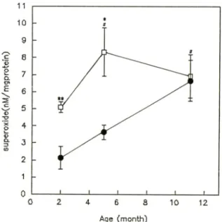
Fig. 1 — Changes in superoxide generation (postnuclear fraction) in male SAM-R/1(#) and SAM-P/2(D) at 2,5 and 11 months of age. Data are meansi SEM from 5 animals. Statistical signi
ficance: **p<0.05 vs. 2 month of SAM-R/1 or SAM-P/2; *p<0.05 and **p<0.01 vs. SAM-R/
등의 범 에 준하여 2.5 mM l-chloro-2, 4-dinitroben
zene 1 m/, 5 mM glutathione 0.5 ml, 0.25 mM phos
phate buffer(pH 6.5)를 각각 취 한 반응액을 25°C 에 서 5분간 preincubation 시킨 후 효소원을 가하여 340 nm에서 3분간 흡광도외 변화를 관찰하여 측정하였다.
6) Catalase 활성 측정*지一50 mM phosphate buf- fer(pH 7.0) 1.5 m/에 효소원 100 m/를 가하고 30 mM H2O2용액의 3배 희석액을 Im / 가하여서 240nm에서 흡광도 번화를 2분간 관찰하여 측정하였다.
7) Superoxide dismutase 활성의 측정*선^—7.5 mM xanthine 50 |j/와 10 mM hydroxylamine hydrochlo
ride 50|j/에 농도별 희석 시료 0.5 m/, blank 로써 65 mM P.B. (pH 7.8) 0.5 m/를 취 해 37에 서 10분간 pre
incubation 시켰다. 0.42 unit/m/의 xanthine oxidase를 0,2 m/가한 후 20분간 incubation 시키고 sulfanil
amide 용액 1 m/와 naphthylethylenediamine 1 m/를 가하여 실온에서 20분간 방처 후 540nm에서 흡광 도를 측정하여 총 SOD활성을 구한 뒤 4m M KCN을 0.2 ml 넣고 측정 한 Mn-SOD 값을 제 하여 Cu, Zn- SOD 값을 구하였다.
실험결과
노화에 따른 SAM-R/1 과 SAM-P/2의 간장중 su
peroxide generation의 변화一 Superoxide genera- tion은 SAM-R/1 의 경우 2개월령의 2,13±0.66nM/
mg protein, 11개월령이 6.67± 1,20 nM/mg protein을 나타내어 SAM-R/1 11개월령에서 2개월령에 비해 68
% 유의성있게 증가하였다(p<0.05). SAM-P/2의 경우 도 2개 월령 이 5.09± 0.32 nM/mg protein, 5개 월령 이 8.33± 0.41 nM/mg protein을 나타내어 SAM-P/2 5개 월령에서 2개월령에 비해 약 38% 유외성있는 증 를 나타내었다(p<0.05).
SAM-R/1 과 SAM-P/2를 각 연령별로 비교헤 보면 SAM-R/1 2개 월령(2.13± 0.66 nM/mg protein)에 비 헤 SAM-P/2 2개 월령(5.09± 0.32 nM/mg protein)에 서 58
% 유의성있게 중가하였으며(pCO.Ol), SAM-R/1 5개 월 령 (3.63 ± 0.42 nM/mg protein) 에 대 해 SAM-P/2 5개월령(8.33± 1.41 nM/mg protein)도 56% 유의성있 는 증가를 나타내었다.(p<0.05, Fig. 1).
노화에 띠론 SAM-R/1 과 SAM-P/2의 간장 중 su
peroxide dismutasell'성의 변화一 Cu, Zn-superoxide
dismutase는 SAM-R/1 의 경우 11개월령이 45.60±
4.72 NU/mg protein, 5개 월령 이 56.13± 2.76 NU/mg protein, 2개 월령이 62.31± 5.04 NU/mg protein을 나 타내어, SAM-R/1 11개월령은 2개월령에 비해 27%
유의성있는 감소를 나타내었다(p<0.01). SAM-R/1 과 SAM-P/2를 각 연령별로 비교해 보았을 때, 5개월령 에서 SAM-R/1(56.13± 27.6 NU/mg protein)에 대해 SAM-P/2(43.73± 2.56 NU/mg protein)는 23% 유외성 있게 감소하였으며(p<0.05), 2개월령에서도 SAM-R/1 (62.31 ± 5.04 NU/mg protein)에 대해 SAM-P/2(36.31
± 7.76 NU/mg protein)는 42% 유외 성 있는 감소를 나타내었대p<0.01, Fig. 2).
Mn-superoxide dismutase는 SAM-R/1 의 경우 11 개월령이 18.58± 1.04 NU/mg protein 5개월령이 16.87
± 0.13 NU/mg protein, 2개월령이 17.38± 0.38 NU/
mg protein을 나타내었으며, SAM-P/2의 경우 11개 월령이 17.38± 0.83 NU/mg protein, 5개월령이 18.33
±0,90 NU/mg protein, 2개 월령 이 16.87± 0.63 NU/
mg protein을 나타내었으나 유외성있는 번화는 관찰 되지 않았다.
Pharm, Soc. Korea 정해영• 김윤경
876543
/su^dplxal&ns
Nonprotein bound-SH는 SAM-R/1 의 경우 11 개월 령 이 3.90± 0.60 최 /g tissue, 5개 월령 이 4.06+0.14 nM/g tissue, 2개월령이 3.94± 0,06 |iM/g tissue를 나타내어 2, 5개월령에 비헤 11개월령에서 감소하는 경향을 나타내었으나 유의성있는 번화는 관찰되지 않았다. SAM-P/2의 경우 11개월령이 2.78± 0.08나M /g tissue, 5개월령이 3.41± 0.20 |iM/g tissue, 2개월 령이 3.71 ± 0.14 |iM/g tissue를 나타내어 SAM-P/2 11개월령에서 2개월령에 대해 25% 유의성있는 감소 를 나타내었으며(p<0.01), 5개월령에 대해서도 18%
10
0
2 10 12
Age (m o n th )
Fig. 2—Cu/Zn-superoxide dismutase(SOD) activities male SAM-R/1(») and SAM-P/2(D) at 2,5 and 11 months of age. Data are means± SEM from 5 animals. Statistical significance; *p<0.01 vs.
2 month of SAM-R/1: *p<0.05 and *p<0.01 vs. SAM-R/1.
노화에 따른 SAM-R/1 과 SAM-P/2의 간장 중 protein bound-SH, nonprotein bound-SH, gluta- thione의 변화一 Protein bound-SH는 SAM-R/1 5개 월령이 19.05± 0.20 |iM/g tissus, SAM-P/2 5개월령이 14.12± 0.78 (iM/g tissue를 나타내어, 5개월령에서 SAM-R/1 에 비헤 SAM-P/2는 protein bound-SH외 26% 유외성있는 감소를 나타내었으며(p<0.01), 11개 월령에서도 SAM-R/1 은 18.49± 0.78 |jM/g tissue, SAM-P/2는 15.79± 1.24 jiM/g tissue로서 SAM-R/1 에 대해 SAM-P/2에서 15% 유의성있는 감소룔 나타내 었다(p<0.05, Table I).
유외성있는 감소를 관찰할 수 었었nKp<0.05). SAM- R/1 과 SAM-P/2를 각 연련별로 비교헤 보았을때, 5 개 월령 에 서 SAM-R/1(4.06± 0.14 |jM/g tissue)에 대 해 SAM-P/2(3.41± 0.20 |iM/g tissue)는 16% 유의성 었게 감소하였으며 (p<0.05), 11개월령 에서도 SAM-R/1 (3.94± 0.06 MM/g tissue)에 대 해 SAM-P/2(2.78± 0.08 nM/g tissue)는 29% 유의성있는 감소를 나타내었다 (p<0.01, Table I).
Glutathione외 앙은 SAM-P/2의 경우 11개월령이 2.75± 0.09 |iM/g tissue, 5개월령이 3.36+0.20 |iM/g tissue, 2개월령이 3.60± 0.16 |iM/g tissue를 나타내어 SAM-P/2 11개월령에서 2개월령에 대해 24% 유의성 있는 감소를 나타내었으며(p<0.05), 5개월령에 대해 서도 18% 유외성있는 감소를 관찰할 수 있었다{p<
0.01). SAM-R/1 과 SAM-P/2를 각 연령별로 비교해 보았을때, 5개월령에서 SAM-R/1(4.01± 0.13 |jM/g ti
ssue) 에 대 해 SAM-P/2(3.36± 0.20 |iM/g tissue)는 16
% 유외성있게 감소하였으며, 11 개월령에서도 SAM- R/l(3.85± 0.06 |iM/g tissue)에 대해 SAM-P/2(2.75±
0.09 jiM/g tissue)는 29% 유외 성 있는 감소를 나타내 었다{p<0.01, Fig. 3).
노화에 따른 SAM-R/1 과 SAM-P/2의 간장 중 ca-
Table I —Hepatic protein-bound and nonprotein - bound sulfhydryl concentration as a function of age in male SAM-R/1 and SAM-P/2
Age (months)
Protein bound-SH (|iM/g tissue)
SAM-R/1 SAM-P/2
Nonprotein bound-SH (|iM/g tissue)
SAM-R/1 SAM-P/2
2 17.52(± 0.36) 17.97(± 0.35) 3,94(± 0.06) 3.7K+0.14)
5 19.05(± 0.20) 14.12(±0.89)**^ 4.06(± 0.14) 3.41(± 0.20)*
11 18.49(± 0.78) 15.79(± 1.24)** 3.94(± 0.06) 2.78(± 0.08)**
Data are means± SEM from 6 animals.
Statistical significance; *p<0.05 and **p<0.01 vs. 2 month of SAM-P/2: *p<0.05 and * *p<0.01 vs. SAM-R/1
Vol. 36, No. 5, 1992
노화촉진 생쥐에서 노화에 따른 활성산소 생성 및 항산화능외 변화
0
0
0
0
0
0
0 8
7
6
5
4
3
2
{uiHOJdBUJ/nNXK>s-cr5
0,0 10 12
Age (m o n th )
Fig. 3 —Glutathione in male SAM-R/1(#) and SAM-P/2 (□) at 2,5 and 11 months of age. Data are means± SEM from 5 animals. Statistical signi
ficance; *p<0.01 vs. 2 month of SAM-P/2:
*p<0.05 and **p<0.01 vs. SAM-R/1.
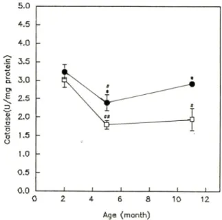
talase황성의 변화一Catalase는 SAM-R/1 의 경우 11 개월령이 2.91± 0.01 U/mg protein, 5개월령이 2.39±
0.02 U/mg protein, 2개월령이 3.29+ 0.02 U/mg pro- tein을 나타내어, SAM-R/1 5개월령에서 2개월령에 비해 27% 유의성있는 감소를 나타내었다(p<0.05).
SAM-P/2의 경우 11 개월령이 1.94± 0.30 U/mg pro
tein, 5개월령이 1.79± 0.12 U/mg protein, 2개월령이 3.01 ± 0.02 U/mg protein을 나타내 어 SAM-P/2 11개 월령에서 2개월령에 버해 36% 유의성있는 감소를 나타내었으며(p<0.05), 5개월령의 경우에도 41% 유 의성있게 감소하였다(p<0.01). SAM-R/1 과 SAM-P/2 를 각 연령별로 비교해 보았을때, 5개월령에서 SAM- R/l{2.39± 0.22 U/mg protein)에 대 해 SAM-P/2(1.79
±0.12 U/mg protein)는 25% 유의성있게 감소하였으 며(p<0.05), 11개월령에서도 SAM-R/1(2.91±0.07U/
mg protein)에 대 헤 SAM-P/2(1.94± 0.30 U/mg pro
tein)-^ 33% 유의성있는 감소를 나타내었다(P<0.05, Fig. 4).
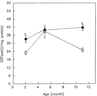
노화에 따른 SAM-R/1 과 SAM-P/2의 간장 중 glu- tathione S-transferase 황성의 번화一 Glutathione S- transferase는 SAM-R/1 외 경우 11개월령이 41.60±
4 6 8
Age (m o n th )
10 12
Fig. 4—Catalase activities in male SAM-R/1 (•) and SAM-P/2(n) at 2,5 and 11 months of age. Data are means ± SEM from 5 animals. Statistical significance: **p<0.05 and ***p<0.01 vs. 2 mo
nth of SAM-R/1 or SAM-P/2: *p<0.05 vs.
SAM-R/1.
2.27 U/mg protein, 5개월령이 39.85± 1.60 U/mg pro
tein, 2개월령이 33.12± 2.54 U/mg protein을 나타내 었으며, SAM-P/2의 경우 11개월령이 24.98±U5U /mg protein, 5개월령이 38.62± 4.78U/mg protein, 2개월령이 22.93소 1.22U/mg protein을 나타내었다.
SAM-R/1 과 SAM-P/2룔 각 연령별로 비교해 보았 을때 2개월령 에서 SAM-R/1 는 33.12± 2.54 U/mg protein, SAM-P/2는 22.93± 1.22 U/mg protein을 나 타내어 SAM-R/1 에 대해 SAM-P/2는 31% 유의성있게 감소하였으며(p<0.01), 11개월령에서도 SAM-R/1 은 41.6± 2.27 U/mg protein, 이 에 대헤 SAM-P/2는 24.98± 1.55 U/mg protein으로서 SAM-R/1 에 버 해 40
% 유외성있는 감소를 나타내었다(p<0.01, Fig.5).
고 찰
대사활성이 높을수록 수명이 짧다는 사실은 수중의 포유동물의 수명이 대사율과 반비례 관계가 성립한 다는 사실로부터 알 수 었으며, 또한 산소 소비량이 증가할수록 노화가 촉진된다는 보고들^^년■부터 활성
/. Pharm. Soc. Korea
464 정해영 • 김윤경
0 5 0 5 0 5 0 5 0 5 5 4 4 3 3 2 2 1 1 0
765432
^9nssn
. e>/2rf}&uo!qvDvllo
0 2 4 6 8 10 12 Age (m o n th )
Fig. 5 —Glutathione S-transferase activities in male SAM-R/K#) and SAM-P/2(n) at 2,5 and 11 months of age. Data are means± SEM from 5 animals. Statistical significance; *p<0.01 vs.
SAM-R/1.
산소가 관여할 것이라는 가능성이 시사되었는데, 노 화에 외해 각조직에 활성산소 반응산물이 증가한다는 사실은 이미 잘 알려겨 있으며 대표적으로 lipofuscin, 과산화 지질의 측적을 둘 수 있다.^> Sawada 등은 aging rotifer에서, Farmer 등은페 housefly, Musca
에서 노화에 따라 superoxide radical 형성이 증가함을 보고한 바 있으며, 본 실험에서도 가령에 따라 웅성 SAM-R/1, SAM-P/2외 간장중 postnuclear fraction에서 superoxide 형성이 증가하였을 뿐 아니 라 SAM-R/1 에 버헤 SAM-P/2에서 더 높은 형성능을 나타내어 노화에 따라 superoxide 형성이 중가함을 관찰할 수 었었나.
노화에 따른 활성산소 반응산물외 측적증가는 활 성산소를 제거할 수 있는 항산화계인 SOD, catalase, glutathione peroxidase, glutathione, protein bound- SH, nonprotein bound-SH 등의 번화와 연관되는데, superoxide는 생체내 SOD에 외해 hydrogen pero- xide로 되고 이는 catalase나 glutathione peroxi- dase에 의해 제거된다고 알려져 있다.
SOD는 metalloproteins으로서 활성 부위 에 존재 하 는 금속의 중류에 따라 Cu, Zn-SOD, Mn-SOD, Fe- SOD로 분류되는데 그 기능은 동일하며, 동물조직에
Vol. 36, No. 5, 1992
서는 Cu, Zn-SOD와 Mn-SOD만이 발견된다고 한다.
Cu, Zn-SOD는 cytoplasm에 Mn-SOD는 mitochon
dria 에 주로 존재하는데, 설치류의 경우 Mn-SOD는 전체 SOD의 약 8% 정도를 차지한다고 알려져있다."단>
De Haan 등은해 흰쥐 뇌에서, Ischiropoulos 등은^효^ 흰쥐외 페에서, Gupta은^^^ 흰쥐외 뇌에서 노화에 따라 SOD 활성이 증가하였다고 보고하였고, Ansari 동은2라 변화가 없다고 보고하였으나, 본 실험에서 SOD 활 성은 SAM-R/1 파 SAM-P/2 모두 가령에 따라 감소 하였으며 SAM-R/1 보다 SAM-P/2에서 더 낮은 활성 을 나타내었는데, 이는 Semsei등이^ > 흰쥐 뇌에 대해, Cand등이즈> 흰쥐 간장에 대해 보고한 결과와 일처 하였다. Mn-SOD외 경우 Irene등은2"» 노화에 따라 중가함을 보고하였으나 본실험에서는 유의성있는 변 화를 관찰할 수 없었다. Cu, Zn-SOD는 superoxide 제거파정에서 생기는 부산물인 hydrogen peroxide에 외해 비가역적으로 불활성화될 수 있으며, 노화 적 혈구에서 보이는 Cu, Zn-SOD의 glycation증가에 외 헤서도 그 활성이 감소한다는 보고가 있 다 또 한 활성부위에 존재하는 Cu와 Zn의 결핍시에도 활성의 현격한 감소가 보이는데, 소아 적혈구에서 Zn농도가 연령의존적으로 나타났다는 보고가 있다.^>
세포내 hydrogen peroxide를 제거하는 효소인 ca
talase 는 생 체내 모든 주요기관에 걸쳐 존재하나 특히 간장이나 적혈구에 많으며, 대개 peroxisome 내 존 재하는 것으로 알려겨 있다.^^^ Catalase 활성의 경우 Semsei등^^과 Sohal등이체 노화에 따라 감소함을 보 고하였으며, Perez등과씨 Ansari동^^ 번화가 없음을 보고하였으나 본실험에서는 가령에 따라 감소하는 결과를 나타내었다.
Farooqui^*등은 흰쥐 간에서 노화에 따라 gluta
thione 의 앙이 감소하여 lipid peroxide가 증가한다고 보고한 바 었으며, Perez등은^^ 흰쥐 페에서 노화에 따라 glutathione의 앙과 lipid peroxidation 정도가 변화가 없옴을 보고하였는데 본 실험에서는 가령에 띠*sf glutathioneprotein bound-SH, nonprotein bound-SH외 앙은 감소하였으며, glutathione S-tran- sferase외 활성도 감소하는 경향을 나타내었다.
Glutathione S-transferase는 세포질 glutathione S- transferase과 mitochondria 및 소포체막 glutathione S-transferase으로 대별되는데 앙 glutathione S-tran- sferase는 생체 거외 전체조직에 함유되어 있지만, 노화촉진 생쥐에서 노화에 따른 활성산소 생성 및 항산화능의 변화
2
6
0
4
8 4
3
3
2
1
^E/roosDiso
정해영 * 김윤경
간에서 최고의 함량을 나타내며 부신, 정도 등에도 앙 glutathione S-transferase 가 고농도•로 분포한다.좌 세포질 glutathione S-transferase의 체내 중요한 역 할외 하나는 천 전자성의 발암성 활성대사물의 해득 작용으로서,페 발암물질은 생체내에서 cytochrome P- 450체이나 phosphotransferase^가 등의 각종 약물대사 효소에 의해서 활성화되어 DNA룰 손상하지만, gluta
thione S-transferase은 이들 소수성잔기를 가지는 활성대사물(R-X)을 기질로 해서, 이것에 glutathione 을 공유결합시켜 R-SG로서 안정화시켜서 최종적으로 N-acetyl conjugate로 뇨중 배설시키는 최초단계외 반응을 촉매한다는 사실이 일반적으로 널러 알려져 있다. 소수성 화합물 및 소수성이 높은 잔기를 가지는 화합물을 잔기로 하는 glutathione S-transferase는 다포화지방산의 peroxide(PUFA-OOH)를 기질로해서, GSSG의 생성과 동반하여 alcoh이체(PUFA-OH)로 전 환시키는 작용도 있다.^» Se 결핍증 흰쥐의 간에서 볼 수 있는 Se함유 glutathione peroxidase의 결손 시에 세포절 glutathione S-tranferase 활성이 증대하 는 것은 glutathione S-transferase가 glutathione- peroxidase 의 대체 역활을 하고 있다고 볼 수 있으며, 또한 모르모트의 간에서 PUFA-OOH의 소거는 gluta- thione S-transferase에 의헤 행해지고 있다.체 세포질 중에 있는 glutathione-peroxidase나 glutathione S- transferase에 외 한 PUFA-OOH외 환원은 mitochon- dria나 소포체막을 구성하는 인지질외 PUFA잔기가 hydroperoxide가 된 경우에 지절파산화의 연쇄반응을 막기위해 phospholipase A2에 의해서 PUFA-OOH가 가수분해되어서 세포질 중에 들어가서 일어난다고 생각되고 있다.^>
본 연구에서 SAM-R/1 에 비해 SAM-P/2에서 간장 중 superoxide생성능이 현저히 증가되어 있고, 이에 대헤 SOD, catalase, glutathione S-transferase활성은 대체로 감소하에 있으며, protein bound-SH, nonpro
tein bound-SH, glutathione도 낮온 농도에 있으므로^
SAM-R/1 에 비해 SAM-P/2에서 활성산소에 의해 조 직손상외 증기를 가져와서 이것이 노화과정을 더욱 촉진시키는 한 요언일 것으로 사료되었다.
간장중 superoxide생성능 중가 요인에 대해서는 더욱 연구가 필요할 것으로 생각되며, 본 연구설에 서는 간장조직의 각 분획별 superoxide생성능 중가 유래에 대해 검토중에 있다.
결 론
가령에 따른 정산노화 생쥐 SAM-R/1 과 촉진노화 생쥐 SAM-P/2의 간장 중 superoxide 생성능과 이에 대한 방어 계로서 SOD, catalase, glutathione, gluta
thione S-transferase, protein bound-SH, nonprotein bound-SH의 번화를 검토하였다.
Superoxide 생성능은 SAM-R/1, SAM-P/2에서 가 령에 따라 유의성있게 증가하였으며. SAM-R/1 보다 SAM-P/2에서 더 높은 생성능을 나타내었다. SOD 활성은 SAM-R/1, SAM-P/2에서 가령에 따라 현저허 감소하였으며, SAM-R/1 에 비해 SAM-P/2에서 더 낮 은 활성을 나타내었다. Catalase 활성도 가령에 따라 감소하였으며, SAM-P/2에서 더욱 감소하였다. 또한, glutathione S-transferase 활성은 가령에 따라서는 명 확한 변화를 나타내지 않았으나, SAM-R/1 에 대해 SAM-P/2는 유의성있는 감소를 나타내었다. Gluta
thione^ ^ 힘 protein bound-SH, nonprotein bound-SH외 경우도 가령에 따라 감소하는 경향을 나타내었으며 SAM-P/2에서 보다 명확히 감소하였 다.
이상의 결과로부터 SAM-R/1 에서 가령에 따른 간장 중 superoxide 생성능은 뚜렷하게 증가하였고 이를 제거하는 항산화능은 감소하였다. 또한, SAM-R/1 의 경우보다 SAM-P/2 에서 더욱 높은 superoxide 생성 능과 낮은 항산화능을 나타내어, 이러한 요인이 SAM- P/2에서 노화과정을 더욱 촉진시킨 것으로 사료되었 다.
감사의 말씀
노화촉진 생쥐 SAM-R/1 및 SAM-R/2를 분양하여
주신 Takeda교수님께 감사드럽니다.
문 헌
1) Balin, A. K.: Testing the free radical theory of aging. In: R. C. Adelman and G. S. Roth, eds., Testing the Theories of Aging. CRC Press, Boca Raton, Fla., pp. 37-56(1982).
2) Holliday, R.: Cross linking theory. Science 213, 505 (1980).
3) Olsonm, C. B.: Accumulation of waste product
Pharm. Soc. Korea
노화촉진 생쥐에서 노화에 따른 활성산소 생성 및 항산화능외 번화 467
theory. Mech. Ageing Dev., 41, 17(1987).
4)
헬]fli ■ 클복, m M m m ,pp.
145-162(1977).
5) Harman, D.: A theory based on free radical and radiation chemistry. J, Gerontol, 11, 298(1956).
6) Corfran, R. S., Kumar, V. and Robbins, S. L: Rob
bins Pathologic Basis of Disease, W. B. Saunders,
Philadelphia, pp. 1-38(1989)7) McCord, J. M. and Fridovich, I.: Superoxide dis
mutase. An enzymic function for erythro cuprein (hemo cuprein). / BioL Chem., 244, 6049(1969).
8) Chan, P. C. and Bielski, B. H. J.: Enzyme catalyzed free radical reactions with nicotinamide adenine nucleotide. / Biol. Chem., 249, 1317(1974).
9) Sedlak, J. and Lindsay, R. H.: Estimation of total protein-bound and nonprotein-bound sulfhydryl groups in tissue with Eliman's reagent. Anal Bio
chem., 25, 192(1968).
10) Higash, T.: Critical review on the determination of glutathione in biological preparations. Proteins,
Nucleic Acid and Enzyme 33, 1370(1988).
11) Gaitonide, M. K.: A spectrophotometric method for the direct determination of cysteine in the pre
sence of other naturally occurring amino acids.
Biochem. J., 104, 627(1967).
12) Habig, W. H., Pabst, M. J. and Jakoby, W. B.; Glu
tathione S-transferase. J. Biol Chem., 249, 7130 (1974).
13) Chance, B. and Maehly, A. C.; Assay of Catalase
and Peroxidase. Vol. II, Academic Press, pp. 764-
775(1955).14) Oyanagui, Y.: Reevaluation of assay methods and establishment of kit for superoxide dismutase acti
vity. Anal. Biochem., 42, 290(1948).
15) Osamu, I.: Lipid Peroxidation and Nutrition, Japa
nese Society of Nutrition and Food Science, To
kyo, pp. 143-214(1986).
16) Uysal, M., Seckin, S., Kocak-Toker, N. and Oz, H.:
Increased hepatic lipid peroxidation in aged mice.
Mech. Ageing Dev., 85, 48(1989).
17) Sawada, M. and Carlson, J. C.: Biochemical cha
nges associated with the mechanism controlling superoxide radical formation in the aging rotifer.
]. Cell Biochem., 153, 44(1990).
18) Farmer, K. J. and Sohal, R. S.: Relationship bet
ween superoxide anion radical generation and aging in the housefly, Musca Domestica. Free Radic.
Biol Med.. 23, 7(1989).
19) Jakoby, W. B.: Enzymatic Basis of Detoxication,
Academic Press, New York, pp. 318-320(1980).
20) De Haan, J. B., Newman, J.D. and Kola, I.: Cu/Zn superoxide dismutase mRNA and enzyme activity and susceptibility to lipid peroxidation increases with aging in murine brains. Brain Res., 179, 13 (1992).
21) Ischiropoulos, H., Nadziejko, C. E. and Kikkawa, Y.: Effect of aging on pulmonary superoxide dis
mutase. Mech. Aging Dev., 11, 52(1990).
22) Gupta, A., Hasan, M., Chancier, R. and Kapoor, N. K.: Age-related elevation of lipid peroxidation products: diminution of superoxide dismutase ac
tivity in the central nervous system of rats. Geron
tology 305, 37(1991).
23) Ansari, K. A., Kaplan, E. and Shoeman, D.: Age- related changes in lipid peroxidation and protec
tive enzymes in the central nervous system. Gro
wth Dev. Aging 117, 53(1989).
24) Semsei, L, Rao, G. and Richardson, A.: Expression of superoxide dismutase and catalase in rat brain as a function of age. Mech. Ageing Dev., 13, 58 (1991).
25) Cand, F. and Verdetti, J.: Superoxide dismutase, glutathione peroxidase, catalase and lipid peroxi
dation in the major organs of the aging rats. Free
Radic. BioL Med., 59, 7(1989).
26) Irene, C. P., Trivier, J.M., Annie, N., Sinet P.M.
and Marc, T.: Age-correlated modifications of Co- pper-Zinc superoxide dismutase and glutathione- related enzyme activities erythrocytes. Clin.
Chem., 38. 66(1992).
27) Sinet, P. M. and Garber, P.: Inactivation of the human Cu, Zn superoxide dismutase during expo
sure to 'C V and H2O2. Arch. Biochem. Biophys,, 212. 411(1981).
28) Salo, D. C., Pacifini, R. E. and Davies, K. J. A.:
Superoxide dismutase is preferentially degraded by a proteolytic system from red blood following oxidative modification by hydrogen peroxide. Free
Vol 36, No. 5, 1992
정해영 • 김윤경
Rad. Biol. Med., 5, 335(1988).
29) Arai, K., Magushi, S., Fujii, S. et al.: Glycation and inactivation of human Cu, Zn superoxide dismu
tase. Identification of the in vitro glycated sites.
J. Biol Chem., 262, 16969(1987).
30) Bettger, W. JL, Fesh, T. J. and OT)ell, B. L: Effects of dietary copper and zinc on erythrocyte stability and superoxide dismutase activity. Proc. Soc. Exp.
Biol. Med, 158, 279(1979).
31) Barry, H. and John, M. C. G.: Free Radicals in
Biology and Medicine, Clarendon Press, Oxford,
pp. 86-123(1989).32) Semsei, I., Roc, G. and Richardson, A.: Changes in the expression of superoxide dismutase and ca
talase as a function of age and dietary restriction.
Biochem. Biophys. Res. Commun., 620, 164(1989).
33) Sohal, R. S., Arnold, L. and Orr, W. C.: Effect of age on superoxide dismutase, catalase, glutathione reductase, inorganic peroxides, TBA-reactive ma
terial, GSH/GSSG, NADPH/NADP^ and NADH/
NAD 구 in drosophila melangaster. Mech, Ageing
Dev., 223, 56(1990).
34) Perez, R., Lopez, M. and Barja de Quiroga, G.:
Aging and lung antioxidant enzyme, glutathione and lipid peroxidation in the rat. Free Radic. Biol.
Med., 35. 10(1991).
35) Farooqui, M. Y., Day, W. W. and Zamorano, D.
M.; Glutathione and lipid peroxidation in the aging rat. Comp. Biochem. Physiol., [B], 177, 88(1987).
36) rbfflfiSIS, Glutathione S-transferase iso
zyme, glutathione •그느4공 > 7.
mm,
33, 1564(1988).37) Watabe, T., Ishizuka, T., Isobe, M. and Ozawa, N.:
7-hydroxymethylsulfate ester as an active metabo
lite of 7,12-dimethylbenz[a]anthracene. Science 215, 403(1982).
38) Sevanian, A., Muakkash-kelly, S. F. and Montest- raque, S.: The influence of phospholipase ki and glutathione peroxidase on the elimination of mem
brane lipid peroxide. Arch, Biochem. Biophys., 223, 441(1983).
39) Sevanian, A. and Kim, E.: Phospholipase ki depe
ndent release of fatty acids from peroxidized me
mbranes. J. Free Radical Biol. Med., 1, 263(1985).
J. Pharm. Soc. Korea