A THESIS
FOR THE DEGREE OF DOCTOR OF PHILOSOPHY
CHARACTERIZATION OF ANTIOXIDANT GENES FROM ABALONE (Haliotis discus discus) cDNA
Prashani Mudika Jayasooriya
Department of Marine Biotechnology GRADUATE SCHOOL
CHEJU NATIONAL UNIVERSITY
CHARACTERIZATION OF ANTIOXIDANT GENES FROM ABALONE (Haliotis discus discus) cDNA
Prashani Mudika Jayasooriya
(Supervised by Professor Sang-Chul Chung )
A thesis submitted in partial fulfillment of the requirement of the degree of Doctor of Philosophy
2005. 12
This thesis has been examined and approved by
………
Chairperson of the supervising committee You-Jin Jeon, Professor of Marine Biotechnology
………
Choon-Bok Song, Professor of Marine Biotechnology
………
In-Kyu Yeo, Professor of Marine Biotechnology
………
Moon-Soo Heo, Professor of Marine Biotechnology
………
Sang-Chul Chung, Professor of Marine Biotechnology
……… Date
Department of Marine Biotechnology GRADUATE SCHOOL CHEJU NATIONAL UNIVERSITY
CONTENTS
요약문 iv
List of Figures vi
Introduction 1
Part I.
Cloning, expression, purification and characterization of catalase from abalone (Haliotis discus discus) cDNA
Abstract. 7
Materials and methods
Sequencing of the gene encoding aCAT 8 Cloning the coding sequence of aCAT into
pMAL expression vector
8
Overxpression of catalase 9
Purification of catalase 10
Gel electrophoresis 10
Catalase activity assay and determination of
protein concentration 11
Analysis of nucleotide and amino acid sequences
12
Results 13
Discussion
Cloning and analysis of aCAT 24
Overexpression of aCAT in E. coli BL 21 DE3 26 Optimal temperature and thermal stability 27
Optimal pH 28
Phylogenetic analysis 29
Part II
Molecular cloning, expression, purification and characterization of Cu,Zn-Superoxide dismutase from Abalone, Haliotis discus discus cDNA
Abstract 31
Materials and methods
Sequencing of the gene encoding Cu,Zn-SOD 32 Cloning the coding sequence of Cu,Zn-SOD
into the pMAL expression vector
32
Overexpression of aCu,Zn-SOD 33
Cu,Zn-SOD activity assay and determination
of protein concentration 34
Analysis of nucleotide and amino acid sequences
35
Results 37
Discussion
Isolation of full length cDNA encoding Cu,Zn-SOD from H. discus discus
46 Overexpression of a Cu,Zn-SOD in E. coli
K12(TB1)
46 Deduced Amino acid sequence comparison 48 Optimal temperature and Thermal stability 49
Optimal pH 50
Phylogenetic analysis 50
Part III
Cloning, expression, purification and characterization of Mn-superoxide dismutase from abalone (Haliotis discus discus) cDNA
Abstract 52
Materials and methods
Sequencing of the gene encoding abalone Mn-SOD:
54 Cloning the coding sequence of aMn-SOD into
pMAL expression vector
54
Overxpression of aMn-SOD 55
Purification of aMn-SOD 56
Mn-SOD activity assay and determination of protein concentration
56 Analysis of nucleotide and amino acid
sequences
57
Results 58
Discussion
Cloning and analyzing abalone Mn-SOD 68 Overexpression of aMn-SOD in E. coli
K12(TB1) 69
Optimal temperature and thermal stability 70
Phylogenetic analysis 71
Summary 73
References 76
요약문
항산화 효소들은 체내에서 생명 유지를 위해 산화 환원 반응을 조절하는 중 요한 역할을 한다. 이 논문에서는 까막전복으로부터 만들어진 cDNA library 로부터 중요한 항산화 효소로 알려진 catalase (aCAT), Cu,Zn-superoxide dismutase (aCu,Zn-SOD) 와 Mn-superoxide dismutase (aMn-SOD)에 대한 코딩 유전자의 서열을 분석하였다. 첫번째로, 전체 길이를 확인한 후 그 서열들은 기존에 data base 를 통해 비교되어졌고 구조와 기능이 유사한 다른 생물의 효소들과의 비교를 통해 보존된 서열이 확인되어졌다.
두 번째로, PCR 을 통해 코딩 서열을 증폭시키고 그 산물을 pMAL-c2X vector 내로 삽입시킨 후 E. coli BL21(DE3)나 K12(TB1)에 형질전환 시켜졌다. 그 각각의 재조합 단백질들은 IPTG 를 이용해 발현이 유도되어졌다. 그리고 발현된 단백질들은 최적 온도와 pH 그리고 열 안정성이 각각 진단되어졌다. 각각의 효소들 에 대한 계통 수는 neighbour-joining 의 방법에 의해 시행되어졌다.
H2O2에서 H2O 와 O2로의 반응을 촉매하는 catalase 는 세포질내의 H2O2 의 독성을 제거하는 중요한 효소로 알려져 있다. 그 catalase 는 501 개의 아미노산 을 코딩하는 1503 bp 의 ORF 로 구성되며 전복에서 true catalase 그룹인 것으로 확인 되어졌다. 그 catalase 의 specific activity 는 30,000 U/mg 이었다. pH 는 5.0 에서 10.5 까지 안정했고 열안정성은 70 oC 이하에서 안정하게 나타났다. 알려 져있는 다른 22 개의 catalase 아미노산 서열과 비교했을 때 173 개의 잔기가 보존 된 서열로 나타났고 34.5% 의 homology 를 보였다. 그리고 aCAT 는 Pacific white shrimp 의 CAT 와 가장 높은 유사성을 나타냈다.
Superoxide dismutase (SOD)는 superoxide (O2-)과 반응하는 하나뿐인 항산화 효소이다. SOD 는 위치와 활성 금속이온에 따라 구분되어진다. aCu,Zn-SOD 는 154 개의 아미노산을 코딩하는 465 개의 염기로 구성되고 pI 값이 5.5, 분 자량이 16 kDa 이다. aCu,Zn-SOD 의 최적 온도는 37 oC 이며 그것은 70 oC 에서 활성을 잃어버렸다. 그리고 산성 pH 인 3.5 – 6.5 범위에서 활성을 가졌다. 47 개의 다른 종의 SOD 와 비교했을 때 48 개의 아미노산이 보존되었고 29%의 homology 를 보였다. Cu 와 Zn 이온의 부착을 위한 모든 잔기들이 aCu,Zn-SOD 에 보존되어있 었다. 계통수로 볼 때 aCu,Zn-SOD 는 H. diversicolor 의 것과 가까웠고 이것은 다 른 전복의 일종으로 두 종간의 관계가 진화적으로 가까웠음을 보여준다. Mn-SOD 는 세 포 질 내 로 생 산 되 어 지 지 만 미 토 콘 드 리 아 의 post-translationally 에 중요하다. aMn-SOD 는 226 개의 아미노산을 코딩하는 690 개 의 염기로 구성되며 25 개의 아미노산으로 구성된 신호서열을 포함한다. 발현된 aMn-SOD 의 분자량은 25 kDa 이고 최적온도는 37 oC 이다. aMn-SOD 도 aCu,Zn-SOD 와 마찬가지로 산성 pH 에서 활성을 가졌다. 그 효소는 80 oC 에서 활 성을 잃었다. aMn-SOD 의 아미노산 서열은 35 개의 다른 서열과 비교했을 때 fresh water snail (Biomphalaria labrata)의 Mn-SOD 와 가장 가까웠다.
이 연구에서는 까막전복으로부터 CAT, Cu,Zn-SOD 와 Mn-SOD 의 유전 자를 분석하고 클로닝하여 과잉발현을 유도하였으며 그 발현된 단백질을 정제하여 각각의 특성을 분석하였다. 각각의 염기서열분석은 무척추동물의 항산화 효소와 그 들의 진화 상에서 비교에 의한 게놈 연구에 기여할 수 있다.
List of Figures
Fig. 1 Nucleotide sequence and the deduced amino acid sequence of aCAT.
Fig. 2 Analysis of catalase (aCAT) protein expressed in E. coli BL21 (DE3) cells following purification in a 12% denaturing polyacrylamide gel.
Fig. 3 Multiple sequence alignments of (human, human erythrocyte, Bos, B. taurus, Xenopus, X. laevis, Danio, D. rerio and Litopenaeus, L. vannamei) known catalase amino acid sequences. Fig. 4 Optimal temperature of aCAT.
Fig. 5 Optimal pH of aCAT. Fig. 6 Thermal stability of aCAT.
Fig. 7 Neighbor-joining tree based on catalase amino acid sequences obtained from 23 species.
Fig. 8 Nucleotide sequence and the deduced amino acid sequence of aCu,Zn-SOD.
Fig. 9 Analysis of aCu,Zn-SOD protein expressed in E. coli K12(TB1) cells following purification in a 12% denaturing polyacrylamide gel.
Fig. 10 Sequence comparison of aCu,Zn-SOD with S. scrofa (P04178),
B. taurus, (P00442), H. sapiens (P00441), G. gallus (NP990395)
and H. diversicolor supertext (AAY18806). Fig. 11 Optimal temperature of aCu,Zn-SOD. Fig. 12 Optimal pH of aCu,Zn-SOD.
Fig. 13 Thermal stability of aCu,Zn-SOD.
Fig. 14 Neighbor-joining tree based on Cu,Zn-SOD amino acid sequences obtained from 47 species.
Fig. 15 Nucleotide sequence and the deduced amino acid sequence of aMn-SOD.
Fig. 16 Analysis of aMn-SOD protein expressed in E. coli K12(TB1) cells following purification in a 12% denaturing polyacrylamide gel.
Fig. 17 Multiple sequence alignments of aMn-SOD with 9 other (human erythrocyte- CAA42066, P. pimaeus- CAH93471, B. Taurus- 151918, R. norvegicus-NP08747, M. undulates-AAO72712, G.
gallus- NP989542, E. coioides-AAW29024, D. rerio- NP956270
and B. glabrata- AA583981) known Mn-SOD amino acid sequences.
Fig. 18 Optimal temperature of aMn-SOD. Fig. 19 Optimal pH of aMn-SOD.
Fig. 21 Neighbor-joining tree based on Mn-SOD amino acid sequences obtained from 35 species.
INTRODUCTION
Although oxygen is essential to life as a part of normal metabolism, its access can give rise to a variety of reactive oxygen species (ROS). Under normal conditions, the body is well equipped with a variety of mechanisms that serve to inactivate the extra ROS. However, under certain conditions, where these mechanisms are faulty or the body has been exposed to environmental chemicals, irradiation, iron loading, and other similar factors, the elevated ROS levels can cause a variety of diseases and could even lead to death. They not only damage biological molecules like protein and DNA but also lead to cell death and thereby become a cause to a tremendous number of diseases including cancer, alzheimer and arthritis. ROS are formed in the body as toxic by-products of oxidative metabolism (Ken et al., 2003). These include superoxides, hydrogen peroxide (H2O2), hydroxyl
radicals and singlet oxygen. Cells have developed multilayered interdependent antioxidant system as a defense against oxidative injury. This system comprises enzymatic and non-enzymatic components. Superoxide dismutase, catalase and glutathione peroxidase are the enzymes that catalyze the dismutation of the ROS. Antioxidant enzymes play a major role in protecting organisms from the potentially deleterious effects of ROS. They contribute towards strengthening the defense mechanism in cells, and are studied in detail in several aquatic organisms (Nakano et al., 1995).
Catalase is an antioxidant enzyme in the cellular protection system and catalyzes the decomposition of hydrogen peroxide to oxygen and water
(Thuy et al., 2004). It is also found in all aerobic cells. The level of catalase expression is highly tissue specific. The highest levels are found in liver, kidney and blood while the lowest levels are found in connective tissues and brain (Chen et al., 2004). Presence of catalase is important in the prevention of toxic wastes, which are harmful to cells. Human acatalasemia is a genetic example of catalase deficiency, which has been observed in Japanese individuals (Ogata, 1991).
Most catalases exist as tetramers of approximately 65 kDa subunits (Bravo et al., 1999) and classified under three sub classes namely typical, catalase peroxidases and manganese catalases. Each of the four subunits contains Fe3+ prosthetic heme groups (protoporphyrin IX), which is exposed
through a 26 Ao long and 17 Ao wide funnel shaped channel. The heme group is proved to be responsible for the enzymatic activity of catalase. Catalytic mechanism of catalase is a two-step reaction (Deisseroth and Dounce, 1970). In the first step, the heme Fe3+ reduces a hydrogen peroxide molecule to water and generates a covalent Fe4+ =O oxyferryl species with a porphyrin π-cation radical, which is referred as compound I and in the second step, compound I oxidizes a second peroxide molecule to molecular oxygen and releases the ferryl oxygen species as water (Chance, 1949). Catalase enzyme complex also binds the reductant NADPH (Fita and Rossmann, 1985) yet, hydrogen peroxide is the source of both oxidative and reductive potential during the normal catalytic cycle.
Catalase cDNAs have been isolated from number of eukaryotic organisms including human, rat, cattle, yeast, mice, and sweet potato (Hass et al., 1991). Most of these catalases share a high degree of amino acid sequence identity. In comparison with catalases from mammalian and bacterial sources, there is little information available on mollusk catalase, especially on its biochemical properties. Gerhard et al. confirmed that other than amphibia (Dadras et al., 1996), little has been reported about the structure or regulation of catalase in non-mammalian vertebrates (Gerhard et al., 2000).
Superoxide dismutases (SOD) are metalloenzymes in aerobic organisms, which play another crucial role in protecting organisms against the toxic wastes caused by ROS, in particular superoxide radicals (O2.-) (Liu
et al., 2002). It catalyses the dismutation of superoxide radicals to molecular oxygen and hydrogen peroxide (Wang et al., 2005). Although O2.- is a mild
reactant, it can cause direct or indirect damage to the membranes and DNA, when it is protonated to hydroxyl radical (HO.) or dismutated to H2O2
(Birnboim and Kanabus-Kamnika, 1985). The reported medical applications of SOD include anti-inflammation, prevention of oncogenesis & tumor growth, and protection against reperfusion damage of ischemic tissue (Garcia –Gonzalez and Ochoa, 1999; Kondo et al., 1999). It has also been proposed that bacterial Cu,Zn-SOD is involved in pathogenic infections and survival at the stationary phase of growth while being resistant to oxidative stress (Kho et al., 2004). Moreover, Fridovich (1986) reported that organisms lacking
SODs exhibit a decreased growth rate, short life span, hypersensitivity towards redox cycling compounds (such as paraquat and quinines) and accelerated spontaneous mutagenesis resulting high death rates.
SODs are classified into several forms and can be distinguished by their primary structure, cellular compartmentalization, primary function and the metal required for activity (Wright et al., 2002). Three distinct groups depending on the metals identified in their active sites are Cu,Zn-SOD, Mn-SOD, and Fe-SOD. Cu,Zn-SODs are widely distributed in eukaryotes, whereas Mn-SOD and Fe-SOD enzymes are predominantly found in mitochondria or prokaryotes (Fridovich, 1986). Recently a variety of other forms of SODs have been identified in bacteria. These include a nickel-containing isozyme (Kim et al, 1998a) and hybrid isoforms nickel-containing iron and zinc (Kim et al., 1998b). Among Cu,Zn-SOD, cytosolic Cu,Zn-SOD and glycosylated extracellular Cu,Zn-SOD has been found in eukaryotes (Jeong et al., 2001) and is also reported to have a widespread distribution in a variety of cells.
The genomic sequence for Cu,Zn-SOD has been identified in the rat (Kim et al., 1993), mouse (Benedetto et al., 1991) and human (Levanon et al., 1985). Fukuhara et al. (2002) have carried out a comprehensive study on structure, molecular evolution, and gene expression of Mn- and Cu,Zn-SOD on eight primate species. Cu,Zn-SOD coding sequences have been cloned from many diverse organisms and expressed in various systems such as
(Hallewell et al., 1991), Drosophila melanogaster (Reveillaud et al., 1991) and even in transgenic mice (Ceballos et al., 1991). Further, molecular cloning of Cu,Zn-SOD from three different mollusk species have been reported by Geret et al. (2004). Although coding sequence for Cu,Zn-SOD of
Haliotis diversicolor supertexta ( AAY18806) is available, limited studies on cloning of Cu,Zn- SOD from Mollusks led this study to clone, sequence and express aSOD in E. coli, and compare them with other known Cu,Zn-SOD sequences to identify conserved regions that may be useful in elucidating its structure-functional relationship.
Out of the SODs found in eyukaryotes (cytosolic Cu,Zn-SOD, glycosylated extracellular Cu,Zn-SOD and mitochondrial SOD), Mn-SOD is particularly important as it is located in mitochondria and represents the first line of defense against superoxide radicals produced as byproducts of oxidative phosphorylation (Beyer et al., 1991). In eukaryotic cells, Mn-SOD is synthesized in the cytosol and imported post-translationally into the mitochondrial matrix (Bannister et al., 1987). Mitochondria is an important site for the single-electron reduction of O2 to O2-. Therefore, Mn-SOD is
thought to be a major scavenger of damaging ROS metabolites in the mitochondrial matrix (Ken et al., 2005).
Mn-SOD is a homotetramer of about 23 kDa subunits and localized in mitochondrial matrix of aerobic cells. Interestingly, its amino acid sequence is distinctly different from the amino acid sequence of Cu,Zn-SOD (Bannister et al., 1987). Mn-SOD has been shown to play a major role in
promoting cellular differentiating and tumorgenesis (St.Clair et al., 1991) and in protecting cells against hyperoxia-induced pulmonary toxicity (Wispe et al., 1992). Further, overexpression of the protein has well-documented anti-apoptotic effects (Pani et al., 2000; Bernard et al., 2001; Drane et al., 2001) and its deletion has lead to neurodegeneration and cardiomyopathy in mice (Lebovitz et al., 1996; Melov et al., 1998). Many scientists have summarized the role of Mn-SOD in protection against ROS and their physiological function in several excellent reviews (Fridovich, 1995; Bannister et al., 1987; McCord and Fridovich, 1988; Oberley and Buettner, 1979). The complete genomic structure of Mn-SOD for human (Church et al., 1992; Wan et al., 1994), rat (Ho et al., 1991), and mouse (DiSilvestre et al., 1995) has been determined. Also, partial identification and characterization of bovine Mn-SOD has described by Meyrick and Magnuson (1994). However, available information on Mn-SOD from invertebrates is not sufficient.
In this study, cDNA coding for three antioxidant genes; catalase, Cu,Zn-SOD and Mn-SOD from disk-abalone (Haliotis discus discus) were sequenced, cloned, expressed and characterized. The isolated sequences were compared with that of other species available in the public database and attempts were made to build structure-functional relationship of the amino acid sequences.
Part I
Cloning, expression, purification and characterization of catalase from abalone (Haliotis discus discus) cDNA
1. ABSTRACT
Catalase is an antioxidant enzyme, which plays a crucial role within the cellular protection system. It facilitates the degradation of hydrogen peroxide, which is a reactive oxygen species, into oxygen and water. Biochemical information on mollusk catalase is however, insufficient. A gene coding for putative catalase of the disk abalone (Haliotis discus discus) was selected from the cDNA library (derived from digestive gland) and the full-length was sequenced using 3 internal primers. The full-length cDNA contained 2,733 bp whilst the coding sequence was 1,503 bp. The catalase coding sequence was amplified with two designed primers and cloned into pMAL-c2X. The recombinant abalone catalase was expressed in E. coli in the soluble form (belonging to typical catalases) and the molecular weight was reported to be 56 kDa. The specific activity of expressed catalase was 30,000 U/mg towards hydrogen peroxide and was stable in a broad range of pH (5.0-10.5). The enzyme has its optimum activity at 37 oC and was inactivated when heated at 70 oC for 20 min. Phylogenetic studies revealed that aCAT is closer to catalase of pacific white shrimp among the available catalase amino acid sequences in the public database.
2. MATERIALS AND METHODS
Cloning and sequencing of the gene encoding aCAT:
A clone with expected function of catalase (aCAT) was selected from abalone cDNA library. The plasmid DNA of the putative aCAT was isolated by the AccuprepTM plasmid extraction kit (Bioneer Co., Korea). The
full-length sequence was determined by three sequencing reactions from 5’ end using three primers (GTTCTACACTGAAGACGGC, CGGCGCATATGGTGACAGG and CGCTCCAAGAATGAGTATGTG). After deriving the full length, the sequence was compared against the National Center for Biotechnology Information (NCBI) databases by BLAST-X
Cloning the coding sequence of aCAT into pMAL expression vector:
Having checked the restriction enzyme sites of the aCAT sequence, a pair of primers was designed for cloning the coding sequence of the aCAT into expression vector, pMAL-c2X (New England Biolabs, USA). The sense
amplification primer was designed as 5’- GAGAGAGATCTAGAATGGCGACCAGGGATAAGGC-3’ having a Xba
I site and an antisense primer 5’-GAGAGAGAAAGCTTCTATGGCTCCACTTTCAAGGCGT-3’ containing a Hind III site. In a total of 50 µl of PCR reaction, 5 units of Ex Taq polymerase (Takara Korea Biomedical Inc., Korea), 5 µl of 10X Ex Taq buffer, 4 µl of 2.5 mM dNTP, 50 ng of template, 50 pmol of each primer were used. After initial incubation at 94 oC for 2 min, 25 cycles were carried
out with 30 sec denaturation at 94 oC, 30 sec of annealing at 55 oC, and 90 sec of elongation at 72 oC, followed by a final extension at 72 oC for 5 min. The PCR product was analysed using 1% agarose gel and ethidium bromide staining. Thereafter, it was purified by the AccuprepTM gel purification kit (Bioneer Co., Korea) and digested with Hind III and Xba I restriction enzymes. The expression vector, pMAL-c2X, was digested with the same restriction enzymes as the PCR product and dephosphorylated with calf intestine phosphatase (NEB, USA) according to the vendor’s protocol. Thereafter, the vector and PCR product was purified by a 1% agarose gel using Qiaex-II gel purification Kit (QIAGEN Inc., USA).
Ligation was carried out at 16 oC, overnight with 100 ng of
pMAL-c2X vector, 70 ng of PCR product, 1 µl of 10X ligation buffer and 0.5 µl 1X T4DNA ligase (Takara Korea Biochemical Inc., Korea). The ligated product was transformed into XL1 cells. The correct recombinant confirmed by colony cracking, restriction enzyme digestion and sequencing was transformed into the competent cells; E. coli BL21 (DE3).
Overxpression of catalase:
The recombinant enzyme was overexpressed in E. coli BL21 (DE3) cells in the presence of isopropyl-ß-thiogalactopyranoside (IPTG). A volume containing 10 ml of starter culture was inoculated into 100 ml Luria broth with 100 µl ampicillin (100 mg/ml) and 10 mM glucose (2% final concentration) and kept at 37 oC with 200 rpm until OD600 approached 0.5.
mM IPTG at the final concentration. After 3 hrs of induction, the cells were cooled on ice for 30 min and harvested by centrifugation at 4000 rpm for 20 min at 4 oC. The cells were re-suspended with 5 ml column buffer (Tri-HCl, pH 7.4, 200 mM NaCl, 0.5 M EDTA) and frozen in liquid nitrogen and stored in -70 oC freezer.
Purification of catalase:
After thawing, the bacterial cells were placed in an ice-water bath and sonicated in short pulses of 10 sec for 6 times. Having centrifuged at 9000 x g for 30 min at 4 oC, the supernatant was diluted with 1:5 column buffer. The pMALTM protein fusion and purification system was followed. In brief, amylose resin was poured into a 1 x 5 cm column and washed with 8 x column volumes of column buffer. The diluted crude extract was loaded at a flow rate of 1 ml/hr. The column was then washed with 12 x column volumes of column buffer and the fusion protein was eluted with elution buffer (column buffer + 10 mM maltose). The elute was collected in 500 µl fractions. The eluted protein content was measured by UV absorbance at 280 nm.
Gel electrophoresis :
SDS-PAGE was performed according to the standard procedure for discontinuous SDS-PAGE. The stacking and separating gels were prepared at 5 and 12% respectively and the gel was stained with Coomassie blue.
Catalase activity assay and determination of protein concentration:
Catalase activity was assayed spectrophotometrically at 25 oC by the method of Muller (1985). The purified enzyme (20 µl) dissolved in 100 µl phosphate-buffer saline (0.1 M, pH 5.0) was mixed with 20 µl of H2O2
(10 mM) in a 96 Microwell Plate, and incubated at 37 oC for 5 min. After the
incubation period, 30 µl 2,2-Azino-bis (3-ethylbenzthiazolin)-6-sulfonic acid (1.25 mM) and 30 µl peroxidase (1 unit/ml) were added to the mixture and was incubated again at 37 oC for 10 min. The incubation of ABTS with peroxidase resulted in the production of the radical cation of ABTS, which is blue-green and can be read using the enzyme linked immunosorbent assay (ELISA) reader at 405 nm. The enzyme assay was carried out in triplicate and the mean velocities were reported. One unit of activity was defined as 1 µmol H2O2 decomposed/min under the assay conditions. Protein
concentration was determined by the procedure of Lowrey et al. (1951) using bovine serum albumin as the standard.
In order to conduct optimal temperature, each reaction was carried out 30, 37, 40, 50, 60, 70, 80, 90 and 100 oC and relative activity was determined. The optimal pH of the aCAT, was determined with acetate buffer (pH 3.5 - 5.5), phosphate buffer (pH 6.5 and pH 7.5) and glycine-NaOH buffer (pH 8.5 - 10.5).
Analysis of nucleotide and amino acid sequences:
Nucleotide sequence analysis was performed with the DNAssit program (version 2.2). The NCBI BLAST program (http://www.ncbi.nlm.nih.gov) was used to search for nucleotide and protein sequences similar to the aCAT. Protein sequence analysis was performed with the CLUSTAL W Multiple Sequence Alignment Program (version 1.8, 1999). Phylogenetic relationship was determined by MEGA 3.0 (Kumar, et al., 2004) program. The phylogenetic tree was constructed using the Neighbour-joining method.
3. RESULTS
CACTCTCCTACCCATGCAAGTAGCTTGGTCTGCGTAACATCGACAAGCCCTCAACGAACT 60 GATCCTGGTTTATGCAACCGGTAATTTGACCTGAATAAGCTATACGTCAGAGGAAAGTTCTGCGCGTTTCTTCACTTCTTAATC 144 ATGGCGACCAGGGATAAGGCGTCCGAGCAGCTAAATGAATTCAGCAAAGGACAGAAGAAACCGGATGTCCTCACAACAGGCACA 228 M--A--T--R--D--K--A--S--E--Q--L--N--E--F--S--K--G--Q--K--K--P--D--V--L--T--T--G--T—
I---N Terminal domain--- GGTGCACCTGTGGGCCGTAAGACAGCCACAATGACTGTGGGACCACAGGGGCCTGTGTTGTTGCAGGACTTCGTGTTCACGGAC 321 G--A--P--V--G--R--K--T--A--T--M--T--V--G--P--Q--G--P--V--L--L--Q--D--F--V--F--T--D— --- GAGATGGCGCATTTCAACAGAGAGAGGATCCCTGAGAGAGTCGTGCATGCTAAAGGAGCAGGGGCGTTCGGCTACTTGGAAATA 396 E--M--A--H--F--N--R--E--R--I--P--E--R--V--V--H--A--K--G--A--G--A--F--G--Y--L--E--I— ---I--- ACACACGACATCACCAAGTATTGTAAAGCAAAGGTATTTGAACGTGTTGGCAAGAAGACGCCACTTGCTATCAGGTTTTCAACT 480 T--H--D--I--T--K--Y--C--K--A--K--V--F--E--R--V--G--K--K--T--P--L--A--I--R--F--S--T— ---β barrel domain--- GTAGGTGGTGAGAAGGGGTCGGCGGACACCGCCAGGGACCCCCCGGGGGTTCGCCATAAGTTCTACACTGAAGACGGCAACTGG 564
V--G--G--E--K--G--S--A--D--T--A--R--D--P--P--G--V--R--H--K--F--Y--T--E--D--G--N--W—
--- GACCTGGTGGGCAATAACACTCCCaTCTTCTTCATAAGGGACCCTATGCTGTTCCCCAGCTTCATCCACACCCAGAAGAGAAAC 648 D--L--V--G--N--N--T--P--I--F--F--I--R--D--P--M--L--F--P--S--F--I--H--T--Q--K--R--N— --- CCCGTTACCAACCTGAAGGACCCCGATATGTTCTGGGACTTCATCACGCTGCGTCCTGAGACCACCCACCAGGTGGCCTTCCTC 732 P--V--T--N--L--K--D--P--D--M--F--W--D--F--I--T--L--R--P--E--T--T--H--Q--V--A--F--L— --- TTCTCGAACCGCGGGACCCCAGATGGTTATCGTCACATGAACGGCTATGGCAGCCACACTTTCAAGATGGTCAACGCCAAGGGG 816 F--S--N--R--G--T--P--D--G--Y--R--H--M--N--G--Y--G--S--H--T--F--K--M--V--N--A--K--G— --- GAGTGTGTGTACTGCAAGTTTCACTTCAAGACAAACCAAGGCATCAAGAACTTGACAGGAGCCCAGGCTGACAAGCTGGCCAGC 900 E--C--V--Y--C--K--F--H--F--K--T--N--Q--G--I--K--N--L--T--G--A--Q--A--D--K--L--A--S— --- GTGGACCCCGACTACGCCACACGTGATCTGTACAACGCCATCGCCGAGGGCAAGTACCCATCCTGGTCTGTCTTCATACAAGTG 984 V--D--P--D--Y--A--T--R--D--L--Y--N--A--I--A--E--G--K--Y--P--S--W--S--V--F--I--Q--V— ---
ATGAACGTCAAGGATGCTGAGAAGCTCAAGTGGAACCCTTTCGACCTCACCAAGGTGTGGCCCCATGGAGAATACCCCCTCATC 1068 M--N--V--K--D--A--E--K--L--K--W--N--P--F--D--L--T--K--V--W--P--H--G--E--Y--P--L--I— --- CCTGTTGGTCGCATGGTACTTGACAAGAACCCCAAGAACTACTTTGCTGACGTGGAACAGATCGCCTTCTCCCCGGCGCATATG 1152 P--V--G--R--M--V--L--D--K--N--P--K--N--Y--F--A--D--V--E--Q--I--A--F--S--P--A--H--M— ---I--- GTGACAGGTATTGAGGCCAGCCCCGACAAGATGCTGCAGGGTCGCCTCTATTCGTACTCGGACACCCACCGGCATCGTCTCGGC 1236 V--T--G--I--E--A--S--P--D--K--M--L--Q--G--R--L--Y--S--Y--S--D--T--H--R--H--R--L--G— ---Connection domain --- AGCAACTACCTGCAACTTCCCGTCAACTGCCCCTACAACACCCGCCTCAGCAACTACCAGAGAGACGGCCCTCAGTGTGTGGAC 1320 S--N--Y--L--Q--L--P--V--N--C--P--Y--N--T--R--L--S--N--Y--Q--R--D--G--P--Q--C--V--D— --- AACAACCAAGGTGGCGCTCCTAATTATTTCCCCAACAGTTTCTCCGGCCCCCAAGAGGAATCCAAGTGCATGGAGTGCCCTTTC 1404 N--N--Q--G--G--A--P--N--Y--F--P--N--S--F--S--G--P--Q--E--E--S--K--C--M--E--C--P--F— --- AAGCTCTCTGGAGACGTCGCCAGATACAGCACAGAGGATGAAGACAACTTCAGCCAAACCGGCATCTTCTGGAAGAAGGTCCTG 1488 K--L--S--G--D--V--A--R--Y--S--T--E--D--E--D--N--F--S--Q--T--G--I--F--W--K--K--V--L— ---I--- CCGCCGGGTGAACGGGACCATCTGATCAACAACCTGGCAGGACATATCATCAACGCCCAGGAGTTCATCCAGAAGCGTGCTGTC 1572 P--P--G--E--R--D--H--L--I--N--N--L--A--G--H--I--I--N--A--Q--E--F--I--Q--K--R--A--V— ---C terminal domain--- GCCAACTTTGGCAAGGCGGACCCCGAGTTCGGCCGTCGCCTGCAGGCTGCTCTCAACGCCTTGAAAGTGGAGCCATAGATTCGG 1656 A--N--F--G--K--A--D--P--E--F--G--R--R--L--Q--A--A--L--N--A--L--K--V--E--P* --- TTGCAGGCAAATCCGGAAGTACAGCCTTGTGTACAGAACAGTCTCCCAGCGTGTATATTGAACTATGCTGACAGATGAATAACT 1740 GATTTGTTACACAGTACACATCGTTGTATTCTGCTTTGCTTTGTATATCGTGTTTAGTTCCACAAATAGCAAGATGGAGAGATT 1824 AATAAGCATACAGTGTAACCATTATGTTTAAAAAGCACTCAAGGAGGCTAAAACGATGAGGGCATATTTTTGAACAGTTACATT 1908 TCTGTTCACGAATGTTTATTAAATTAATGAAAGGCTTTGTTTCTTTGGGAAGGCGCTCCAAGAATGAGTATGTGCAATTATTGA 1992 TCATAAATCTGCGTGAAATTATACTGGAAGGGGAATTTAGCTTAATTTTTGCTATTGTAATAGTATGAGAATTTTCTAGTTCAC 2076 GTTATTTGTATGTGACTTCAACTAAGGTCAGAAATGCAATTAAGCTTAATAAGCGTTCACATTAAAGATAATGTAGCTCTTGTT 2160 AAACCTCTGGACATTTGGTTGAATTACATTTAATATATTGGCAACTATTCAGTGCAAGCGAAGTGCGTCGATCATATCCACTAG 2244 CCCGGCATTATTCTTAAAAGTTGTATTTTACACTCAATGTAAGTATGTTTGGACATAATGTTGATGTTTTGCGTCACACTTCTG 2328 TTATACCTTAGGTGTAACCGAGAAGACAGAATTAAGCCATCAGCTAAAACACTACATACAAGATCAAGGCACAATTGATTTTTT 2412 TTTTTAAATTCTGCCTTGTTTTGTGAAAATATAACATTTATTTCTTGAAAATTTCATGCAACATGAAAACTTGAAAAAAAATCG 2496 AAAATGCAGTGTTTGCATACGCAAGAATGAAAATGTTGGTCAACAGAGTACTATTGTTATATGTTGCCCGTTATTTGCTGCGCT 2580
CTTAATAATCAATACATGGTTGGATAGATATTGATTGAGAAGTAGATGCATGAGCAAGTTCACGTATACAGTGATGCGGAAATT 2664 CCGCATAGAAGCATGTATTCTTCAGCTTATCGCATATTTCATATGGAAATGGGAATTTCACACATTCAATGTACTTAACTGGAT 2748 TCTATAATAATATGTATATTCAATTATTCAAGAGAGGTGATGATAAGACTACAGCTGTAGTGCTACACTTCGGTCCATATTAAG 2832 ATGCaataaaATATTTTGAAACAAAAAAAAAA
Fig. 1: Nucleotide sequence and the deduced amino acid sequence of aCAT.
The coding sequence (from 13 to 1515) is in bold letters. Amino acid sequence corresponding to mature protein consists of 501 amino acids. The poly (A) tail is in bold simple case and the polyadenylation signal is indicated by bold capital letters. The neucleotide from 1 to 12 and 1515 to 2720 indicate 5' UTR and 3' UTR respectively. The residues related to heme binding are shaded; those concerned to the NADPH are in bold italics. The residues related to probable catalytic site are boxed. The identified domains are indicated with broken line.
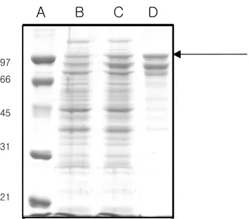
Fig. 2: Analysis of aCAT protein expressed in E. coli BL21(DE3) cells
following purification in a 12% denaturing polyacrylamide gel. A: Biorad low range protein (precision) standard; B: before induction with IPTG; C: after cells were induced with 1 mM IPTG and grown at 20 oC for 3 hrs; D: Recombinant protein purified under native conditions by pMALTM Protein Fusion and purification system
97 66 45 31 21
A B C D
human ---SRDPASDQMQHWKEQRAAQKADVLTTGAGNPVGDKLNVITVGPRGPLLVQDVVFTDE 57 B.taurus -ADNRDPASDQMKHWKEQRAAQKPDVLTTGGGNPVGDKLNSLTVGPRGPLLVQDVVFTDE 59 X. laevis MADKRDNAADQMKLWKNGRGSQKPDVLTTGGGNPISDKLNLLTVGPRGPLLVQDVVFTDE 60 D. reiro MADDREKSTDQMKLWKEGRGSQRPDVLTTGAGVPIGDKLNAMTAGPRGPLLVQDVVFTDE 60 H. discus -MATRDKASEQLNEFSKGQ--KKPDVLTTGTGAPVGRKTATMTVGPQGPVLLQDFVFTDE 57 L. vannamei --MPRDKCAEQLNDFKKQQ--TAPDNLTTSHGCPLADKLNSLTVGPRGPILLQDIQLLDE 56 *: .::*:: :.: : .* ***. * *:. * :*.**:**:*:**. : ** human MAHFDRERIPERVVHAKGAGAFGYFEVTHDITKYSKAKVFEHIGKKTPIAVRFSTVAGES 117 B.taurus MAHFDRERIPERVVHAKGAGAFGYFEVTHDITRYSKAKVFEHIGKRTPIAVRFSTVAGES 119 X. laevis MAHFDRERIPERVVHAKGAGAFGYCEVTHDITKYSKAKVFENIGKRTPIAVRFSTVAGEA 120 D. reiro MAHFDRERIPERVVHAKGAGAFGYFEVTHDITRYSKAKVFEHVGKTTPIAVRFSTVAGEA 120 H. discus MAHFNRERIPERVVHAKGAGAFGYLEITHDITKYCKAKVFERVGKKTPLAIRFSTVGGEK 117 L. vannamei MAHFDRERIPERVVHAKGAGAFGYFEVTHDISKYCKAALFSEIGKRTPIAVRYSTVGGES 116 ****:******************* *:****::*.** :*..:** **:*:*:***.** human GSADTVRDPRGFAVKFYTEDGNWDLVGNNTPIFFIRDPILFPSFIHSQKRNPQTHLKDPD 177 B.taurus GSADTVRDPRGFAVKFYTEDGNWDLVGNNTPIFFIRDALLFPSFIHSQKRNPQTHLKDPD 179 X. laevis GSSDTVRDPRGFAVKMYTEDGNWDLTGNNTPVFFIRDAMLFPSFIHSQKRNPQTHLKDPD 180 D. reiro GSSDTVRDPRGFAVKFYTDEGNWDLTGNNTPIFFIRDTLLFPSFIHSQKRNPQTHLKDPD 180 H. discus GSADTARDPPGVRHKFYTEDGNWDLVGNNTPIFFIRDPMLFPSFIHTQKRNPVTNLKDPD 177 L. vannamei GSTDTARDPRGFAVKFYTEEGNWDLVGNNTPIFFIRDPILFPSFIHTQKRNPATHLKDCD 176 **:**.*** *. *:**::*****.*****:*****.:*******:***** *:*** * human MVWDFWSLRPESLHQVSFLFSDRGIPDGHRHMNGYGSHTFKLVNANGEAVYCKFHYKTDQ 237 B.taurus MVWDFWSLRPESLHQVSFLFSDRGIPDGHRHMDGYGSHTFKLVNADGEAVYCKFHYKTDQ 239 X. laevis MVWDFWSLRPESLHQVSFLFSDRGIPDGHRHMNGYGSHTFKLVNAKDEAVYCKFHYKTDQ 240 D. reiro MVWDFWSLRPESLHQVSFLFSDRGIPDGYRHMNGYGSHTFKLVNAQGQPVYCKFHYKTNQ 240 H. discus MFWDFITLRPETTHQVAFLFSNRGTPDGYRHMNGYGSHTFKMVNAKGECVYCKFHFKTNQ 237 L. vannamei MFWDFISLRPETTHQVSFLFSDRGTPDGYRHMNGYGSRTSKLVNEKGEAVYCKFHYKTDQ 236 *.*** :****: ***:****:** ***:***:****:* *:** ..: ******:**:* human GIKNLSVEDAARLSQEDPDYGIRDLFNAIATGKYPSWTFYIQVMTFNQAETFPFNPFDLT 297 B.taurus GIKNLSVEDAARLAHEDPDYGLRDLFNAIATGNYPSWTLYIQVMTFSEAEIFPFNPFDLT 299 X. laevis CIQNLTVDEANRLAASDPDYGIHDLYEAITTGNYPSWSFYIQVMTFEQAERFKFNPFDLT 300
D. reiro GIKNIPVEEADRLAATDPDYSIRDLYNAIANGNFPSWTFYIQVMTFEQAENWKWNPFDLT 300 H. discus GIKNLTGAQADKLASVDPDYATRDLYNAIAEGKYPSWSVFIQVMNVKDAEKLKWNPFDLT 297 L. vannamei GIKCLSSKKADELAGSDPDYATRDLYNAISSGDYPSYTMCIQVMTFEEAEKWKFNPFDLT 296 *: :. .* .*: ****. :**::**: *.:**::. ****...:** :****** human KVWPHKDYPLIPVGKLVLNRNPVNYFAEVEQIAFDPSNMPPGIEASPDKMLQGRLFAYPD 357 B.taurus KVWPHGDYPLIPVGKLVLNRNPVNYFAEVEQLAFDPSNMPPGIEPSPDKMLQGRLFAYPD 359 X. laevis KIWPHGDYPLIPVGKLVLNRNPTNYFAEVEQLAFDPSNMPPGIEPSPDKMLQGRLFSYPD 360 D. reiro KVWSHKEFPLIPVGRFVLNRNPVNYFAEVEQLAFDPSNMPPGIEPSPDKMLQGRLFSYPD 360 H. discus KVWPHGEYPLIPVGRMVLDKNPKNYFADVEQIAFSPAHMVTGIEASPDKMLQGRLYSYSD 357 L. vannamei KVWPHGEFPLIPVGRLTFDRNPKNYFAEVEQIAFSSANMVPGIEASPDKMLQGRLFSYND 356 *:*.* ::******::.:::** ****:***:**..::* .***.**********::* * human THRHRLGPNYLHIPVNCPYRARVANYQRDGPMCMQDNQGGAPNYYPNSFGAPEQQPSALE 417 B.taurus THRHRLGPNYLQIPVNCPYRARVANYQRDGPMCMMDNQGGAPNYYPNSFSAPEHQPSALE 419 X. laevis THRHRLGPNYLQLPVNCPYRTRVANYQRDRPMCFTDNQGGAPNYYPNSFCAPENQPQVRE 420 D. reiro THRHRLGANYLQLPVNCPYRTRVANYQRDGPMCMHDNQGGAPNYYPNSFSAPDVQPRFLE 420 H. discus THRHRLGSNYLQLPVNCPYNTRLSNYQRDGPQCVDNNQGGAPNYFPNSFSGPQEESKCME 417 L. vannamei THRHRLGANYTQIPVNCPYRARTRNYQRDGPMCVDGNQESAPNYFPNSFSGPQDCRKHTA 416 *******.** ::******.:* ***** * *. .** .****:**** .*: human HSIQYSGEVRRFNTANDDNVTQVRAFYVNVLNEEQRKRLCENIAGHLKDAQIFIQKKAVK 477 B.taurus HRTHFSGDVQRFNSANDDNVTQVRTFYLKVLNEEQRKRLCENIAGHLKDAQLFIQKKAVK 479 X. laevis HRFQVSADVARYNSSDEDNVSQVRDFYVKVLSEEQRLRLCENIAGHLKDAQLFIQKRAVK 480 D. reiro SKCKVSPDVARYNSADDDNVTQVRTFFTQVLNEAERERLCQNMAGHLKGAQLFIQKRMVQ 480 H. discus CPFKLSGDVARYSTEDEDNFSQTGIFWKKVLPPGERDHLINNLAGHIINAQEFIQKRAVA 477 L. vannamei PKFSVSADVDRYNSADEDNFTQVGIFYRQVLNEAERQRLVENIAGHMVGAQEFIQDRAIK 476 * :* *:.: ::**.:*. *: :** :* :* :*:***: .** ***.: : human NFTEVHPDYGSHIQALLDKYN--- 498 B.taurus NFSDVHPEYGSRIQALLDKYNEEKPKN--- 506 X. laevis NFTDVHPEYGARIQALLDKYNAEGAKKKTVKTYTQHSSYATSKDKANL 528 D. reiro NLMAVHSDYGNRVQALLDKHNAEGKKN-TVHVYSRGGASAVAAASKM- 526 H. discus NFGKADPEFGRRLQAALNALKVEP--- 501 L. vannamei NFTQADPEYGANIRRAIDKIKMSQASSKT--- 505
*: ...::* .:: :: :
Fig. 3: Multiple sequence alignments of (human, human erythrocyte; B. taurus, cow; X. laevis, African clawed frog; D. rerio, zebrafish and L. vannamei, pacific white shrimp) known catalase amino acid
sequences. The alignment program Clustal W automatically introduces gaps (dash) to maximize similarity among the primary structures of these catalases. All amino acid residues identical to corresponding ones of aCAT are represented with asterisks. Conserved substitutions depending on functionality are indicated with colon and the semi-conserved residues with a dot. The putative catalytic site is marked with a box.
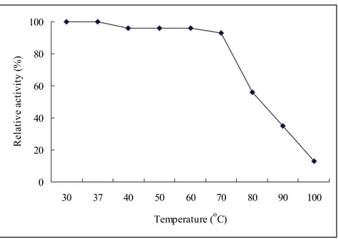
0 20 40 60 80 100 30 37 40 50 60 70 80 90 100 Temperature (oC) R ela ti ve a ctiv ity (% )
Fig. 4: Optimal temperature of aCAT. Enzyme activity was carried out at
different temperatures (30, 37, 40, 50, 60, 70, 80, 90 and 100 oC) and relative activity was determined.
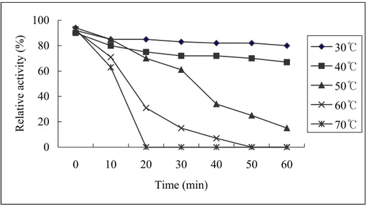
0 20 40 60 80 100 0 10 20 30 40 50 60 Time (min) Relative activity (%) 30℃ 40℃ 50℃ 60℃ 70℃
Fig. 5: Thermal stability of aCAT. Enzyme stability at different temperatures
was assessed by heating aliquots of aCAT at 30, 40, 50, 60 and 70 oC for 0, 10, 20, 30, 40, 50 and 60 mins. The residual enzyme activity was determined
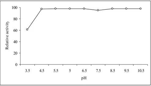
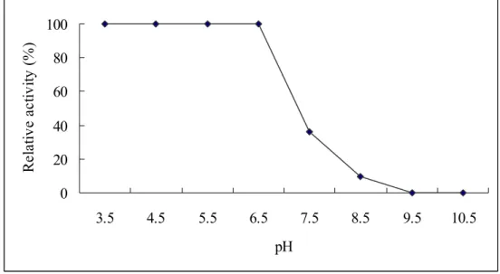
Fig. 6: Optimal pH of aCAT. The enzyme activity was carried out in buffers
with different pH values and relative activity was determined. 0 20 40 60 80 100 3.5 4.5 5.5 5 6.5 7.5 8.5 9.5 10.5 pH Re la tiv e a ct iv it y
Fig. 7: Neighbor-joining tree based on catalase amino acid sequences
obtained from 23 species. (MEGA 3.0, Kumar et al., 2004). Bootstrap values (1000 replications) are displayed over internal nodes.
4. DISCUSSION
Cloning and analysis of aCAT:
In this study, we present evidence for the cloning of a gene encoding catalase (aCAT) from H. discus discus digestive gland cDNA. Many studies have been already carried out on cloning and characterization of catalases from different species (Thuy et al., 2004; Hass et al., 1991; Ni et al., 2001; Nakamura et al., 2000; Moreira et al., 2004; Kwon and An, 2001). Some studies have also been carried out to assess the importance of catalase in the antioxidant defense system by over expressing catalase in cell lines and transgenic mice (Chen et al., 2004). Cloning and sequencing of the aCAT cDNA revealed a coding region of 1503 nucleotides and 12 bp 5’ untranslated region and 1216 bp 3’ untranslated region (Fig.1).
Comparison of deduced amino acid sequence of aCAT with 22 known catalases by CLUSTAL W (1.82) multiple sequence alignment program revealed that 173 amino acid residues conserved in all 22 species. This accounts to 34.5% similarity at amino acid level. The alignment of aCAT with catalases from human erythrocyte, Bos Taurus (cow), Xenopus
laevis (african clawed frog), Danio rerio (zebrafish) and Litopenaeus vannamei (pacific white shrimp) is shown in Fig. 3. The Comparison of
aCAT with above 5 sequences revealed that 264 amino acid residues were conserved in all catalasesaccounting for 52.69% identity at amino acid level. Since the number of residues in the nucleotide sequence is 3 times higher than the amino acid sequence it codes, the nucleotide sequence may hold 3
times more mutations than that of the amino acid sequence, while maintaining the same conservation. Zamocky and Koller (1999) reported that a so-called consensus sequence contains 84 residues highly conserved in many catalases. The alignment reveals that aCAT shares about 66, 65, 66, 69, and 65% identity with human erythrocyte, B. taurus, X. laevis, D. rerio and L. vannamei catalases respectively. Moreover, aCAT also contained the conserved H72, present in all heme catalases (Fig. 3). This residue has been
reported to allow the proper binding and reduction of a peroxide molecule (Thuy et al., 2004). According to the Moreira et al. (2004), the catalytic site is composed of amino acids F-D-R-E-R-I-P-E-R-V-V-H-A-K-G-A and G, which is conserved in aCAT at positions 61-79 in the amino acid sequence. Moreover, this active site is conserved in all other five catalase sequences we considered in Fig. 3. NADPH is reported to be tightly bound to several catalases, although the role of the dinucleotide remains unclear (Fita and Rossmann, 1985). In this study, we identified 29 amino acid residues responsible for binding of the NADPH. Eight of them were also conserved in the sequence of human erythrocyte CAT which the structure was recently determined (Yamamoto et al., 2005). Studies on the role of NADPH in the BLC suggests that NADPH could be both preventing and reversing the accumulation of intermediate compound (known as compound II) in the catalytic action (Kirkman et al., 1987). Experiments with Saccharomyces
mechanism, binding of NADPH stabilizes the quaternary structure of the protein (Zamocky and Koller, 1999).
Fita and Rossmann (1985) have determined the three-dimensional crystal structure of the mammalian catalase for BLC. The deduced amino acid sequence of aCAT shows 65% of identity with that of BLC allowing us to suggest that the three dimensional structure of aCAT be similar to that of BLC. Further, the amino acid residues interacting with heme and NDPH are highly conserved in both sequences.
Overexpression of aCAT in E. coli BL 21 DE3:
Cloning of coding sequence to pMAL-c2X plasmid resulted in expression of soluble aCAT when induced with IPTG in E. coli BL 21(DE3) cells. According to structural and functional similarities, catalases can be divided into three subgroups namely typical (true catalases), catalase peroxidase and manganese catalase or non-heme catalase (Zamocky and Koller, 1999). The catalase identified from abalone gonad cDNA in this study belongs to typical catalase. The largest subgroup; typical catalases are homotetrameric heme proteins varying the molecular weight from 200- 340 kDa. In agreement, the aCAT isolated, coded for 501 amino acid expecting a size of 56 kDa with a pI of 8.8 pH, containing lysine and glycine as the major constituents (Expert Protein Analysis System, proteomics server from the Swiss Institute of Bioinformatics). Fig. 2 confirms the expected size of the protein as 98 kDa together with the fusion protein where maltose binding protein (MBP) contributes to 42 kDa molecular weight. When
aCAT was expressed in E. coli BL21(DE3) cells, and analysed on 12% SDS-PAGE, a distinct band could be observed after induction with IPTG and purification with amylose resin column compared to the samples uninduced (Fig. 2). The reason for the other two bands present after the purification could be due to the degradation of the MPB-aCAT fusion protein. Western blotting using mono-clonal antibody against MBP confirmed that the other two bands are MBP-aCAT variants (data not shown). Thuy et al. (2004) reported that for most industrial applications, especially in the textile industry, bovine liver catalase (BLC) is used. The overexpressed of aCAT in E. coli suggests the possibility of using this system as a source of catalase in industrial work. Since the purified catalase showed 30,000 U/mg activities both with and without cleaving the fusion protein with factor Xa (data not shown), protein without cleaving was used for further experiment in this study.
Optimal temperature and thermal stability:
The rate at which an enzyme works is influenced by several factors including the temperature. It is important to determine an optimal temperature for enzymatic action. The Fig. 4 demonstrates that aCAT was active following exposure up to 70 oC and then decrease the activity. The higher thermal stability is probably due to its long β- barrel domain containing the catalytic site with hem moiety. Some catalases reported to have elongated C-terminal domains, which contribute to increase stability against chemicals and under high temperature. Penicillium vitale typical
catalase and HPII from E. coli reported to contain such domains with a “falvodxin-like” topology (Zamocky and Koller, 1999). Studies on BLC – structure revealed that it includes a β-barrel, which consists of two four stranded anti-parallel β-sheets that twist to form a closed cylindrical surface making the heat inactivation of BLC at higher temperature a difficult task. Thuy et al. (2004) suggest that thermo-stability of catalase from Halomonas sp. SK1 overexpressed in E. coli is due to elongated C-terminal peptide. Contrasting study is reported by Nakano et al. (1995) where catalase from seaweed Porphyra yezoensis had activity only up to 50 oC.
The thermal stability of aCAT is shown in Fig 5. It can be observed that irreversible thermal inactivation occurred when heating the protein at 50, 60 and 70 oC for about 30 mins. On the other hand heat inactivation was not observed when heated at 30 and 40 oC even for 1 hr. Observed thermal stability at 30 and 40 oC could be a result of the fusion protein or due to its
nature.
Optimal pH:
In agreement with the studies conducted by Hass et al. (1991), where recombinant catalase from Listeria seeligeri showed activity in a broad spectrum of pH, aCAT was also active in a wide range of pH (Fig. 6) making the overexpressed protein in this study easy to use in the industry. Mostly the industrial wastes are alkaline, specially in the textile industry where commercial catalases are highly used. In this case, aCAT cloned in a bacterium that survives under extreme conditions may be a suitable
replacement as it shows high activity in a broad range of pH. When characterizing catalase from Porphyra yezoensis, Nakano et al. (1995) have also observed the high activity of catalase from the seaweed over a very wide pH range (6.0-11.0). However, they have observed decrease in activity below pH 6.0 where we observed the decrement below pH 4.5. The reason could be due to the former is from plant and latter from animal source.
Phylogenetic analysis:
Catalase, primarily responsible for the metabolism of hydrogen peroxide, is a key antioxidant enzyme that is present throughout phylogeny, from bacteria to humans. BLAST program (Basic Local Alignment Search Tool), was used to search for all complete protein sequences of catalases. Twenty-two eukaryote sequences were considered to visualize the relationship between aCAT in terms of amino acid sequence similarity and phylogenetic tree was constructed. Fig. 7 shows the deduced phylogeny of aCAT, as calculated from neighbor-joining tree based analysis on amino acid sequences. The aCAT sequence was closer to pacific white shrimp revealing 84% boot-strap value.
Oxidative stress has been defined as “a disturbance in the pro-oxidant-antioxidant balance” in favor of the former, leading to potential damage (Kaizer et al., 2005). Physiological responses to oxidative stress in mammals have been studied by many researchers (Thuy et al., 2004; Bouzyk et al., 2000). Shull et al. (1991) reported that catalase mRNA is induced by oxidative stress in lung epithelial cells. Even though it was not
considered studying about the role of catalase in abalone in this study, most probably aCAT is playing a crucial role in healing the oxidative stress by facilitating the degradation of H2O2. Further research should be carried out
to study the role of aCAT in the abalone’s internal defense system.
In conclusion, gene encoding the abalone (H. discus discus) catalase was sequenced, and the recombinant aCAT was successfully overexpressed in E.coli BL23(DE3) cells and characterized for the first time. This enzyme suggestively belonged to typical catalase can be used in the industry as well as the information on the aCAT sequence can be used in comparative studies on marine invertibrate catalases which are just flourishing to be investigated.
Part II
Molecular cloning, expression, purification and characterization of Cu,Zn-superoxide dismutase from Abalone, Haliotis discus discus cDNA
1. ABSTRACT
Cu,Zn-superoxide dismutase (Cu,Zn-SOD) is a metalloenzyme that catalyzes dismutation of harmful superoxide radicals into H2O2 and O2. This
work reports the sequencing, cloning, expression and characterizing of Cu,Zn-superoxide dismutase (aCu,Zn-SOD) encoding gene from the disk-abalone (Haliotis discus discus) cDNA library. The full-length cDNA contained 1027 bp, with an ORF of 465 bp coding for 154 amino acids with a pI value of 5.5. The expression of gene in E.coli K12 (TB1) resulted in a soluble protein of 16 kDa. The purified protein exhibited 2461 Unit/mg activity when induced with 0.5 mM of IPTG. The optimum temperature of the enzyme was 37 oC and it was active in a range of acidic pH (from 3.5 to 6.5). The enzyme was heat inactivated after 70 oC. When compared with 47 other Cu,Zn-SODs, it was revealed that 48 amino acid residues were conserved in all 47 species, accounting for 29% identity. In comparison with known Cu,Zn-SODs, where structural studies have been carried out, the residues maintaining the active site geometry were conserved in aCu,Zn-SOD amino acid sequence (Gly45, Gly62, Pro75 and Gly83). Findings of this study will contribute more to the future studies of comparative genomics on invertebrate Cu,Zn-SOD.
2. MATERIALS AND METHODS
Cloning and sequencing of the gene encoding aCu,Zn-SOD:
A clone with an expected function of Cu,Zn-SOD was selected from the abalone cDNA library. The plasmid DNA of the putative aCu,Zn-SOD was isolated by the AccuprepTM plasmid extraction kit (Bioneer Co., Korea).
The full-length sequence was determined by sequencing reactions from 3’ end using oligo dT primer. After deriving the full length, the sequence was compared with the Cu,Zn-SOD sequences in the database of the National Center for Biotechnology Information (NCBI), BLAST-X.
Cloning the coding sequence of aCu,Zn-SOD into the pMAL expression vector:
Having checked the restriction enzyme sites of the aCu,Zn-SOD sequence, a pair of primers was designed for cloning the coding sequence of the aCu,Zn-SOD into the expression vector, pMAL-c2X (New England Biolabs, USA). The sense amplification primer was designed as 5’-gagagaGAATTCATGTCTATCAAAGCAGTTTGTGTGC -3’ having EcoR I site and antisense primer 5’-gagagaAAGCTTTCACTTGGTGATGCCGATCA -3’ containing Hind III site. In a total of 50 µl of PCR reaction, 5 units of Ex Taq polymerase (Takara Korea Biomedical Inc., Korea), 5 µl of 10X Ex Taq buffer, 4 µl of 2.5 mM dNTP, 50 ng of template and 50 pmol of each primer were used. After initial incubation at 94 oC for 2 min, 25 cycles were carried out with 30 sec denaturation at 94 oC, 30 sec of annealing at 55 oC, and 30 sec of
elongation at 72 oC, followed by a final extension at 72 oC for 5 min. The PCR product was analysed using 1% agarose gel and ethidium bromide staining. Thereafter it was purified by the AccuprepTM gel purification kit (Bioneer Co., Korea) and digested with Eco RI and Hind III restriction enzymes. The expression vector, pMAL-c2X, was digested with the same restriction enzymes as the PCR product and dephosphorylated with calf intestine phosphatase (NEB, USA) according to the vendor’s protocol. Thereafter the vector and the PCR product was purified by a 1% agarose gel using the Qiaex-II gel purification kit (QIAGEN Inc., USA).
Ligation was carried out at 16 oC, overnight with 100 ng of pMAL-c2X vector, 70 ng of PCR product, 1 µl of 10X ligation buffer and 0.5 µl 1X T4DNA ligase (Takara Korea Biochemical Inc., Korea). The ligated product was transformed into XL1 cells. The correct recombinant (confirmed by colony cracking, restriction enzyme digestion and sequencing), was transformed into competent cells E. coli K12 (TB1).
Overexpression of aCu,Zn-SOD:
The recombinant enzyme was overexpressed in E coli K12(TB1) cells in the presence of isopropyl-ß-thiogalactopyranoside (IPTG). A volume of 10 ml of starter culture was inoculated into 100 ml Luria broth with 100 µl ampicillin (100 mg/ml) and 10 mM glucose (2% final concentration) and kept at 37 oC with 200 rpm until OD600 approached 0.5.The culture was then
shifted to 20 oC for 15 min prior to the induction with 0.5 mM IPTGat the final concentration. After 3 hrs of induction, the cells were cooled on ice for
30 min and harvested by centrifugation at 4000 rpm for 20 min at 4 oC. The cells were re-suspended with 5 ml column buffer (Tri-HCl, pH 7.4 + NaCl) and freezed in –70 oC.
Purification of aCu,Zn-SOD :
After thawing, the bacterial cells were placed in an ice-water bath and sonicated six times in short pulses of 10 sec. Having centrifuged at 9000 x g for 30 min at 4 oC, the supernatant was diluted with a 1:5 column buffer. The pMALTM protein fusion and purification system was followed. In brief, amylose resin was poured into a 1 x 5 cm column and washed with 8 x column volumes of column buffer. The diluted crude extract was loaded at a flow rate of 1 ml/hr. The column was then washed with 12 x column volumes of column buffer and the fusion protein was eluted with elution buffer (column buffer + 10 mM maltose). The elute was collected in 500 µl fractions. The eluted protein content was measured by UV absorbance at 280 nm. A slight modification was done to the column buffer by excluding EDTA as SOD disassociates with EDTA. Discontinuous SDS-PAGE was performed according to its standard procedure. The stacking and separating gels were prepared at 5 and 12% respectively and the gel was stained with Coomassie blue.
Cu,Zn-SOD activity assay and determination of protein concentration:
Activity of aCu,Zn-SOD was detrmined by the xanthine oxidase method according to the procedures described by Nagai et al. (2003). The reaction mixture consisted of 0.48 ml of 0.05 M sodium carbonate buffer (pH
10.5), 0.02 ml of 3 mM xanthine, 0.02 ml of 3 mM EDTA, 0.02 ml of 0.15% bovine serum albumin, 0.02 ml of 0.75 mM NBT and 0.02 ml of aCu,Zn-SODsample. After incubation at 25 °C for 10 minutes, the reaction was initiated by adding 6 mU xanthine oxidase and maintaining the temperature at 25 °C for 20 min. The reaction was stopped by adding 0.02 ml of 6 mM CuCl2. The absorbance was recorded in a microplate reader (Sunrise; Tecan
Co. Ltd., Austria) at 560 nm. One Unit was defined as the amount of enzyme required to reduce the reaction by 50%. Specific activity was defined as Unit/mg protein. The protein concentration was determined by the procedure of Lowrey et al. (1951) using bovine serum albumin as the standard.
In order to determine the optimal temperature, each reaction was carried out at 25, 30, 37, 40, 50, 60, 70 and 80 oC and the relative activity was determined. To determine the optimal pH of the aCu,Zn-SOD each reaction was carried out in acetate buffer pH 3.5 - 5.5, phosphate buffer pH 6.5 - 7.5 and glycine-NaOH buffer pH 8.5 - 10.5 and the relative activity was determined.
Analysis of nucleotide and amino acid sequences:
Nucleotide sequence analysis was performed with the DNAssit program (version 2.2). The NCBI BLAST program (http://www.ncbi.nlm.nih.gov) was used to search for nucleotide and protein sequences homologous to the aCu,Zn-SOD. The protein sequences were aligned with the CLUSTAL W multiple sequence alignment program (version 1.8). Phylogenetic relationship was determined by reconstructing a protein phylogeny using
MEGA3.1 program (Kumar et al., 2004). Neighbor-Joining algorithm with the PAM matrix model was applied in constructing in the phylogenetic tree. The tree topology was evaluated by the bootstrapping method (1000 replications).
3. RESULTS GGGGATTACAGTGCAATTTCTCGGCGGTCTCGGCTACAACAAGCACTTTCCGGTGAAATATTCAGCTCTTGAAAC 75 ATGTCTATCAAAGCAGTTTGTGTGCTGAGAGGTGATTCGGAAGTCAAGGGAACAGTATTCTTCTCACAGGGAGATGCAGAC 156 M—-S—-I—-K--A—-V—-C—-V—-L—-R—-G—-D—-S—-E--V--K—-G--T--V--F—-F--S--Q--G—-D--A--D- AGTCCAGTGAAAGTGACGGGCTCCATCACGGGCCTGACGGAGGGCAAACATGGCTTCCACGTTCATCAGTTCGGGGACAAC 237 -S--P--V--K--V--T--G--S--I--T--G--L--T--E--G--K--H—-G--F--H--V--H--Q--F--G—-D--N ACGAATGGCTGTACCAGTGCCGGGTCCCACTTCAACCCTTTCGGCAAGACCCATGGAGCGCCAGAAGACGAAAACAGACAT 319 --T--N--G--C--T--S--A--G--S--H--F—-N--P--F--G--K--T--H--G--A--P--E--D--E--N--R-- GCTGGTGACCTTGGCAACGTTACTGCTGACGCATCAGGAGTAGCAAACATCGACATCGAGGACAAGATCATAAGTTTGACT 399 H--A--G--D--L--G--N--V--T--A--D--A--S--G--V--A--N--I--D--I--E--D--K—-I—-I—-S—-L- GGGGACAAATCAATCATTGGCAGAACTATTGTTGTCCATGCTGGAGTGGATGACCTGGGCAAGGGAGGCAATGAAGAAAGC 480 -T--G--D--K--S--I--I--G--R--T--I--V--V--H--A--G--V--D--D--L--G--K--G--G—-N--E--E CTGAAGACAGGGAACGCTGGTGGTCGTCAGGCCTGTGGGGTGATCGGCATCACCAAGTGACCAAAGTGTTGAATCAGTTGC 561 --S--L--K--T--G--N--A--G--G--R--Q--A--C--G--V—-I--G--I--T--K-- GTCAGCACCATGTGGTGTCTTACCGCTAGCATAGGTGGGAATTTGTTGTACCAGATGAAGTTTATTTGTTGTTTGTTGGG 641 CATTATTGCAAAATAACTATTCCAAACGTTAACTTTTTTTCAAATTGTGGACGTTTCATGTAGAAAATAAATCTATGGAA 721 AATAAGACTGACAAGCCATTTTCTAATTCATAAGTTGGAGGGTTTTTTTTGTTCAGCTGAACTACCGGTAGAATATTTGT 901 GTTTAATATGCTCTAGGTCAGACACGTTACTCTTCATTCCAAGACTATCTAGACTGATTGTATATCCCTGAGCTAGCTAT 992 TTGCTGTTGAATCATTGATACTGGAATTCCATAATGTGGAACAATGTTGCACCTTTTGGTGTGGGGTGATCACTGTCTATC 962
TAAGAGTACAAAGAGaataaaTCCTACTGTTGTATAAAAAAAAAAAAAAAAAAAAAAAAAAAA 1025
Fig. 8: Nucleotide sequence and the deduced amino acid sequence of
aCu,Zn-SOD. The coding sequence (from 76 to 537) is in bold letters. Amino acid sequence corresponding to protein consists of 154 amino acids. The poly (A) tail is in bold italics and the polyadenylation signal is indicated by bold simple case. The neucleotide from 1 to 75 and 538 to 999 indicate 5' UTR and 3' UTR respectively. The residues that interact with copper and zinc are underlined and boxed respectively. Five well conserved active sites are shaded.
Fig. 9: Analysis of aCu,Zn-SOD protein expressed in E. coli K12(TB1) cells
following purification in a 12% denaturing polyacrylamide gel. Cells were grown at 30 oC and induced with 0.5 mM IPTG. Recombinant
protein was purified under native conditions by pMALTM Protein Fusion and purification system.
250 100 50 37 25 75
S. scrofa -AT-KAVCVLKGDGPVQGTIYFELKGEK-TVLVTGTIKGLAEGDHGFHVHQFGDNTQGCT 57 B. taurus MAT-KAVCVLKGDGPVQGTIHFEAKGD--TVVVTGSITGLTEGDHGFHVHQFGDNTQGCT 57 H. sapiens MAT-KAVCVLKGDGPVQGIINFEQKESNGPVKVWGSIKGLTEGLHGFHVHEFGDNTAGCT 59 G. gallus MATLKAVCVMKGDAPVEGVIHFQQQGSG-PVKVTGKITGLSDGDHGFHVHEFGDNTNGCT 59 H. discus -MSIKAVCVLRGDSEVKGTVFFSQGDADSPVKVTGSITGLTEGKHGFHVHQFGDNTNGCT 59 H. diversicolor -MSVKAVCVLKGAGEVEGTIHFSQTEADGPVTVTGKISGLEGGLHGFHVHEFGDATNGCM 59 : *****::* . *:* : *. ..* * *.*.** * ******:*** * ** S. scrofa SAGPHFNPESKKHGGPKDQERHVGDLGNVTAGKDGVATVYIEDSVIALSGDHSIIGRTMV 117 B. Taurus SAGPHFNPLSKKHGGPKDEERHVGDLGNVTADKNGVAIVDIVDPLISLSGEYSIIGRTMV 117 H. sapiens SAGPHFNPLSRKHGGPKDEERHVGDLGNVTADKDGVADVSIEDSVISLSGDHCIIGRTLV 119 G. gallus SAGAHFNPEGKQHGGPKDADRHVGDLGNVTA-KGGVAEVEIEDSVISLTGPHCIIGRTMV 118 H. discus SAGSHFNPFGKTHGAPEDENRHAGDLGNVTADASGVANIDIEDKIISLTGDKSIIGRTIV 119 H. diversicolor SAGPHYNPFGKTHGAPEDENRHAGDLGNVLANADGVADIKIDDRIISLTGVRSIIGRTIV 119 ***.*:** .: **.*:* :**.****** *. .*** : * * :*:*:* .*****:* S. scrofa VHEKPDDLGRGGNEESTKTGNAGSRLACGVIGITQ- 152 B. Taurus VHEKPDDLGRGGNEESTKTGNAGSRLACGVIGIAK- 152 H. sapiens VHEKADDLGKGGNEESTKTGNAGSRLACGVIGIAQ- 154 G. gallus VHAKSDDLGRGGDNESKLTGNAGPRLACGVIGIAKC 154 H. discus VHAGVDDLGKGGNEESLKTGNAGGRQACGVIGITK- 154 H. diversicolor VHAGKDDLGKGGNEESLKTGNAGGRLACGVVGITK- 154 ** ****:**::** ***** * ****:**::
Fig. 10: Sequence comparison of aCu,Zn-SOD with S. scrofa (P04178), B. taurus, (P00442), H. sapiens (P00441), G. gallus (NP990395) and H. diversicolor supertext (AAY18806). The alignment program Clustal
W automatically introduces gaps (dash) to maximize similarity among the primary structures of these Cu,Zn-SODs. All amino acid residues identical to corresponding ones of H. discus discus are represented with asterisk.
0 20 40 60 80 100 25 30 37 40 50 60 70 80 Temperature (oC) R ela tiv e a ctiv ity (% )
Fig. 11: Optimal temperature of aCu,Zn-SOD. Enzyme activity was carried
out at different temperatures (25, 30, 37, 40, 50, 60, 70 and 80 oC) and relative activity was determined by xanthine oxidase method.
0 20 40 60 80 100 0 10 20 30 40 50 60 Time (min) R ela tiv e a ctivity ( % ) 30℃ 40℃ 50℃ 60℃ 70℃
Fig. 12: Thermal stability of aCu,Zn-SOD. Enzyme activity at different
temperatures was assessed by heating aliquots of enzyme at 30, 40, 50, 60 and 70 oC for 0, 10, 20, 30, 40, 50 and 60 min. The residual enzyme activity was determined
0 20 40 60 80 100 3.5 4.5 5.5 6.5 7.5 8.5 9.5 10.5 pH R ela tiv e a ctiv ity ( % )
Fig. 13: Optimal pH of aCu,Zn-SOD. The enzyme activity was carried out
in buffers with different pH values and relative activity was determined by xanthine oxidase method.
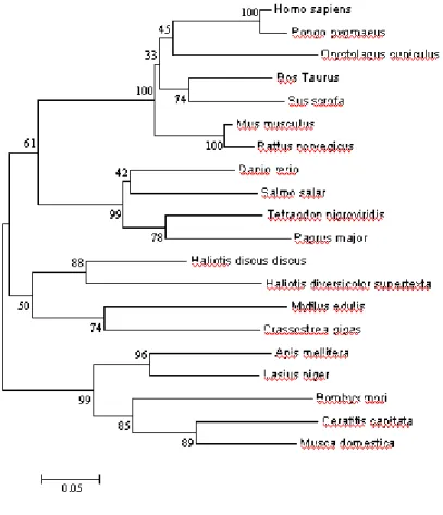
Fig. 14: A phylogenetic tree of CuZn-SOD proteins constructed by the
Neighbor-Joining method with the PAM matrix model (MEGA 3.1, Kumar et al., 2004). Numbers at the nodes are Bootstrap values representing their robustness (1000 replications). Twenty species considered in the tree construction were Homo sapiens(NP000445),
Pongo pygmaeus(Q8HXQ4), Oryctolagus cuniculus(CAA80357),
Bos Taurus (NP777040), Sus scrofa (PO4178), Mus musculus (NPO3564), Rattus norvegicus(NP058746), Danio rerio(NP571369), Salmo salar (AAW59361), Tetraodon nigroviridis(CAG00454), Pagrus major(AA015363), Haliotis diversicolor supertexta(AAY18806), Mytilus edulis(CAE46443), Crassostrea gigas (CAD42722), Apis mellifera(AAP93581), Lasius niger(AAQ81639), Bombyx mori (AAR97568), Ceratitis capitata(P28755) and Musca domestica(AAR23787).
4. DISCUSSION
Isolation of full length cDNA encoding Cu,Zn-SOD from H. discus
discus:
By cloning and sequencing the putative aCu,Zn-SOD, a coding region of 465 bp nucleotides coding for a polypeptide of 154 amino acids, with an 74 bp 5’ untranslated region and an 487 bp 3’ untranslated region was identified (Fig. 8) with a predicted molecular weight of 16 kDa and a theoretical pI of 5.5. The length of the coding region is similar to most of the other Cu,Zn-SOD coding regions reported so far (Castellanos-Gonzalez et al., 2002). The major constituent of the protein was glycine accounting for 16% (Expert Protein Analysis System, proteomics server from the Swiss Institutes of Bioinformatics), which is common for aCu,Zn-SODs (Buettner, 1998). Comparison of aCu,Zn-SOD with other 47 known Cu,Zn-SOD sequences in the public database revealed 48 amino acid residues conserved in all Cu,Zn-SODs accounting to 29% homology at amino acid level.
Overexpression of a Cu,Zn-SODin E. coli K12(TB1):
When aCu,Zn SOD was expressed in E. coli K12(TB1) and analysed using 12% Sodium-dodecyl sulfate polyacrylamide gel electrophoresis (SDS-PAGE), a distinct band with a high intensity emerged in IPTG induced sample (Fig. 9). The purified recombinant enzyme was obtained following the pMAL protein purification system, which made the purification profile simple and efficient. The homogeneity of the purified product was examined
by SDS-PAGE. The result derived by subtracting the value obtained from this analysis from that of corresponding to the fusion protein (42.5 kDa) coincided with the expected molecular weight of 16 kDa. The enzymatic activity of purified enzymes was 2461 Unit/mg when tested by the xanthine oxidase method described by Nagai et al. (1985). This value is moderate when compared with that of other recombinant Cu,Zn-SODs expressed. Activity observedin aCu,Zn-SOD is higher to the activity of zebrafish Cu,Zn-SOD expressed in AD494(DE3)pLysS (Ken et al., 2003) but lower to that of reported value for papaya Cu,Zn-SODs (Lin et al., 1998). When comparing the amino acid sequence between papaya with both zebrafish Cu,Zn-SOD and aCu,Zn-SOD, it was observed that Cys residue at position 7 in H. discus discus is replaced with Ala in plant. Hallewell et al. (1991) reported that buried Cys would interfere with the protein folding by forming an incorrect disulfide bond. Ken et al. (2003) suggests that further studies should be carried out using site-directed mutagenesis to replace Cys-7 to Ala-7 to observe the change of activity. In the recent history of expressing recombinant Cu,Zn-SODs, some studies have carried out enriching the growth media with external Cu and Zn sources to enhance the activity (Liu et al., 2002). According to the general accepted theory, the negative charge elsewhere on the protein surface reinforces the attraction by the positive channel around the copper. The enzyme-catalyzed dismutation by Cu,Zn-SOD is believed to proceed by subsequent reduction and oxidation of Cu ion acting as an electron carrier. The Zn atom is completely buried within the
protein structure and is suggested to play a structural role helping the protein stability (Ozturk-Urek and Tarhan, 2001).
Deduced Amino acid sequence comparison:
The deduced amino acid sequence from the cDNA of H. discus
discus was compared with those of the H. diversicolor supertext
(AAY18806), S. scrofa (P04178), G. gallus (NP990395), B. taurus, (P00442) and H. sapiens (P00441) using the Clustal W program (Fig. 10). The alignment reveals that the H. discus dicus shares 78, 67, 66, 71 and 68% homology with H. diversicolor supertext, S. scrofa, G. gallus, B. taurus, and
H. sapiens respectively. The close evolutional relationship between two
abalone species was demonstrated by the highest homology between the two sequences. Altogether 82 amino acid residues were conserved among all 6 amino acid sequences accounting to 53% homology at amino acid level. Further, comparison of aCu,Zn SOD with three aquatic molluscs; Ruditapes
decussates, Dreissena polymorpha and Bathymodiolus azoricus and
mammalian Cu,Zn SODs (Geret et al., 2004) showed several residues maintaining the active site geometry being conserved (Gly45, Gly62, Pro75 and Gly83). The metal binding sites His47, -49, 64, -121 for copper and His64, 72, 81 and Asp84 for Zinc were also conserved in aCu,Zn-SOD. Two Cys residues which is believed to contribute towards forming the intra-chain disulfide bridge in mammalian Cu,Zn-SOD were found at Cys58 and Cys147. Three- dimensional structure predicted for aCu,Zn-SOD by Deep view –
spdbv 3.7, confirmed the potential of this disulfied bond formation between Cys58 and Cys147 (data not shown).
Optimal temperature and Thermal stability:
Temperature is a crucial factor that influences the rate at which an enzyme works. Thus, it is important to determine an optimal temperature for an enzymatic action. As shown in Fig. 11, aCu,Zn-SOD has its optimal activity at 37 oC. When exposed to temperatures varying from 25, 30, 37, 40, 50, 60, 70 and 80 oC, the enzymatic activity gradually decreased from 37to 60 oC and then showed irreversible heat inactivation at 70 oC. Buettner (1998) has reported that Cu,Zn-SOD is disassociated by SDS and β-mercaptoethanol or EDTA and heat at 40-55 oC . Temperature to break apart
50% of Cu,Zn-SOD activity in 10 min is reported as 67 oC. It is also reported that Cu,Zn-SOD is stable to repeated freeze thaw cycles and prolonged refrigeration (4 oC) (Buettner,1998).
The thermal stability of aCu,Zn-SOD was detected with irreversible thermal denaturation at 70 oC after 20 minutes. During continuous incubation at varying temperatures from 30 to 70 oC, the residual enzymatic activity was assayed by means of the xanthine oxidase method. The result is plotted in Fig. 12. Similar observations were reported by Liu et al. (2002) for recombinant duck Cu,Zn-SOD where it had lower thermal stability compared to the control bovine Cu,Zn-SOD used in the experiment. Site directed mutagenesis studies indicated that free cystein residues might be the major molecular determinants for different protein stabilities among various