성상세포종양에서 혈관내피증식인자의 발현
- 종양주변부 부종 및 미세혈관과의 상관관계 -
원광대학교 의과대학 신경외과학교실
김태영·박종태·문성근·한원철
*
= Abstract =
Expression of Vascular Endothelial Growth Factor in Astrocytic Tumors
- - - Correlation to Peritumoral Brain Edema and Microvasculature - - - - - Tae Young Kim, M.D., Jong Tae Park, M.D.,
Seong Keun Moon, M.D., Weon Cheol Han, M.D.*
Department of Neurosurgery and Anatomical Pathology,* School of Medicine, Wonkwang University, Iksan, Korea
bjectives:It has been known that vascular endothelial growth factor(VEGF), as an endothelial cell-specific mitogen, induces angiogenesis, and possesses vascular permeability and procoagulant properties. Peritumoral brain edema(PTBE) is a common accompaniment of malignant gliomas. It results from microvascular extra- vasation of plasma and proteins through the interendothelial spaces. The correlation between pathological grading, PTBE, neovascularization, and the expression of VEGF were analyzed in 31 patients with astrocytic tumors.
Methods:Astrocytic tumor samples(8 astrocytomas, 14 anaplastic astrocytomas, and 9 glioblastomas) from 31 pa- tients(21 males and 10 females:average age 37±24 years) who underwent surgery were examined retrospectively for the expression of VEGF and CD31(microvasculature) immunohistochemically. The extent of PTBE was examined by using preoperative CT or MRI as an edema index(EI). In addition to VEGF and CD31, several causative factors including tumor size, histologic type were compared with EI.
Results:Only one of 8 astrocytomas, and majority of high grade(21 of 23 anaplastic astrocytomas and glioblasto- mas) tumors demonstrated PTBE(p<0.05). The majority of high grade tumors showed higher expression of VEGF (p<0.01). High grade tumors showed even higher CD31 expression(p<0.05), however, there was no close correlation between expression of VEGF and CD31. The EI was increased significantly, just as VEGF(p<0.01), but CD31 expre- ssion was not correlated with high EI.
Conclusion:These data suggest that VEGF expression is closely correlated with PTBE and histological grading in astrocytic tumors. Microvasculature(CD31) in tumors is highly correlated with histological grading, however, shows no correlation with the expression of VEGF and PTBE.
KEY WORDS:Astrocytic tumors・Vascular endothelial growth factor・Brain edema・Microvasculature・Edema Index.
서 론
뇌종양에서 자주 관찰되는 종양 주변부 부종은 혈관인성 부종으로 알려져 있으며, 부종이 심한 경우에는 임상 증상의 악화뿐만이 아니라 심한 경우에는 죽음을 초래하게 된다.
이러한 부종은 혈뇌장벽의 개방으로 인하여 혈관으로부터 혈장이 혈관밖으로 빠져나감으로써 발생하게 된다. 이러한 현상을 유발하는데는 종양세포로부터 어떤 물질이 분비되어 종양 혈관에 형태학적 변화를 초래한다는 사실이 보고되어
왔으며
2)3)7)14-16)21), 최근에는 혈관내피성장인자(vascular
endothelial growth factor:VEGF)가 그러한 대표적인 물
OOOO
질로 연구되고 있다.
Guinea pig의 간암에서 복수로부터 종양세포에서 분비된 단백질을 분비하여 이를 혈관투과인자(vascualr permea- bility factor:VPF)라 하였다. 그후 이와는 개별적으로 혈 관내피세포 증식을 초래하는 다른 성장인자가 뇌하수체에서 기인한 folliculostriate cell에서 분리되어 이를 혈관내피성장 인자라고 하였다. 그러나 후에 이 두 인자의 아미노산 배열 을 조사한 결과 동일한 구조를 가졌음이 밝혀졌으며, 혈관 내피세포의 증식과 혈관투과에 관여한다는 사실이 알려졌다.
혈관내피성장인자는 45-kDa heparin-binding glycopro- tein으로, pletelet-derived growth factor나 placental growth factor와 구조적으로 비슷하며, 인간에서는 아미노 산의 수에 따라 VEGF-121, VEGF-165, VEGF-189, VEGF-206의 4가지 형태가 있고, 이중 VEGF-165가 중 추신경계 종양이나 세포에 잘 나타난다고 한다
2)4)11)21).
혈관내피성장인자는 강력한 혈관 신생인자로서 발생기나 생리적 또는 병적 상태에서 혈관 신생에 관여하고, 특히 종 양의 혈관 신생에 중요한 역할을 한다고 알려져 있다. 특이 적으로 혈관내피세포에 유사분열물질(mitogen) 및 주화성 인자(chemotactic factor)로 작용하며, 큰 혈관보다는 작은 소정맥이나 모세혈관 같은 미세혈관에서 혈관내피세포에 손 상을 주지 않고 혈장단백이 혈관 밖으로 빠져나가도록 정상 혈관의 투과를 증가시킨다. 미세혈관의 투과를 증가시키는 능 력은 히스타민과 비교하여 볼 때 약 1000배정도 강력한 것 으로 알려져 있다
6).
교모세포종에서 혈관내피증식인자가 과발현되는 사실은 여 러 보고에서 밝혀져 있으며, 또한 신경교종에서 병리 등급 의 악성도가 높을수록 과발현된다고 한다
5)6)8)14)16)22)23). 병 리조직학적인 소견으로 볼 때 혈관 신생은 종양의 악성도와 밀접한 관계를 갖고 있음을 알 수 있다. 뇌수막종에서 혈관 내피성장인자의 표현과 종양주변부 부종을 직접 비교한 연 구는 많으나, 성상세포종양에서는 이를 비교한 연구가 없어 본 연구를 계획하였으며, 방법은 종양의 등급에 따른 혈관 내피성장인자의 표현정도 및 종양내의 미세혈관을 조사하고, 이와 종양주변부 부종과의 관계를 비교함으로써 각 인자가 종양주변부 부종에 어떠한 영향을 미치는가를 분석하여 보 았다.
대상 및 방법
수술로 확진된 성상세포종양 환자중 파라핀 포매되어 보 관된 조직에서 이용 가능한 31례를 대상으로 하였다. 남자 는 21례, 여자는 10례였으며 평균 연령은 37±24세였다.
조직학적 분류는 World Health Organization(WHO) 분류 에 의하여 시행하였으며, 면역 조직화학 염색을 시행하여 혈 관내피성장인자 및 미세혈관 표현 양상을 측정하였고, 각 환자의 CT 및 MRI를 이용하여 종양주변부 부종과 종양의 용적을 측정한 후 부종지수(edema index)를 계산하였다.
1. 혈관내피성장인자 및 미세혈관의 면역화학조직염색 파라핀에 포매된 조직을 4um 두께로 박절하여 통상의 탈 파라핀 및 함수과정을 거쳐, 3% 과산화 수소로 5분간 처리 하여 endogenous peroxidase의 작용을 차단하였다. 미세 혈관의 표현을 보기 위해서는 CD31을 이용하였다. Pepsin 으로 전처리한후 Immuno/DNA 완충액으로 4회 세척하고 protein blocker 용액으로 10분간 처리하였다. 일차항체인 CD31(Dako Patts,Glostrup, Denmark)), VEGF(Calbio- chem, La Jolla, CA)을 하룻밤 부착시킨 다음 2차 항체에 10분간 부착시킨 후 AEC로 발색시키고 Mayer’s hema- toxylin으로 대조염색을 시행 후 universal mount로 봉입하 여 검경하였다. 양성대조군은 CD31은 대장암 조직으로, 혈 관내피성장인자는 실험군에 포함되지 않은 교모세포종으로 하였으며, 음성대조군은 일차항체 대신 식염수를 처리하고 나머지 과정은 동일한 방법으로 염색하였다. 혈관내피성장인 자의 표현 양상은 200배의 시야에서 염색된 정도를 4등급 (0:염색 안됨, +1:희미한 염색, +2:중등도 염색, +3:
강한 염색)으로 구분하였으며, 두 명의 관찰자의 의견이 일 치하는 경우 그 등급으로 결정하였다. 미세혈관의 수는 CD31 에 양성인 미세혈관을 100배 시야에서 관찰하여 그 수를 기 록하였으며, 적혈구가 7개 이상 포함되어 있는 혈관은 미세 혈관에서 제외하였다.
2. 종양 및 종양 주변부 부종 용적의 측정
CT 및 MRI를 이용하여 타원형 구체의 용적을 계산하는 식을 이용하였다. 종양의 전후 및 좌우 장경(a and b)을 측 정하고, 각 단면의 두께를 계산하여 종양의 두께(c)를 구한 다음 4/3×πabc로 종양의 용적을 계산하였다
20)22). 종양 주 변부 부종의 용적도 모양의 변화는 심하지만 타원형 구체에 준하여 같은 방법으로 용적을 구하였으며, 종양 주변부 부종 의 용적을 종양 용적으로 나눈 값을 구하여 이를 부종 지수 (edema index)로 하였다. 즉 부종 지수 1이라 함은 전혀 부 종이 없는 상태를 의미한다.
3. 통계적 분석
자료의 통계 분석을 위하여 Pearson correlation을 사용
하였으며, p-value가 0.05 미만일 경우 유의성이 있는 것으
로 판정하였다.
결 과
부종지수를 나이, 성별, 종양 용적, 조직학적 악성도, 미 세 혈관의 정도, 혈관내피성장인자의 발현도와 비교한 결과 고령인 경우(p=0.001), 종양의 악성도가 증가할 때(p=0.
022), 혈관내피성장인자의 발현도가 높을수록(p=0.003) 부종 지수가 증가함을 알 수 있었다(Table 1).
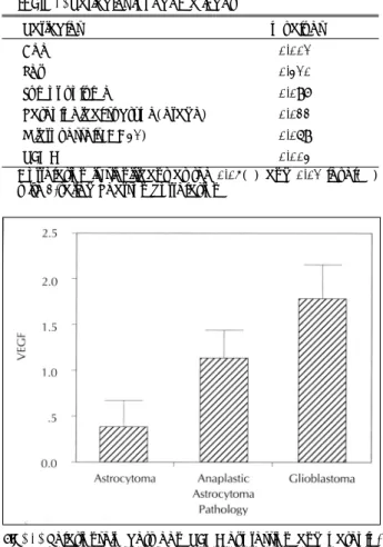
1. 조직학적 등급과 혈관내피성장인자 표현의 상관관계 성상세포종에서는 0.38±0.18, 역형성 성상세포종은 1.
14±0.18, 교모세포종은 1.78±0.22로 종양의 악성도가 증 가할수록 혈관내피성장인자의 표현도가 증가함을 알 수 있 었다(p=0.022)(Fig. 1).
2. 조직학적 등급과 미세혈관과의 상관관계
성상세포종은 17.25±2.24, 역형성 성상세포종은 45.14
±7.35, 교모세포종은 59.67±22.04로 악성도가 증가할수 록 미세혈관이 많음을 알 수 있었으며, 이는 일반적인 조직
소견에도 합당함을 보여주고 있었다(p=0.037)(Fig. 2).
3. 부종 지수와 혈관내피성장인자 표현의 상관관계 혈관내피성장인자의 발현이 전혀 없는 경우(0)는 7례 모 두에서 전혀 부종을 동반하지 않았다. +1 등급에서는 2.
95±0.61, +2 등급은 3.79±0.89, +3 등급인 경우는 7.
2±0.00으로 혈관내피성장인자의 발현도가 증가할수록 종 양 주변부 부종의 정도가 심해짐을 알 수 있었다(p=0.003) (Fig. 3).
4. 조직학적 등급과 부종지수와의 상관관계
성상세포종은 1.18±0.18, 역형성 성상세포종은 3.26±0.
69, 교모세포종은 3.84±0.87로 종양의 악성도가 증가할수
Table 1. Variables for edema index
Variables p values
Age 0.001**
Sex 0.310 Tumor volume 0.097
Pathological subtype(grade) 0.022*
Microvessels(CD31) 0.069
VEGF 0.003**
Correlation is significant at the 0.05(*) and 0.01 level(**) with 2-tailed Pearson correlation
Fig. 1. Relationship between VEGF expression and patholo- gical type. There was a significant difference(p<0.05).
The VEGF expression was related to the histological grade of astrocytoma.
Fig. 2. Relationship between microvessels(CD31) and patholog- ical type. A positive relationship between microvessel counting and tumor grade was observed(p< 0.05).
Fig. 3. Correlation between edema index(EI) and the expre-
ssion of vascular endothelial growth factor(VEGF). EI is
significantly increased in proportion to VEGF express-
ion levels(p=0.006).
록 부종의 정도가 심해짐을 알 수 있었고, 이는 일반적으로 알려진 사실과 부합하였다(p=0.022)(Fig. 4).
5. 미세혈관과 혈관내피성장인자의 표현도 및 부종지수와 의 상관관계
미세혈관의 수와 혈관내피성장인자의 표현도를 비교한 결 과 두 인자간에 의미있는 상관관계는 없었으며(p=0.112), 부종 지수와도 유의성있는 상관 관계는 없었다(p=0.369).
고 찰
혈관내피성장인자는 신경 교종에 있어서 종양의 혈관신생 을 유발하는 중요한 인자이며, 혈관 삼투 인자로 작용하여
종양 주변부 부종을 유발하는데 관여하고 있다고 알려져 있 다. 따라서 종양의 형성과 성장에 중요한 역할을 하기 때문 에, 혈관내피성장인자는 신경 교종의 성장을 촉진하여 환자 를 죽음에 이르게끔 한다는 것을 예측할 수 있다.
성상세포종양에서 역형성 탈분화는 세포 역형성(cellular anaplasia), 혈관 증식, 괴사 등에 의하여 나타나며, 이러한 조직 소견이 보이는 경우 예후는 좋지 않게 된다. 즉 악성 화 할수록 혈관 증식이나 종양 괴사가 진행되는데, 혈관 증식에 가장 중요한 역할을 하는 혈관내피성장인자는 성 장세포종양의 악성화에 관여하는 중요한 인자라고 알려져 있다
3)6)8)11)13)14)22). 혈관내피성장인자의 up-regulation을 초래하는 원인은 확실히 밝혀져 있지 않다. 그러나 여러 보 고에 의하면 대부분의 경우에서 혈관내피성장인자가 종양 괴사주변에 있는 세포에서 강하게 표현되는 것으로 알려져 있으며, 특히 저산소증에 노출된 경우 더욱 분비가 촉진된다 고 하기 때문에, 혈관내피성장인자의 분비는 종양괴사 주변 부의 저산소증에 의하여 촉진된다고 생각되고 있다
8)11)13)23). 본 실험의 결과에서도 종양의 악성도가 진행할수록 혈관내 피성장인자의 표현이 강하게 나타남을 알 수 있었으며, 또한 종양 주변부 부종이 증가함을 보여주었다.
저등급의 성상세포종양에서의 혈관신생에 관여하는 인자 에 대해서는 아직 잘 알려져 있지 않다. 본 실험에서도 저등 급의 성상세포종 8례중 5례에서는 전혀 혈관내피성장인자 가 발현되지 않았으며, 종양 주변부 부종도 동반하지 않았 다. 나머지 3례는 혈관내피성장인자가 약하게 염색되었으나 (1+) 그중 2례는 전혀 주변부 부종이 없었으며, 오직 1례 에서 만이 부종지수 2.4의 종양 주변부 부종을 동반하고 있 었다. 이러한 사실을 고려할 때 저등급의 성상세포종 모두 가 혈관내피성장인자를 분비하지 않는 것은 아니며, 종양의 크기나 주위 환경의 변화에 따라 분비가 촉진될 수 있음을 알 수 있고, 경우에 따라서는 심한 부종을 유발할 수도 있다 는 것을 고려해야 하며, 또한 혈관 신생에 관여하는 다른 인 자들(basic and acidic fibroblast growth factors, epid- ermal growth factor, platelet-derived growth factor, transforming growth factor alpha) 및 혈관신생 억제자 (thrombospondin-1)등의 소실 등이 이러한 과정에 관여했 을 가능성을 고려해야 할 것으로 생각되었다
1)5)11)13).
대부분의 악성 종양에서는 신생혈관의 증식이 관찰된 다
3)5)6)9)14)17). 본 실험에서도 종양의 등급이 증가할수록 혈 관 증식이 증가함을 알 수 있었다. 그러나 혈관 증식과 혈관 내피성장인자의 발현, 혈관 증식과 부종지수와의 상관관계는 통계적으로 유의성있는 결과를 보여 주지 않았다. 이러한 결 과를 해석하는데는 논란이 있을 수 있지만, 혈관의 증식이
Fig. 4. Relationship between edema index(EI) and pathologi- cal type. There was a significant difference(p<0.05).
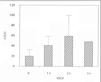
Fig. 5. Group showing the average level of CD31 for each
VEGF expression. There was no significant correlation
(p=0.3379).
항상 혈관내피성장인자의 과발현을 동반하는 것은 아니고, 본 실험에서는 모든 혈관을 측정한 것이 아니라 직경이 작 은 미세혈관(적혈구 6개 이하를 함유한 미세혈관)만을 측 정했기 때문에 이런 상반된 결과가 나왔을 가능성을 배제할 수 없을 것으로 생각된다. 또한 신경교종에서도 저등급의 회돌기 세포종의 경우 많은 모세혈관을 가지고 있음에도 불 구하고 혈관내피성장인자의 발현은 거의 없거나 아주 약하 게 발현되었다는 보고도 있기 때문에 종양내의 혈관의 수와 혈관내피성장인자가 항상 비례하는 것은 아닐 가능성이 있
다
3)5)17). 또한 경우에 따라서는 혈관 신생에 관여하는 다른
cytokines이나 성장인자들이 관여했을 가능성도 고려해야 할 것으로 생각된다
1)5)11)21).
종양 주변부 부종을 유발하는데 혈관내피성장인자가 관여 한다는 사실은 여러 보고에 의하여 제시되어 왔다
4)9)12)14)20). 또한 주변부 부종이 심한 뇌종양에서 혈관내피성장인자 mRNA가 up-regulation되어 나타나는 것도 이러한 가능성 을 제시하고 있다
3)16)22)23). 그러나 아직까지도 혈관내피성장 인자가 어떻게 혈관투과성을 유발하는지는 잘 알려져 있지 않다. 종양 주변부 부종은 혈관인성 부종으로서, 뇌의 모세혈 관에 구조적인 변화가 발생하여 혈뇌장벽이 깨짐으로써 나 타나며, 이러한 구조적인 변화로서는 창(fenestration), 음작 용포(pinocytic vesicle) 수의 증가, 기저막의 손상, 세포사 이 연접의 확장(widened intracellular junctions), 미토콘드 리아 수의 감소 등이 제시되고 있다
4)7)9)10)12)18)19). 최근 들 어 이러한 혈관의 구조적인 변화를 유발하는 물질이 종양세 포로부터 분비된다는 증거가 제시되기 시작하였으며, 이러한 물질 중 혈관내피성장인자가 주목을 받게 되었다
3)7)15-17)20). Roberts 등
17)18)은 혈관내피성장인자를 투여하는 경우 소정 맥과 모세혈관에서 혈관내피세포에 창(fenestration)을 일으 킨다고 하였으며, 종양세포를 VEGF-165로 transfection 시킨 결과 혈관에서 창 내피세포를 관찰할 수 있었다고 보고 하였다. 창 내피세포는 신장의 사구체(glomeruli), 맥락총, 태반 등의 혈관에서 관찰되며 이를 통하여 물질이 쉽게 이동 하게 되는데, 이러한 조직에서는 혈관내피성장인자가 발현 되어 나타나고 있다. 따라서 혈관내피성장인자가 증가하여 종양 혈관에 이러한 창 내피세포가 형성되면 혈관 내부로부 터 혈관외 유출이 많아질수록 종양 주변부 부종을 더 심하게 동반한다는 사실은 이미 CT나 MRI 같은 영상을 통하여 잘 알 수 있다. 본 실험에서도 이러한 결과를 확인할 수 있었으 며, 부종이 심한 종양일수록 혈관내피성장인자 표현이 강하 게 나타남을 알 수 있었다. 이러한 결과를 볼 때 성상세포종 양에서 종양 주변부 부종을 유발하는데 혈관내피성장인자 표현이 밀접한 관계를 가지고 있음을 확인할 수 있었다.
•
논문접수일:2000년 6월 5일•
심사완료일:2000년 10월 13일•
책임저자:김 태 영570-711 전북 익산시 신용동 344-2 원광대학교 의과대학 신경외학교실
전화:063) 850-1267, 전송:063) 852-2606 E-mail:[email protected]
References
1) Abdulrauf SI, Edvardsen K, Ho KL, Yang XY, Rock JP, Ro- senblum ML:Vascular endothelial growth factor expression
and vascular density as prognostic markers of survival in patients with low-grade astrocytoma. J Neurosurg 88
:512- 520, 1998
2) Abe T, Okamura K, Ono M, Mori T, Hori S, Kuwano M:
Induction of vascular endothelial tubular morphogenesis by human glioma cells
:a model system for tumor angiogenesis.
J Clin Invest 92
:54-61, 1993
3) Berkman RA, Merrill MJ, Reinhold WC, Monacci WT, Saxena A, Clark WC:Expression of the vascular permeability fac-
tor/vascular endothelial growth factor gene in central nervous system neoplasms. J Clin Invest 91
:153-159, 1993
4) Brock TA, Dvorak HF, Senger DR:Tumor-secreted vascular
permeability factor increases cytosolic CA
2+and von Wille- brand factor release in human endothelial cells. Am J Pathol 138
:213-221, 1991
5) Chan AS, Leung SY, Wong M, Yuen ST, Cheung N, Fan YW, et al:Expression of vascular endothelial growth factor and
its receptors in the anaplastic progression of astrocytoma, oli- godendroglioma, and ependymoma. Am J Surg Pathol 22
:816-826, 1998
6) Connolly DT, Heuvelman DM, Nelson R, Olander JV, Eppley BL, Delfino JJ, et al:Tumor vascular permeability factor
stimulates endothelial cell growth and angiogenesis. J Clin Invest 84
:1470-1478, 1989
7) Criscuolo GR, Lelkes PI, Rotrosen D, Oldfield EH:Cytosolic
calcium changes in endothelial cells induced by a protein pro- duct of human gliomas containing vascular permeability factor activity. J Neurosurg 71
:884-891, 1989
8) Criscuolo GR, Merrill MJ, Oldfield EH:Further characte-
rization of malignant glioma-derived vascular permeability factor. J Neurosurg 69
:254-262, 1988
9) Dvorak HF, Brown LF, Detmar M, Dvorak AM:Vascular
permeability factor/vascular endothelial growth factor, micro- vascular hyperpermeability, and angiogenesis. Am J Pathol 146
:1029-1039, 1995
10) Esser S, Wolburg K, Wolburg H, Breier G, Kurzchalia T, Risau W:Vascular endothelial growth factor induces endothelial
fenestrations in vitro. J Cell Biol 140
:947-59, 1998
11) Ferrara N:Role of vascular endothelial growth factor in theregulation of angiogenesis. Kidney Int 56
:794-814, 1999
12) Kohn S, Nagy JA, Dvorak HF, Dvorak AM:Pathways of ma-
cromolecular tracer transport across venules and small veins.
Structural basis for the hyperpermeability of tumor blood ve- ssels. Lab Invest 67
:596-607, 1992
13) Leon SP, Folkerth RD, Black PMcL:Microvessel density is a
prognostic indicator for patients with astroglial brain tumors.
Cancer 77
:362-372, 1996
14) Machein MR, Kullmer J, Fiebich BL, Plate KH, Warnke PC:
Vascular endothelial growth factor expression, vascular volume, and, capillary permeability in human brain tumors. Neuro- surgery 44
:732-740, 1999
15) Plate KH, Breier G, Weich HA, Mennel HD, Risau W:Vas-
cular endothelial growth factor and glioma angiogenesis
:coordinate induction of VEGF receptors, distribution of VEGF protein and possible in vivo regulatory mechanisms. Int J Can- cer 59
:520-529, 1994
16) Plate KH, Breier G, Weich HA, Risau W:Vascular endoth-
elial growth factor is a potent angiogenesis factor in human gliomas in vivo. Nature 359
:845-848, 1992
17) Pietsch T, Valter MM, Wolf HK, von Deimling A, Huang HJ, Cavenee WK, et al:Expression and distribution of vascular
endothelial growth factor protein in human brain tumors. Acta Neuropathol
(Berl
)93
:109-117, 1997
18) Roberts WG, Palade GE:Increased microvascular permea-
bility and endothelial fenestration induced by vascular endo- thelial growth factor. J Cell Sci 108
:2369-2379, 1995
19) Roberts WG, Palade GE:Neovasculature induced by vas-cular endothelial growth factor is fenestrated. Cancer Res 57
:765-772, 1997
20) Strugar JG, Criscuolo GR, Rothbart D, Harrington WN:Vas-
cular endothelial growth/permeability factor expression in human glioma specimens
:correlation with vasogenic brain edema and tumorassociated cysts. J Neurosurg 83
:682- 899, 1995
21) Tsai JC, Goldman CK, Gillespie GY:Vascular endothelial
growth factor in human glioma cell lines
:induced secretion by EGF, PDGF-BB, and bFGF. J Neurosurg 82
:864-873, 1995
22) Weindel K, Moringale J, Marme D, Weich HA:Detection andquantification of vascular endothelial growth factor/vascular permeability factor in brain tumor tissue and cyst fluid
:The key to angiogenesis? Neurosurgery 35
:439-449, 1994
23) Wesseling P, van der Laak J, de Leeuw H, Ruiter DJ, BurgerPC:Quantitative immunohistochemical analysis of the mic-