- 61 - 대한두경부종양학회지, 제33권 제1호, 2017. pp.61-64
Korean Journal of Head & Neck Oncology, Vol.33, No.1
http://dx.doi.org/10.21593/kjhno/2017.33.1.61 ISSN 1229-5183(Print)
이하선에 발생한 임파선종1예
성종엽⋅윤성호⋅강태구⋅이동훈+
전남대학교 의과대학 화순전남대학교병원 이비인후-두경부외과학교실
A Case of Lymphadenoma in the Parotid Gland
Jong Yuap Seong, MD, Sung Ho Yoon, MD, Tae Gu Kang MD and Dong Hoon Lee, MD, PhD+
Department of Otorhinolaryngology-Head and Neck Surgery, Chonnam National University Medical School &
Chonnam National University Hwasun Hospital, Hwasun, South Korea
= Abstract =
Lymphadenoma is a rare benign neoplasm of the salivary gland. Herein, we present a 67-year old woman with lymphadenoma in the parotid gland. Physical and radiologic examinations of parotid lymphadenoma are not specific and preoperative diagnosis is usually difficult. Therefore, clinicians should consider the possibility that lymphadeno- ma may occur in the parotid gland mimicking the other more frequently observed lesions.
Key W ords:Parotid; Lymphadenoma.
R eceived R e v i s e d A ccepted
: February 13, 2017 : April 25, 2017 : May 1, 2017 +Corresponding author: 이동훈
전남 화순군 화순읍일심리 160번지 전남대학교
의과대학 화순전남대학교병원, 이비인후-두경부외과학교실 Tel: (061) 379-8190 Fax: (061) 379-8199
E-mail: [email protected]
서 론
임파선종은 타액선에 발생하는 매우 드문 종양으로 피 지 분화 정도에 따라 피지 임파선종과 비피지 임파선종 으로 구분된다.1)국내에서는 이하선에 발생한 1예의 임 파선종만이 보고되었다.2)최근 저자들은 신경외과 수술 전 시행한 전산화 단층 촬영에서 우연히 이하선 종물을 발견하였고, 수술적인 치료 후에 임파선종을 진단 하였 기에 문헌고찰과 함께 보고하는 바이다.
증 례
67세 여자가 우연히 발견된 우측 이하선 종물을 주소
로 본원에 내원하였다. 과거력상 특별한 기저질환은 없 었으며, 본원 신경외과에서 2014년 3월경 개두술 후 뇌 수막종 제거술을 시행 받은 적이 있었다.
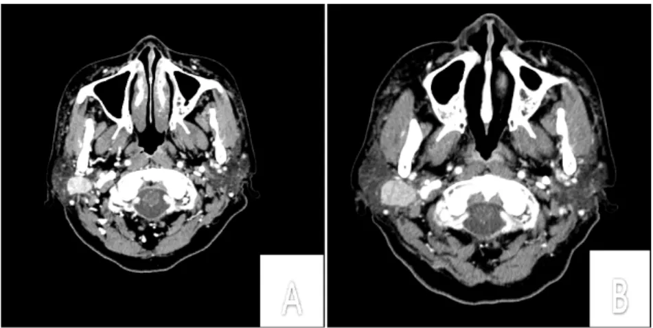
신경외과 수술 당시 시행한 전산화 단층 촬영상 우측 소뇌에 수막종 외에 우측 이하선에 1.8 x 1.2cm 크기의 조영 증강되는 경계가 또렷한 이하선 종양이 발견되었다 (Fig. 1). 환자의 자각 증상이 없어서 우측 소뇌 수막종에 대한 수술만을 시행한 후 주기적인 전산화 단층 촬영을 시행하였다. 2년 후 이 전에 발견되었던 우측 이하선 종 양은 뇌 자기 공명 영상과 경부 전산화 단층 촬영에서 2.2 x 1.5cm으로 크기가 증가된 것이 확인되어, 이하선 천엽에 생긴 다형선종이 의심되어 본과에 수술적인 치료 를 위하여 의뢰되었다 (Fig. 1 & 2).
이학적 검사상 경부에 저명하게 만져지는 종물은 없 었으며, 안면신경 마비 등 신경학적 이상 소견도 관찰되 지 않았다. 추가적인 세침흡인검사를 시행하지 않았으 며, 우측 이하선 양성 종양, 특히 다형선종, 의심 하에 전신 마취 하에 수술적인 치료를 계획하였다.
전신마취 하에 우측 이개 전방부터 귀를 따라 흉쇄유 돌근 전방 경계에 걸쳐 피부 절개 후에 우측 이하선을
- 62 -
Fig. 1. (A) In 2014, Skull base CT axial scan show about 1.8 x 1.2cm sized well-defined enhancing mass in the right parotid gland. (B) In 2016, neck CT axial scan show an increased mass in the right parotid gland.
Fig. 2. Brain MRI scans (A, T1-weighted image; B, T2-weighted image; C, T1-enhanced) show a 2 x 1.5 x 2.3cm well-defined enhancing mass in right parotid gland.
Fig. 3. The tumor is firm and well circumscribed, measured 2.8cm size.
확인하였고, 안면신경을 따라 이하선 천엽 절제술 후 약 2 x 1.5cm 크기의 경계가 비교적 분명하고 다소 부드러운 종양이 관찰되었다. 종양은 주변 조직과 유착 없이 잘 박리 되었으며, 안면신경이나 주변 구조물의 손상 없이 완전히 적출하였다 (Fig. 2).
조직 병리 소견상 단일한 하얗고 노란, 캡슐로 둘러 쌓여 있었으며, 피지세포와 도관 구조로 구성되어 있어 임파선종으로 진단되었으며, 수술 당시 함께 절제한 인 접한 임파선은 특별한 이상 소견이 발견되지 않았다 (Fig. 3). 술 후 안면 신경 등의 다른 합병증 없이3일째 퇴원하였고, 현재 재발 없이 외래에서 추적 관찰 중이다.
고 찰
이하선 종양 중 80%가 양성 종양이며 다형선종이 대 부분을 차지한다.3,4) 임파선종은 타액선에서 발생하는 매우 드문 양성질환으로, 1931년 Hamperl에 의해 처음으 로 기술되었고,5)이하선에서 11~28%, 악하선에서 6%에 서 발견된다고 알려져 있다.6,7)현재까지 국내에는 이하 선에 발생한 임파선종1예만이 보고되었다.2)
임파선종은 병리학적으로 림프 조직에 외배엽기원의 경계가 잘 형성된 종양으로 외배엽은 피지세포와 도관 구조로 구성되어 있다. 임파선종의 발생 원인과 병리학 적인 기원에 대해서는 이하선 내부나 주변의 림프절에
- 63 -
Fig. 4. (A) Small cysts are present within a dense lymphoid stroma which contains germinal center (H & E, x40). (B) Numerous cysts supported by a dense lymphoid population. Epithelial population with squamoid and cuboidal lining is distributed within the neoplasm (H & E, x200).
타액선 조직의 함입이 되었다는 설이 유력하지만 아직 확실히 정립된 바는 없다.1)
이하선 종양은 주로 무증상으로 이학적 검사나 다른 이유로 시행한 방사선 검사에서 우연히 발견되는 경우가 많다.8,9)임파선종도 다른 이하선 종양과 유사하게 무증 상의 커지는 종물을 주된 증상으로 나타낸다.1)본 증례 에서도 환자는 전혀 자각 증상이 없었으며, 신경외과 수 술을 위하여 시행한 전산화 단층 촬영상 우연히 발견 되었다.
이하선 종양의 진단은 이학적 검사, 초음파, 전산화 단 층 촬영, 자기공명영상, 핵의학 검사 등의 방사선 검사, 세침흡인검사, 조직 검사 등을 통하여 이루어 진다.10) 이 하선 종양의 진단에는 세침흡인검사 바늘이 유용하게 쓰일 수 있으며, Banich J. 등에 의해 세침 흡인 검사를 통해 임파선종을 진단한 증례가 보고된 바 있다.11)그러 나, 임파선종은 특별한 임상적, 방사선학적인 특징이 없 으므로 수술 전에 다른 이하선 종양과 감별하는 것이 쉽지 않다.1)본 증례에서도 2년간 신경외과에서 세 차례 전산화 단층 촬영과 자기공명영상을 시행하였으나, 방 사선학적으로는 이하선에 발생한 다형선종일 가능성이 높은 것으로 생각하였다. 가장 확실한 진단 방법은 병리 검사를 통한 진단이지만, 이번 증례에서는 술 전에 세침 흡인 검사를 따로 시행되지 않아 술 전 임파선종의 가능 성을 생각하지 못하였다.
완전한 수술적 절제가 최선의 치료법이며, 또한 정확 하게 진단할 수 있는 방법이다.1,12)예후는 비교적 좋으며 재발이나 악성 변이 가능성은 거의 없다.12)
본 증례는 타과 검사에서 우연히 우측 이하선에 발생 한 종양을 발견하였고, 수술을 통하여 임파선종으로 확 진된 예이다. 수술 전에 방사선학적인 검사를 시행하였
으나 임파선종의 가능성을 생각하지 못하였고, 임파선 종의 진단에 도움이 되는 세침 흡인 검사를 시행하지 않아 진단에 실패하였다. 그러므로, 임파선종도 이하선 에 발생할 수 있음을 염두해 두어야 하겠으며, 이하선 종양이 의심될 경우 세침흡인검사를 포함한 다양한 검사 를 시행하여 정확한 진단 및 치료를 시행해야 할 것이다.
중심 단어:임파선종, 이하선
References
1) Liu G, He J, Zhang C, Fu S, He Y. Lymphadenoma of the salivary gland: Report of 10 cases. Oncol lett 2014;7:1097-101.
2) Kwon GY, Kim EJ, Go JH. Lymphadenoma arising in the parotid gland: A case report. Yonsei Med J 2002;43:536-8.
3) Spiro RH, Koss LG, Hajdu SI, Strong EW. Tumors of minor sali- vary origin. A clinicopathological study of 492 cases. Cancer 1973;31:117-29.
4) Sungur N, Akan IM, Ulusoy MG, Ozdemir R, Kilnc H, Ortak T.
Clnicopathological evaluation of parotid gland tumors: a retro- spective study. J Craniofac Surg 2002;13:26-30.
5) Hamperl H. Beitrage zur normalen und pathologischen Histologie menschlicher Speicheldrusen. Zeit Mikroskopisch Anat Forschung 1931;27:1-55.
6) Limhartova A. Sebaceous glands in salivary gland tissue. Arch Pathol 1974;98:320-4.
7) Meza-Chavez L. Sebaceous glands in normal and neoplastic pa- rotid glands: possible significance of sebaceous glands in respect to origin of tumours of the salivary glands. Am J Pathol 1949;25:627-45.
8) Spiro RH, Huvos AG, Strong EW. Cancers of the parotid gland.
A clinicopatholgic study of 288 primary cases. Am J Surg 1975;130:452-9.
9) Seethala RR, Thompson LD, Gnepp DR, Barnes EL, Skalova A, Montone K, et al. Lymphadenoma of the salivary glnad: clin-
- 64 -
icopathological and immunohistochemical analysis of 33 tumors.
Mod Pathol 2012;25:26-35.
10) Bussu F, Parilla C, Rizzo D, Almadori G, Paludetti G, Galli J.
Clinical approach and treatment of benign and malignant paro- tid masses, personal experience. Acta otorhinolaryngol Ital 2011;31:135-43.
11) Banich J, Reyes CV, Bier-Laning C. Sebaceous lymphadenoma identified by fine needle aspiration biopsy: a case report. Acta Cytol 2007;51:211-3.
12) Rawlinson NJ, Almarzooqi S, Nicol K. Sebaceous lymphadeno- ma of the parotid gland in a 13-year-old girl: a case report, Head neck Pathol 2010;4:144-7.