紫蘇葉 추출물의 抗凝血 활성에 관한 硏究
정경희, 한신희, 길기정*1)
중부대학교 한약자원학과
A Study of Anticoagulation Activity from Perillae Folium Extract
Gyong-Hee Jeoung, Sin-Hee Han, Gi-Jung Kil*
Dept. of Oriental Medicine Resources, Joongbu University
ABSTRACT
Objectives : This research was investigated to find out the effect of the anticoagulant Perillae folium extract.
Methods : To examine an active effect of anticoagulation in Perillae folium extract, the study measured Prothrombin time(PT) and activated partial thromboplastin time(APTT) of human plasma in vitro and measured bleeding time and arterio-venous shunt model in rats in vivo.
Results : Bleeding time of Perillae folium extract in vivo had a significant increase 1.6 times and thrombus weight of Perillae folium extract had a significant reduction of thrombus weight as 68%.
Perillae folium extract had an effect of anticoagulation by operating on extrinsic pathway factor II, V, VII, X and intrinsic pathway factor Ⅷ, Ⅸ, Ⅹ, Ⅵ, Ⅶ in the coagulation system.
Conclusions : Considering the above mentioned results, it is judged that a Perillae folium extract has a control effect of thrombus creation.
Key words: Perillae folium, Anticoagulant, Thrombosis, Prothrombin time, Bleeding time
Introduction
Perillae folium is an annual herbaceous belonging to Lamiaceae family with its origin from China that has Perilla frutescens Britton var. acuta kudo leaves with unique fragrance and also grows wild in various regions of our country.
Leaves sprout face to face with long leafstalk, often purplish color and serrations in the edges,
and ones with strong fragrance and without branches and leafstalk are considered better.
The leaves of Perilla frutescens, a traditional Asian medicinal herb, have been used in the Orient for centuries to treat various conditions including depression1), infection2), inflammation3,4) and allergies5,6). Several studies have also suggested that extract of Perillae folium take in antitumor growth in a murine skin model in vivo7,8). In vitro
* Corresponding author : Gi-Jung Kil, Dept. of Oriental Medicine Resources, Joongbu University, 101 Daehakro Choobumyeon Geumsangun Choongnam, Republic of Korea
․ Tel : 041-750-6225 ․ E-mail : [email protected]
․ Acceptance:2008. 11. 30. ․ Adjustment:2008. 12. 17. ․ Adoption:2008. 12. 22.
studies have reported that Perillae folium components, rosmarinic acid and luteolin, can induce apoptosisin a variety of cancer cell lines9-12). However, the mechanism of the antitumorigenesis effects of whole Perillae folium extract remains unclear.
Formation of blood clots in blood vessels can be divided largely into 3 steps. Step 1 is the process in which thromboplastin is separated from the damaged tissue or cut thrombocyte when bleeding, step 2 is the process in which prothrombin is converted to thrombin under presence of Ca2+, and step 3 is the process in which various coagulation factors are continuously activated to form blood clot by changing fibrinogen in blood plasma into insoluble fibrin using thrombin. Clot is formed by extrinsic pathway that begins from damage in blood vessel walls or surrounding tissues and intrinsic pathway that begins from coagulation factors within blood vessels13-15).
The purpose of this study is to conduct an experiment on anti-coagulation effect of Perillae folium in the screening process for development of anti- coagulation medical supplies and to report its results.
Materials and methods
1. Preparation of Perillae foliumPerillae folium (Perilla frutescens var. acuta Kudo) had been purchased from Hebei region of China, examined by the Department of Oriental Medicine Resources at Joongbu University for careful selection and used as samples while being refrigerated at 4℃.
460 mL of extract obtained by 3 hours of heating of 30 g Perillae folium(PLF) in 1,000 mL distilled water was filtered once using a filter paper (Whatman No. 1) and decompressed and concentrated using a rotary vacuum evaporator. It was then settled in a -80℃ deep freezer for 4 hours and lyophilized by freeze dryer for 12 hours to obtain 6.2 g of powder. The powder was refrigerated (4℃) until used for this experiment and had been diluted to 10 ㎎/mL (PLF 1), 30 ㎎/mL (PLF 2) or 50 ㎎/mL (PLF 3) according to necessity.
Preparation of Human plasma
Fresh frozen plasma for research use was donated by Daejeon, Chungnam Chapter of National Red Cross for human plasma, and separated human plasma was refrigerated with volumes of 10 mL in 15 mL conical tubes to be defrosted to 37℃ before use.
2. Animals
Male Sprague-Dawley rats were purchased from Core Tech Co., Ltd.. 12-week-old male Sprague Dawley (SD) rats with body weight of 350±10 g had been received from Core Tech Co., Ltd. and were used for this experiment after adaptation period of at least one week. Until the day of experiment, they were provided with enough compressed feed (crude protein 22.1% or above, crude fat 8.0% or below, crude fiber 5.0%
or below, crude ash 8.0% or below, calcium 0.6%
or above, phosphorus 0.4% or above, Samyang, Korea) and water. Breeding condition was fixed at 22±2℃ room temperature, 50±10% relative humidity, 12 hours of lighting (07:00~19:00) and 150~300Lux illuminance.
3. Prothrombin time (PT) measurement
For measurement of prothrombin time, Quick's one stage method16) was used. 45 uL of blood plasma and 3 mL of human plasma were put inside a dish and on top of ice. Then, 5 uL of Perillae folium extract (10 ㎎/mL) was put in the well of strip well plate and 5 uL of distilled water was put in the other well as control group.
After heating for 5 minutes at 37℃, 100 uL of PT reagent was added and time for coagulation was measured under extinction of 405nm.
4. Activated Partial Thromboplastin Time (APTT) measurement
3 mL of human plasma was put inside a dish and on top of ice. 5 uL of Perillae folium extract
(10 ㎎/mL) was put in the well of strip well plate, and 5 uL of distilled water was put in the other well as control group. For each group, 45 uL of plasma was added and heated for 2 minutes in a microplate reader.
After adding 50 uL of APTT reagent and heating for 5 minutes, 100 uL of 25mM calcium chloride (CaCl2) was added to measure the time required for coagulation under extinction of 405nm.
5. Bleeding time (BT) measurement
400 ㎎/㎏ Perillae folium extract was oral administered to SD rats once each day for 5 days and medicine was given on the 6th day. After using the method17) of Han et al that used 1 mL abdominal injection of 1.25 g/㎏ ether after 60 minutes for anesthesia, 0.3 cm was cut on the end of the tail. 5 cm of remaining tail was immediately put inside 37.5℃ saline solution and time for hemostasis was measured.
6. Thrombus weight measurement
400 ㎎/㎏ Perillae folium extract was oral administered to SD rats once each day for 5 days and medicine was given on the 6th day. After 1 mL abdominal injection of 1.25 g/㎏ ether was made after 60 minutes for anesthesia, canulation was done on left carotid artery and right jugular vein using polyethylene catheters. Then, 5cm silicon tube filled with a cotton thread was filled up using saline and blood was made to flow through the tube connected to artery and vein to induce shear-stress and thrombus for 15 minutes.
Created thrombus was dried and weighed for evaluation of antiplatelet efficacy.
7. Statistical analysis
Experimental results had been expressed as mean value±standard deviation for 3 repeated trials. For testing of significance of each group, T-test of Student was used with the statistics program SPSS (ver. 12.0, Korea). After comparing
between the values of each experimental group, values with 95% confidence or above (p<0.05) were determined to have significance.
Results and discussion
1. Effects on PT
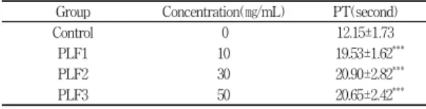
As a result of studying the effects on PT by adding Perillae folium extract to human plasma, control group showed 12.15±1.73 second and 50 ㎎/mL group showed 20.65±2.42 second. In comparison to control group, 50 ㎎/mL group showed twice as much increase in speed of blood flow in PT, showing that PT became longer as concentration of Perillae folium extract increased (Table 1).
Table 1. Effect of Perillae Folium Extract on Prothrombin Time
Group Concentration(㎎/mL) PT(second)
Control 0 12.15±1.73
PLF1 10 19.53±1.62***
PLF2 30 20.90±2.82***
PLF3 50 20.65±2.42***
Control : Untreated, PLF : aqueous extract of Perillae folium.
* : Statistically significant value compared to control by Student's t-test. (***, p ≤ 0.001).
Lengthening of PT means that thrombin creation is delayed by suppressing activation of extrinsic pathway factors that participate in conversion of prothrombin to thrombin. From such result, Perillae folium extract was found to have an impact on extrinsic pathway in coagulation system.
2. Effects on APTT
As a result of studying the effects on APTT by adding Perillae folium extract to human plasma, control group showed 39.98±1.36 sec, 30
㎎/mL group showed 60.29±2.62 sec and 50 ㎎/mL group showed 81.93±4.85 second. In comparison to control group, experimental groups showed twice as much increase in APTT, showing that APTT is dependent on concentration of Perillae folium
extract because APTT increased with increasing concentration (Table 2).
Increasing APTT means that creation of thrombus due to creation of fibrin is suppressed by activation intrinsic pathway factors. Perillae folium extract was found to have an impact on intrinsic pathway in coagulation system.
Table 2. Effect of Perillae Folium Extract on Activated Partial Thromboplastin Time
Group Concentration(㎎/㎖) APTT(second)
Control 0 39.98±1.36
PLF1 10 47.41±5.13
PLF2 30 60.29±2.62**
PLF3 50 81.93±4.85***
Control : Untreated, PLF : aqueous extract of Perillae folium.
* : Statistically significant value compared to control by Student's t-test. (** p ≤ 0.001, *** p ≤ 0.001).
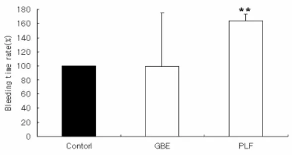
3. Effects on bleeding time
As a result of comparison between effects on bleeding time in rats with Perillae folium extract and rats with Ginkgo biloba extract (GBE), bleeding time was 498.3±75.3 sec for control group and 505.7±62.9 sec for GBE group, showing no significant difference.
Perillae folium extract group showed bleeding time of 811.7±9.60 sec, meaning that bleeding time had a significant increase as about 1.6 times in comparison to control group (Fig. 1).
Measurement of bleeding time in vivo allows estimation of coagulation response in rats and can
Fig. 1. Effect of Perillae folium extract on the bleeding time Rats was treated with oral administration of 400 ㎎/㎏ of GBE and PLF, representatively.
Control : Untreated, GBE : Ginkgo biloba extract, PLF : Perillae folium extract.
be used as the basis for determining the response according to absorption of Perillae folium extract.
The results suggest that Perillae folium extract affected coagulation system through absorption in the body.
4. Effects on thrombus weight
Effects on thrombus weight in rats with Perillae folium extract and rats with GBE were compared.
Thrombus weight was 286.67±32.15 ㎎ for control group, 253.30±15.28 ㎎ for GBE group and 195.08±23.45 ㎎ for Perillae folium extract group, showing a significant different of about 68%
reduction (Fig. 2).
0 20 40 60 80 100 120
Control GBE PLF
Thrombusamountrate(%)
* *
0 20 40 60 80 100 120
Control GBE PLF
Thrombusamountrate(%)
* *
Fig. 2. Inhibition effect of Perillae folium extract on thrombus weight
Rats was treated with oral administration of 400 ㎎/㎏ of GBE and PLF, representatively.
Control : Untreated, GBE : Ginkgo biloba extract, PLF : Perillae folium extract.
Conclusions
In order to investigate anti-coagulation activity of Perillae folium, PT and APTT were measured in vitro in human plasma and effects of medication of Perillae folium extract in rats on bleeding time and thrombus weight were found in vivo to obtain the following conclusion.
1. As a result of measuring PT and APTT in human plasma, PT showed a significant increase in speed of blood flow in 50 ㎎/mL Perillae folium
extract and APTT was significant by more than a factor of 2 in 50 ㎎/mL.
2. BT from Perillae folium extract showed a significant increase of about 61% in vivo, and formation of thrombus weight significantly decreased by about 68%.
3. Perillae folium extract was found to have influence on extrinsic pathway factors II, V, VII, X and intrinsic pathway factors VIII, IX, X, XI, XII in coagulation system.
Meaning of such results is that Perillae folium extract impedes coagulation of blood and shows anti-coagulation effects. A continued study on detailed control passway of blood coagulation factor will be conducted.
References
1. Takeda H, Tsuji M, Matumiya T and Kubo M.
Identification of rosmarinic acid as a novel antidepressive substance in the leaves of Perilla frutescens Britton var. acuta Kudo (Perillae Herba). Japanese J of Psychopharmacology.
2002 ; 22 : 15-22.
2. Kawahata T, Otake T, Mori H, Kojima Y, Oishi I, Oka S, Fukumori Y and Sano K. A novel substance purified from Perilla frutescens Britton inhibits an early stage of HIV-1 replication without blocking viral adsorption.
Antiviral Chemistry & Chemotherapy. 2002 ; 13 : 283-8.
3. Ueda H, Yamazaki C and Yamazaki M. Luteolin as an anti-inflammatory and anti- allergic constituent of Perilla frutescens. Biological &
Pharmaceutical Bulletin. 2002 ; 25 : 1197-202.
4. Banno N, Akihisa T, Tokuda H, Yasukawa K, Higashihara H, Ukiya M, Watanabe K, Kimura Y, Hasegawa J and Nishino H. Triterpene acids from the leaves of Perilla frutescens and their anti-inflammatory and antitumor-promoting effects, Bioscience. Biotechnology and Biochemistry. 2004 ; 68 : 85-90.
5. Makino T, Furuta Y, Wakushima H, Fujii H,
Saito K and Kano Y. Anti-allergic effect of Perilla frutescens and its active constituents.
Phytotherapy Res. 2003 ; 17 : 240-3.
6. Takano H, Osakabe N, Sanbongi C, Yanagisawa R, Inoue K, Yasuda A, Natsume M. Baba S, Ichiishi E and Yoshikawa T. Extract of Perilla frutescens enriched for rosmarinic acid, a polyphenolic phytochemical, inhibits seasonal allergic rhinoconjunctivitis in humans. Experimental Biology and Medicine (Maywood). 2004 ; 229 : 247-54.
7. Ueda H, Yamazaki C and Yamazaki M. Inhibitory effect of Perilla leaf extract and luteolin on mouse skin tumor promotion. Biological &
Pharmaceutical Bulletin. 2003 ; 26 : 560-3.
8. Osakabe N, Yasuda A, Natsume M and Yoshikawa T. Rosmarinic acid inhibits epidermal inflammatory responses: anticarcinogenic effect of Perilla frutescens extract in the murine two- stage skin model. Carcinogenesis. 2004 ; 25 : 549-7.
9. Hur YG, Yun Y and Won J. Rosmarinic acid induces p56lck-dependent apoptosis in Jurkat and peripheral T cells via mitochondrial pathway independent from Fas/Fas ligand interaction. J of Immunology. 2004 ; 172 : 79-87.
10. Kolettas E, Thomas C, Leneti E, Skoufos I, Mbatsi C, Sisoula C, Manos G and Evangelou A. Rosmarinic acid failed to suppress hydrogen peroxide-mediated apoptosis but induced apoptosis of Jurkat cells which was suppressed by Bcl-2.
Molecular and Cellular Biochemistry. 2006 ; 285 : 111-20.
11. Selvendiran K, Koga H, Ueno T, Yoshida T, Maeyama M, Torimura T, Yano H, Kojiro M and Sata M. Luteolin promotes degradation in signal transducer and activator of transcription 3 in human hepatoma cells: an implication for the antitumor potential of flavonoids. Cancer Res. 2006 ; 66 : 4826-34.
12. Lim do Y, Jeong Y, Tyner AL and Park JH.
Induction of cell cycle arrest and apoptosis in HT-29 human colon cancer cells by the dietary compound luteolin. American Journal of Physiology:
Gastrointestinal and Liver Physiology. 2007 ; 292 : G66-G75.
13. Mustard JF and Packham MA. Factors influencing platelet function: adhesion release and aggregation.
Pharmacol Res. 1970 ; 22 : 97-187.
14. Longenecker GL, Swigt IA, Bowen RJ, Beyers BJ and Shah AK, Kinetics of ibuprofen effect on platelet and endothelial prostanoid release.
Clin Pharmacol Ther. 1985 ; 37 : 343-8.
15. Davie EW and Ratnoff OD. Waterfall sequence
for intrinsic blood clotting. Science. 1964 ; 145 : 1310-12.
16. Quick AJ. The prothrombin in haemophilia and in obstructive jaundice. J Biol Chem. 1935 ; 109 : 73-4.
17. Han YN, Baik SK, Kim TH and Han BH.
Antithrombotic activities of saponins from Ilex pubescens. Arch Pharm Res. 1987 ; 10 : 115-22.