Intentional Replantation of a Root-Fractured Tooth with Pulp Canal Obliteration
Mihee Kim, Sangho Lee, Nanyoung Lee
Department of Pediatric Dentistry, College of Dentistry, Chosun University
Root fracture is defined as a fracture involving the dentin, cementum, and pulp. Most fractures occur in the maxillary anterior teeth between the ages of 11 and 20 years old. The treatment for root fracture in permanent teeth involves the reduction and fixation of the displaced coronal segment. When signs of pulp necrosis or inflammatory root resorption are present, root canal therapy should be performed. Since most apical fragments maintain pulp vitality, root canal therapy is typically limited to coronal fragments. However, it’ s too difficult to achieve a proper apical stop on coronal fragment.
Intentional replantation involves performing root apex treatment outside the mouth after intentional extraction of the tooth in a controlled environment and then replanting it. The objective is ‘perfect’root canal therapy.
Intentional replantation may be used in cases of failed typical root canal therapy, problematic endodontic retreatment due to the existing restoration or a calcified root canal, and when apical surgery is contraindicated because of a lack of reasonable approaches.
In this case, intentional replantation was carried out to treat a horizontal root fracture in a maxillary central incisor with a calcified root canal due to previous trauma. We achieved a clinically and functionally satisfactory result.
Key words : Intentional replantation, Horizontal root fracture, Pulp canal obliteration, Retrograde filling Abstract
Ⅰ. Introduction
When an external force is applied to teeth or periodon- tal tissue, damage may occur according to the magnitude and direction of the force. Root fracture is defined as frac- ture involving the dentin, cementum, and pulp. This is relatively rare and comprises 0.5-7% of all cases of trau- ma to permanent teeth. Most root fracture in permanent teeth occurs in maxillary anterior teeth between the ages of 11 and 20 years. The most common general causes are fights or blows with external materials to the tooth
1).
Root fracture can be classified broadly as horizontal and vertical root fracture. A vertical root fracture is rare; the fracture line extends along longitudinal axis of tooth towards the root apex
2). Horizontal root fracture, which is more common, occurs mostly in the middle third of the root and less frequently in the apical and cervical thirds
3,4).
The treatment of root fractures varies depending on the location of the fracture line, the existence of a coro- nal segment, and the length of the residual root seg- ment. Generally, the preferred treatment for a perma-
Corresponding author : Sangho Lee
Department of Pediatric Dentistry, College of Dentistry, Chosun University, 303 Pilmun-daero, Dong-gu, Gwangju, 61452, Korea Tel : +82-62-220-3865 / Fax : +82-62-225-8240 / E-mail : [email protected]
Received July 14, 2015 / Revised September 22, 2015 / Accepted September 2, 2015
nent tooth is reduction and fixation of the displaced coronal segment
2). The duration of fixation should be at least 4 weeks to allow sufficient formation of hard tissue.
Sixty to eighty percentage of root fracture maintain pulp vitality. However, when pulp necrosis occurs, a root canal therapy is necessary
1).
When pulp necrosis progresses, the root of the frac- tured tooth usually maintains pulp vitality
1). Thus, root canal therapy is limited to the coronal fragment.
However, achieving a proper apical stop on the coronal fragment is difficult and perfect sealing of the fragment is difficult
5).
Intentional replantation is a treatment approach that involves root apex treatment outside the oral cavity after intentional extraction of the tooth under the controlled environment and then replants it. The objective is to perform the ‘perfect’root canal therapy. Intentional re- plantation is the last treatment method that can be car- ried out in cases of failed typical root canal therapy, failed nonsurgical endodontic retreatment using a micro- scope due to excessive calcification of the root canal or a large periapical radiolucent lesion, and when tooth ex- traction must be considered because apical surgery is impossible due to the proximity of the relevant anatomi- cal structures
6).
In the present case, intentional replantation was carried out to treat a root fracture in a maxillary central incisor with a calcified root canal due to previous trauma. We achieved a clinically and functionally satisfactory result.
Ⅱ. Case Report 1. History taking and diagnosis
A 13-year-old male presented to the Department of Pediatric Dentistry, Chosun University Dental Hospital
with the chief complaint of breaking and falling out the tooth. He had bumped it against a chin-up bar 1 hour before visiting our clinic. There was no specific medical history, and no laceration or contusion inside or outside his mouth was observed. Through clinical and radi- ographic examinations, the patient was diagnosed with root fracture of the maxillary right central incisor and lateral luxation of the maxillary left central and lateral incisors. The injured tooth had grade 3 mobility. In the case of the maxillary right central incisor with root frac- ture, the root canal of the coronal segment was calcified due to trauma, the details of which were unknown to the patient or his parents. The fracture line was be- tween the cervical and middle third regions of the root, and the gap between the fragments was wide (Fig. 1).
2. Intentional replantation
Multiple considerations guided the treatment plan in the present case. In performing a splint after reposition- ing a coronal fragment, when pulp necrosis occurs, typi- cal endodontic therapy through a calcified root canal is difficult. Moreover, given the severe mobility of the coro- nal segment because the fracture line was located be- tween the cervical and middle thirds of the root, and given the large distance between the fragments, coronal segment could be extracted. Intentional replantation limited to the coronal segment was planned. The coronal segment of the maxillary right central incisor was ex- tracted carefully with dental forceps under local anesthe- sia on the day of the patient’ s visit to the clinic. After extraction, the coronal portion was grabbed by hand to prevent damage to periodontal ligament cells. Cavity preparation was carried out to a depth of 2-3 mm using a #330 high-speed carbide bur (SS White Burs Inc., Lakewood, NJ, USA) for retrograde filling. Retrograde
Fig. 1. (A) Periapical radiograph and (B) Intraoral photograph taken during the patient's first visit to the clinic. Fracture of the root of the maxillary right
central incisor can be seen. The fracture line was between the cervical and middle thirds of the root, and the distance between the fragments was large.
filling was then performed on the prepared cavity using mineral trioxide aggregate (MTA) (ProRoot MTA White;
Dentsply, Konstanz, Germany; Fig. 2A). Following the removal of a blood clot from the extraction socket and ir- rigation, replantation was performed. The periapically treated tooth was carefully replanted in the extraction socket. At this point, repositioning was performed as slowly as possible to allow stagnant blood to escape.
After repositioning the maxillary left central and lateral incisors to their original locations, we performed fixation with composite resin and steel wire from the maxillary left first premolar to the maxillary right canine (Fig. 2B and C). After carrying out splint, extirpation through the crown was tried at the same time. But, it’ s hard to access the orifice of canal due to pulp canal obliteration.
The time required outside the mouth to replant the maxillary right central incisor was approximately 20 minutes. The patient was recommended to eat liquid foods and maintain good oral hygiene.
3. Assessment after surgery and the results of periodic follow-up examinations
After 4 weeks, the resin-wire splint was removed. The maxillary right central incisor, which had been treated with intentional replantation, showed tenderness to per- cussion 1 week after replantation. However, the re- sponse to percussion and mobility disappeared 4 weeks later. The maxillary left central and lateral incisors on the other side showed tenderness to percussion 1 week after splint. After 4 weeks, because of continuous re- sponse to percussion, crown discoloration and a negative response to an electrical pulp test, extirpation and con- ventional root canal treatment were performed (Fig.
3A). At 1, 3, 7, 10, 15, 20, 25 and 30 months after treatment, the maxillary right central incisor exhibited healing with no specific clinical signs or radiographic findings (Fig. 3). Thirty-month follow-up examinations indicated a normal lamina dura with no periapical lesion
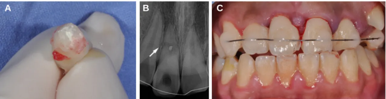
Fig. 2. Operational procedure for intentional replantation. (A) After extraction of the coronal segment of the maxillary right central incisor, cavity preparation was carried out to a depth of 2-3 mm using a #330 high-speed carbide bur. Next, retrograde filling was performed using MTA. (B, C) After replantation of the coronal segment of the maxillary right central incisor, a resin-wire splint was set up from the maxillary left first premolar to the maxillary right canine.
Fig. 3. Periapical radiographs produced (A) 4 weeks, (B) 3 months, (C) 7 months, (D) 10 months, (E) 15 months, (F) 20 months, (G) 25 months, and (H) 30 months after intentional replantation.
The maxillary right central incisor healed well around the fracture line (arrow), and a normal lamina dura was observed around the root. The maxillary right central incisor showed physiological mobility and the patient had no specific clinical symptoms.
A B C
or inflammatory external root resorption, except for blunting of the margin of the fracture. The patient, who was satisfied esthetically, exhibited no specific clinical symptoms and the injured tooth showed physiological mobility (Fig. 4). Ongoing assessments regarding anky- losis, inflammatory external root resorption, and the oc- currence of periapical lesions in connection with the re- planted tooth will be necessary.
Ⅲ. Discussion
Clinical symptoms of root-fractured teeth include ex- trusion of the crown and linguoversion. Tooth mobility increases the closer the fracture is to the crown; in most cases, radiography is necessary to make a diagnosis be- cause it is difficult to distinguish clinically between dis- placement by luxation and fracture
1).
Although the treatment of root-fractured teeth can be differentiated by the location of the fracture line, the presence of a coronal segment, and the length of the residual part of the root, the basic procedure is reduction and fixation of the fractured part
2). If a fracture occurs in the apical third of the root, the tooth usually shows no mobility and the apical fragment usually maintains its vitality. However, in cases where necrosis of the apical fragment is observed, surgical removal is necessary. In the case of the fracture in the cervical third of the root, if the fractured coronal segment is missing, a post crown with periodontal treatment, orthodontic or surgical ex- trusion, can be considered. In cases where other conser- vative treatments are not appropriate, the tooth can be extracted
1,7).
Types of healing in root fracture are various : healing
with calcified tissue, interposition of connective tissue, interposition of bone and connective tissue, interposition of granulation tissue. Through the periodic radiograph follow-up, maxillary right central incisor of this case seems to heal with interposition of bone. At this type of healing, bone and connective tissue are interposed be- tween fragments and tooth has sound periodontal liga- ment and clinically has no mobility. But, coronal frag- ment can be erupted and apical fragment can remain in the jaw due to continuous growth of alveolar bone be- tween fragments. Through this, crown-root ratio of coro- nal fragment becomes unfavorable, prognosis of the tooth can be poor
1).
Intentional replantation, defined by Grossman
8)in 1966, is a method for extracting a tooth and replanting it immediately after treatment to fill the root canal out- side the alveolus. Intentional replantation can be planned in case where there is difficulty in endodontic retreatment due to the existence of an original restora- tion or a calcified root canal, and when apical surgery is contraindicated because of a lack of a suitable approach.
In this case, the root canal of the maxillary right later- al incisor was calcified due to an unidentified previous trauma. Extraction could not be excluded due to the se- vere mobility of the coronal segment because the fracture line was located between the cervical and middle thirds of the root, and the distance between the fragments was large. It seemed that a regular endodontic procedure through the calcified root canal would not be possible;
necrosis occurred after regular treatments, including re- duction and fixation of the fractured coronal segment.
Considering this, we decided to perform intentional re- plantation, limited to the coronal segment.
The precautions or considerations for clinically suc- cessful intentional replantation are as follows: 1. Be careful not to damage the surface of the root or alveolus during extraction or replantation. If extraction is achieved easily, it can have a positive influence on prog- nosis, which is related to root-alveolar bone ankylosis or root resorption. 2. Pay attention to early fixation. If the tooth moves a great deal due to poor fixation, it can have a negative impact on the attachment of periodontal tissue. However, it is not recommended to apply high forces to fix the coronal segment because it can have a traumatic effect on a tooth that has already been dam- aged. Thus, a semi-rigid fixation method (e.g., an acid- etched resin splint) must be used. 3. Minimize the ex- traoral time. As the extraoral time increases, the prog- Fig. 4. An intraoral photograph taken 30 months after treatment. No
discoloration of the maxillary right central incisor, treated with retrograde
filling using MTA, was observed.
nosis worsens. In particular, if a tooth is left dry for more than 30 minutes, the probability of survival is less than 50%. Thus, preoperative preparations must be made carefully and instruments and movements must be organized so as to reduce the extraoral time. 4. The in- take of liquid foods and good oral hygiene management are essential. If possible, an intentional replantation should be performed on the day the tooth was fractured or before the resorption of alveolar bone is severe
6,9,10).
In this case, the crown portion of the root-fractured maxillary right central incisor was extracted carefully with dental forceps to minimize periodontal damage.
Also, it was easy to extract the tooth due to mobility of grade 3. A semi-rigid fixation method using a resin-wire splint was used for early fixation, and the extraoral op- eration was completed within 20 min, which is relatively quick. Intentional replantation was performed on the day of fracture and the patient was instructed to eat liq- uid foods and maintain good oral hygiene. Considering this situation, the postoperative prognosis is expected to be favorable.
MTA has many desirable properties including a good sealing ability, biocompatibility, mechanical strength, ability to increase the healing of tissues surrounding the root, radiopacity, and ability to set up in the presence of blood. MTA, which was developed for surgical root-end filling, has greater apical obturation capabilities than amalgam, IRM, and super-EBA; this can be explained by the fact that it expands during polymerization in a humid environment
11-14). Recently, Yildirim, Gencoglu
5), Kusgoz et al.
15)reported remarkable clinical results about use of MTA to create apical plugs in fractured roots. The biggest weakness of MTA is the possibility of discoloration. However, in the present case, no clinical crown discoloration was observed (Fig. 4).
The maxillary right central incisor of our patient ex- hibited a clinically sound result in terms of mobility, the response to percussion, and a radiographic evaluation at 30 months after treatment. However, periodic long-term follow-up is necessary because there is no previous re- port on intentional replantation limited to a crown seg- ment in root fracture cases.
Ⅳ. Summary
In this case, intentional replantation was performed on a patient whose maxillary right central incisor had been fractured by trauma. We achieved a satisfactory result.
In all trauma cases, early diagnosis and appropriate in- tervention increase the likelihood of a successful result.
Like this case, in the treatment of root fracture with a calcified root canal caused by previous trauma where conventional endodontic treatment is unavailable, inten- tional replantation limited to a crown segment, decided in early diagnosis, is appropriate. Thus, information re- garding the long-term prognosis of this treatment is cur- rently lacking; however, it could be used as a new strat- egy for tooth conservation if it is used appropriately.
References
1. Andreasen FM, Andreasen JO, Andersson L : Textbook and Color Atlas of Traumatic Injuries to Teeth. 4
thed., Blackwell Publishing Ltd, 337-371.
2008.
2. Malhotra N, Kundabala M, Acharaya S : A review of root fractures - diagnosis, treatment and prognosis.
Dent Update, 38:615-616, 619-620, 623-642, 2011.
3. Hovland EJ : Horizontal root fractures. Treatment and repair. Dent Clin North Am, 36:509-525, 1992.
4. Cali kan MK, Pehlivan Y : Prognosis of root-frac- tured permanent incisors. Endod Dent Traumatol, 12:129-136, 1996.
5. Yildirim T, Gen ç o g
̆lu N : Use of mineral trioxide aggregate in the treatment of horizontal roof frac- tures with a 5-year follow up - report of a case. J Endod, 35:292-295, 2009.
6. Choi YH : Short-term clinical outcome of intention- ally replanted posterior molars. J Korean Acad Conserv Dent, 36:12-18, 2011.
7. Feiglin B : Clinical management of transverse root fractures. Dent Clin North Am, 39:53-78, 1995.
8. Hayashi M, Kinomoto Y, Ebisu S, et al. : Short- term evaluation of intentional replantation of verti- cally fractured roots reconstructed with dentin-bond- ed resin. J Endod, 28:120-124, 2002.
9. Jin MU : Clinical evaluation of Intentional replanta- tion. J Kor Dent Assoc, 4:288-296, 2010.
10. Kim SK, Ahn ST, Park JH, et al. : Intentional replantation of the crown-root fractured tooth - a case report. J Korean Acad Pediatr Dent, 37:381- 386, 2010.
11. Arens DE, Adams WR, DeCastro RA : Endodontic surgery-Medical Considerations and Contraindications.
Harper and Row Publishing, 154-157, 1981.
12. Torabinejad M, Rastegar AF, Kettering JD, Pitt
Ford TR : Bacterial leakage of mineral trioxide aggregate as a root-end filling material. J Endod, 21:109-112, 1995.
13. Hatibovic
′-Kofman S, Raimundo L, Andreasen JO, et al. : Fracture resistance and histological findings of immature teeth treated with mineral trioxide aggre- gate. Dent Traumatol, 24:272-276, 2008.
14. Torabinejad M, Chivian N : Clinical applications of mineral trioxide aggregate. J Endod, 25:197-205, 1999.
15. Kusgoz A, Yildirim T, Tanriver M, Yesilyult C :
Treatment of horizontal root fractures using MTA as
apical plug - report of 3 cases. Oral Surg Oral Med
Oral Pathol Oral Radial Endod, 107:e68-72, 2009.
근관협착된 치근파절 치아에서 의도적 재식술 치험례