http://dx.doi.org/10.14405/kjvr.2014.54.1.7
7
<Original Article>
Cutaneous peripheral nerve sheath tumors in 15 dogs
Seung-Bo Ko
1,†, Kyoung-Ok Song
3,†, Sang-Chul Kang
2, Jae-Hoon Kim
1,*
1
College of Veterinary Medicine and Veterinary Medical Research Institute, Jeju National University, Jeju 690-756, Korea
2
Optipharm, Cheongwon 363-954, Korea
3
Jeju Self-Governing Provincial Veterinary Research Institute, Jeju, 695-963, Korea (Received: August 4, 2013; Revised: January 17, 2014; Accepted: January 22, 2014)
Abstract : Peripheral nerve sheath tumors (PNSTs) are heterogeneous tumor groups of peripheral nerves that originate from either Schwann cells or modified Schwann cells, fibroblasts, or perineural cells. In this study, signalment and clinical data such as tumor location and size were evaluated for 15 cases of PNSTs collected from local animal hospitals.
The mean age of dogs with malignant PNST was higher than that of dogs with benign PNST. Additionally, the male to female ratio in dogs with PNST was 1 : 4. In dogs with PNST, the primary sites of involvement were the hindlimb, forelimb, around the mammary glands, the neck, and the abdomen. Histiopathologic examination revealed that eight PNSTs were benign and seven were malignant. The tumor cells were composed of loosely to densely arranged interlacing bundles and wavy spindle cells arranged in short bundles, palisading, and whirling. High mitotic figures, local invasion, multifocal necrosis and atypical multinucleated giant cells were observed in malignant PNST cases.
All PNSTs showed immunoreactivity for vimentin and S-100. However, only 93.3% and 73.3% were immunoreactive for NSE and GFAP, respectively. Overall, these results indicated that immunohistochemical markers such as vimentin, S-100 and NSE could help confirm the diagnosis of canine PNSTs.
Keywords : dog, immunohistochemistry, peripheral nerve sheath tumors, Schwann cell, spindle cell
Introduction
Schwannoma is classically used term when the tumor cells are solely originated from Schwann cells [7]. Peripheral nerve sheath tumors (PNSTs) are heterogeneous group of tumors of peripheral nerves that originated from either Schwann cells or modified Schwann cells, fibroblasts, or perineural cells [10]. These tumors including schwannomas, neurilemmomas, neurofibromas were classified as PNSTs by the World Health Organization (WHO) in 1999 [4]. They are classified as benign PNSTs or malignant PNSTs based on the morphologic features and biological behavior [4, 5]. Histo- pathologically, benign PNSTs are composed of wavy spindle cells arranged in bundles, palisades, and whorls. Malignant PNSTs have similar clinical features to their benign counter- parts, but they show cytologically anaplastic forms and local invasive tendency [9, 10].
Cutaneous PNSTs are uncommon neoplasms in dogs and cats [8]. In dogs, PNSTs are found most commonly unilater- ally in the spinal nerves, with highest frequency in nerve forming the brachial plexus, less in the lumbosacral plexus, and least in subcutaneous sites of distal peripheral nerve [10].
According to previous literature, the combined incidence of benign and malignant neural tumors is reported to be 0.5% in dogs and 1.4% in cats [7, 8]. PNSTs accounted for only 0.9%
(7/748 cases) of total number of cutaneous tumors diagnosed in large scaled survey in Korea [13].
Some literatures demonstrated that immunohistochemical studies have not contributed to the clear diagnostic criteria for PNSTs [4, 5]. The expression of S-100 protein is used to differentiate between spindle cell tumors of neural and non- neural origins in human medicine [15]. In dog and cat, PNSTs in formalin fixed, paraffin embedded tissue some- times can be stained immunohistochemically positive to S- 100 protein, glial fibrillary acidic protein (GFAP), vimentin, collagen IV, or laminin [10].
Two cases of cutaneous PNSTs were previously reported in Korea [16, 17]. These reports described the clinical findings such as radiography and ultrasonography, and final diag- noses were confirmed using cytologic, histopathologic fea- tures and immunohistochemical examinations. Here we describe the clinicopathologic findings in 15 dogs with benign or malignant PNSTs.
*Corresponding author
Tel: +82-64-754-3387, Fax: +82-64-702-9920 E-mail: [email protected]
†The first two authors contributed equally to this work.
Materials and Methods
Case studies
This study was performed using archival biopsy samples from 15 canine cutaneous tumors that had been examined between 2007 and 2010 at the laboratory of Veterinary Pathology in Jeju National University. Eight cases of benign PNSTs and seven malignant PNSTs were diagnosed by gen- eral histopathologic examinations.
Histopathology
Surgically excised tissue samples were immediately fixed in 10% neutral buffered formalin. The samples were pro- cessed routinely for histopathologic examination and tissue sections were stained with hematoxylin and eosin (H&E).
Malignancy of PNSTs was determined based on the local invasion, the degree of cellular anaplasia and pleomorphism, increased mitotic rate, and necrosis.
Immunohistochemistry (IHC)
To clarify the immunohistochemical profiles of PNSTs,
streptavidin-biotin peroxidase complex IHC method was per- formed using several antibodies such as vimentin, S-100, neuron-specific enolase (NSE), and GFAP. The antibody source, type, and working dilution for each antibody were listed in Table 1. For negative controls, the primary antibody was replaced with tris-buffered saline. Positive controls for each antibody were also processed simultaneously.
The distribution of positive tumor cells expressing these each protein was scored as follows: − (negative), + (individ- ual to focal, < 10%), ++ (multifocal. 10~50%), +++ (diffuse,
> 50%) and the intensity of the immunoreactivity was graded as
− (absent), + (weak), ++ (moderate) and +++ (strong staining).
Results
Clinical data
Clinical data including breed of dogs, age, sex, location, and size of tumor mass are summarized in Table 2. There was no breed prevalence in dogs with PNSTs. Three dogs (20%) were of mixed breed, seven dogs (46.7%) were small breeds such as maltese, poodle, and mini schnauzer, and four
Vimentin Monoclonal mouse, V9 1 : 100 DAKO A/S, Denmark
S-100 Polyclonal rabbit, Z0311 1 : 400 DAKO A/S, Denmark
NSE Monoclonal mouse, M0873 1 : 200 DAKO A/S, Denmark
GFAP Polyclonal rabbit, Z0334 1 : 500 DAKO A/S, Denmark
Table 2. Clinical data of canine cutaneous peripheral nerve sheath tumors Number
of dog Breed Age (year) Sex Location Size (diameter)
BPNST
1 Unknown Unknown F Right hindlimb 3 cm
2 Mixed 8 F Neck 3.5 cm
3 German shepherd 10 F Neck 5 cm
4 Maltese 10 F Right forelimb 3 cm
5 Cocker spaniel 10 F Left hindlimb 1 cm
6 Poodle 3 F Abdomen 10 cm
7 Afghan hound 8 F Left hindlimb 2 cm
8 Shih Tzu 8 M Left forelimb 1 cm
MPNST
1 Mixed 11 F Around mammary gland 2.5 cm
2 Maltese 15 F Around mammary gland 10~25 cm
3 Yorkshire terrier 11 M Right hindlimb NI
4 Mixed 15 M Left hindlimb NI
5 Mini Schnauzer 15 F Around mammary gland 8~9 cm
6 Jindo dog 10 F Right hindlimb 6~9 cm
7 Alaskan Malamute 7 F Left forelimb 5 cm
F: female, M: male, NI: no information.
dogs (26.7%) were large breeds including German shepherd, Jindo, and malamute, respectively. The mean age of dogs with malignant PNST (12 years: range, 7 to 15 years) was higher than those with benign PNST (8.1 years: range, 3 to 10 years). The male to female ratio in dogs with PNST was 1 : 4 (three/twelve).
In dogs with PNST, the primary sites of involvement were the hindlimb (6 cases, 40%), forelimb (3 cases, 20%), around mammary glands (3 cases), neck (2 cases, 13.3%), and abdo- men (1 case, 6.7%). The right to left ratio in the limb of dogs was 1 : 1.25 (four/five).
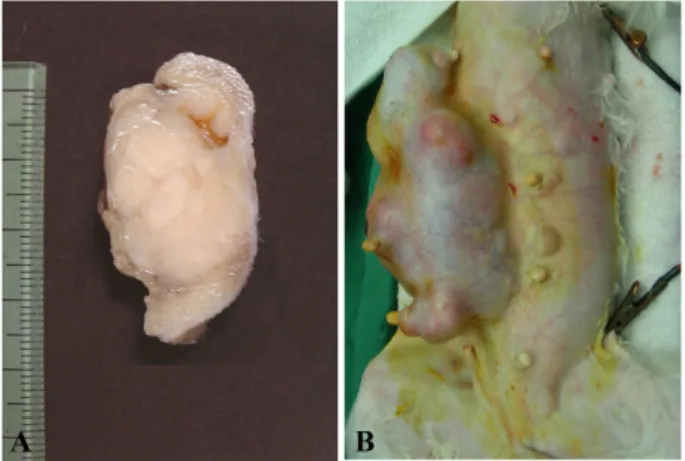
According to the document of clinicians, most benign PNSTs were located in dermis or subcutis without involve- ment of adjacent muscle. These masses were well demar- cated and easily excised from the original sites (Fig. 1A).
However malignant PNST cases were extended into deep subcutis and firmly adhered to underlying soft tissues or muscles (Fig. 1B). Because of muscular invasion of tumor mass, severe deformity of thigh muscle and abnormal gait were observed in dog number 3 and 4 with malignant PNST.
Histopathologic lesions
Based on the histopathologic examinations, 8 cases of benign PNSTs and 7 cases of malignant PNSTs were diag- nosed. Most PNST cases were composed of wavy spindle cells. The mass of benign PNSTs were unencapsulated der- mal or subcutis mass and well circumscribed with surround- ing thin connective tissue. Most tumor cells were loosely to densely arranged interlacing or crisscrossing bundles and produced small amounts of mucinous materials. Morphologi- cally tumor cells were elongate shape or polyhedral pattern and distributed in fibrillar matrix (Fig. 2A). Wavy spindle shaped tumor cells arranged in bundles, palisading, and whorl. Nuclei were central or eccentric with low mitotic fig- ures (usually 0~1 per ×400 high power field), and cytoplasm were strongly eosinophilic or vacuolated. Bi-nucleated or
multi-nucleated giant cells were observed in three cases.
However, most tumor cells in benign PNST cases did not show any invasive tendency and malignancy.
Tumor masses in malignant PNSTs were not circum- scribed by connective tissues and tumor cells exhibited an invasive behavior and high cellular density and located in subcutis. Overall histopathologic features of malignant PNSTs were similar to benign counterparts. Most malignant PNST cases were composed of variable pleomorphic, plump fusi- form spindle cells arranged mostly in interlacing short bun- dles, whorls or fascicles (Fig. 2B). More than four or five mitotic figures per high power field were observed in most cases. Other malignant features such as local invasion (7/7, 100%) to lower tissues, multifocal necrosis (5/7, 71.4%), atypical multinucleated giant cells (5/7, 71.4%), hemorrhage (1/7, 14.3%), and multifocal presence of cartilage-like tis- sues (1/7, 14.3%) were also present in malignant PNSTs (Fig.
2B and 3). Regional lymph node in tissue section had been invaded by tumor cells in dog number 1 (Fig. 3B).
Fig. 1. Well circumscribed solitary firm milky white dermal benign peripheral nerve sheath tumor mass (A, benign case no.
2). Note large irregular malignant peripheral nerve sheath tumor mass around mammary glands (B, malignant case no. 2).
Fig. 2. Tumor cells were elongate shape or polyhedral pattern in benign peripheral nerve sheath tumor (A) and malignant periph- eral nerve sheath tumor (B). Note necrotic foci (N) in tumor mass. H&E stain, (A) ×200, (B) ×100.
Fig. 3. Malignant peripheral nerve sheath tumor had multi- nucleated giant cells (arrow head) and many mitotic figures (arrows) (A). Note the Invasion of tumor cells in regional lymph node (B). H&E stain, (A) ×200, (B) ×100.
Immunohistochemistry
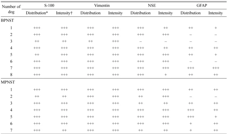
The results of immunohistochemistry are summarized in Table 3. The immunoreactivities for S-100 and vimentin were demonstrated in all PNSTs. All tumor cells showed uni- form and strong positive expression for vimentin and S-100 (Fig. 4). Overall intensity of positive expression for vimentin
was stronger than that of S-100. Immunoreactivity for NSE was found in 93.3% (14 cases) of PNSTs. However, expres- sion of GFAP, another neuronal marker, was observed in 73.3% (11 cases) of PNSTs. In addition, distribution and intensity of immuno-positive cells were lower than NSE application.
dog Distribution* Intensity† Distribution Intensity Distribution Intensity Distribution Intensity BPNST
1 +++ +++ +++ +++ +++ ++ ++ +
2 +++ +++ +++ +++ +++ +++ − −
3 ++ ++ ++ +++ – – – –
4 +++ +++ +++ +++ +++ ++ ++ ++
5 ++ +++ +++ +++ +++ +++ ++ +
6 +++ +++ +++ +++ +++ +++ – –
7 +++ +++ +++ +++ +++ +++ +++ +++
8 +++ +++ +++ +++ +++ + ++ ++
MPNST
1 +++ +++ +++ +++ +++ +++ ++ ++
2 ++ ++ +++ +++ ++ +++ – –
3 +++ +++ +++ +++ ++ ++ ++ ++
4 +++ +++ +++ +++ +++ +++ +++ ++
5 +++ +++ +++ +++ +++ +++ +++ +
6 +++ +++ +++ +++ +++ +++ + ++
7 +++ ++ +++ +++ ++ ++ + ++
* –: negative, +: focal/ individually, ++: multifocal, +++: diffuse. † –: absent, +: weak, ++: moderate, +++: strong.
Fig. 4. Tumor cells demonstrated positive reactions for vimentin (A) and S-100 (B), NSE (C), and GFAP (D). IHC, ×400.
Discussion
According to previous literatures, dogs with PNST are middle-aged or older, the mean age in dogs with benign PNSTs and malignant PNSTs is 7.3~8.3 years and 9 years, respectively [1]. The mean age of dogs with malignant PNST (12 years) was higher than those with benign PNST (8.1 years) in this study. This result is similar to other previous reports. There was no breed predilection in the occurrence of canine PNSTs in this study.
The sex distribution of this studied cases suggested a higher incidence in female (80%). According to previous sur- veys, male predominance (71.4% and 58.8%) was reported in two studies [2, 12] and female predominance was described in one study [5]. Interestingly, three female cases around mammary glands in this study were confirmed as malignant PNST.
Canine PNSTs are most commonly located on the trunk, distal legs or extremities, while feline tumors are mostly observed on the head and neck [8]. Of the 51 PNSTs reported previously in dogs, the limb affected was thoracic in 40 dogs and pelvic in 11 dogs [12]. In contrast, the prevalence of PNSTs in hindlimb was two times higher than those in fore- limb in this study. A retrospective evaluation of canine PNSTs demonstrated the location of tumor mass in each limb. Twenty-six cases had left-limb involvement; 19 had right-limb involvement and six cases showed bilateral loca- tions [2]. The limb affected in this study was left in 5 dogs and right in 4 dogs. The clinical signs in dogs with PNSTs were chronic progressive lameness in affected limb, muscu- lar atrophy, hyporeflexia, palpable mass, and pain on palpa- tion [2, 12]. Severe muscular deformity in hindlimb and neurologic signs were observed in two dogs with malignant PNST in present study. However, the dogs with benign PNSTs did not show any clinical signs without palpable mass on the skin.
Histopathologic finding of these PNSTs revealed different cellular components such as dense sheets of tumor cells arranged in interwoven bundles, streams, or concentric whorls pattern and low cellular density with small dark nuclei in loose fibrous stroma. In this study, local invasion and high mitotic figures in 100% of malignant PNSTs and multifocal necrosis and abnormal multi-nucleated giant cells in 71.4%
of cases were the most common histopathologic features.
Multifocal hemorrhage and cartilage formation were less fre- quent findings. In the dog, malignant PNSTs and hemangio- pericytomas (HP) have similar histomorphologic features [7, 8]. The majority of the cells of malignant PNSTs are charac- terized by interwoven bundles of small, wavy spindles cells with occasionally palisading and whorls. In contrast to hemangiopericytom, whorls are less prominent in PNSTs and are usually around collagenous bundles rather than blood capillaries. Small nerve fibers are occasionally found in or adjacent the PNSTs, this may be a useful indicator for the differentiation of PNST from hemangiopericytoma [5, 9].
Immunohistochemically, the various markers such as S- 100 protein, GFAP, NSE, and myelin basic protein have been widely used to identify cells of nerve sheath origins [10, 11].
This method also applies to differentiated PNSTs from other spindle cell tumors including HP. However immunohis- tochemical studies have not contributed to the definition of clear diagnostic criteria for PNSTs [5]. All tumor cases in this study had strong expression of vimentin with mesenchy- mal origin as previously described [4, 5, 11, 17]. In human medicine, the expression of S-100 proteins is used to differ- entiate between spindle cell tumors of neural and non-neural origins [15]. In veterinary medicine, lack of S-100 expres- sion is frequently described in canine malignant PNSTs [4, 5]. In contrast previous literatures, all PNSTs cases in this study were strong positive for vimentin and S-100 protein. A correlation between the lack of S-100 immuno-reactivity and malignancy has been postulated in malignant epithelioid PNSTs [6, 14]. These studies demonstrated that most spindle cells in epithelioid PNSTs expressed S-100 protein. But in the epithelioid cells, a weak or negative reaction to S-100 protein was observed. Both types of cell strongly expressed vimentin. Therefore the correlation was not as notable in canine malignant PNSTs, indicating that S-100 reactivity might depend on the origin of predominant tumor cells rather than malignancy as previously described [4]. Malignant PNSTs are possibly composed of various cell types such as Schwann cells, perineural cells, or perineural or endoneural fibroblasts and heterogenous differentiation [4, 5, 6]. Diffuse and strong S-100 reactivity in this study indicated that predominant tumor cells in PNSTs were derived from Schwann cells than other cell types.
Positive and negative immunolabelling have been described for both NSE and GFAP, and these procedures are therefore of little value in the diagnosis of malignant PNSTs [6, 14]. In this study, NSE expression (93.3%) in canine PNSTs was more frequently found than GFAP expression (73.3%). These observations are similar to other previous reports [11, 17].
Hence vimentin, S-100 and NSE immunohistochemical stain- ing may be useful to help confirm the diagnosis of canine PNSTs.
Complete excision of benign PNSTs is usually curative, but a few tumors will recur [7]. Malignant PNSTs com- monly recur after excision, but a metastasis is rare. Location of PNSTs may affect the ability to resect a tumor surgically [2]. Although there was no significant difference, PNST of nerve roots tended to have shorter relapse-free intervals and survival times than those of the brachial or lumbosacral plexus. In addition, rapid malignant progression of an ini- tially benign PNST in dog with unilateral limb enlargement was also reported [3]. According to literatures, the prognosis for dogs with PNSTs is poor, therefore early diagnosis and aggressive surgical resection at an early stage, possibly before the PNST has spread to involve the spinal canal or multiple nerves, may improve surgical treatment results [2].
Recently aggressive surgical resection with adjuvant metro-
Acknowledgments
This research was supported by the 2014 scientific promo- tion program funded by Jeju National University, Korea.
References
1. Bradley RL, Withrow SJ, Snyder SP. Nerve sheath tumors in the dog. J Am Anim Hosp Assoc 1982, 18, 915- 921
2. Brehm DM, Vite CH, Steinberg HS, Haviland J, van Winkle T. A retrospective evaluation of 51 cases of peripheral nerve sheath tumors in the dog. J Am Anim Hosp Assoc 1995, 31, 349-359.
3. Brower A, Salamat S, Crawford J, Manley P. Unilateral limb enlargement in a dog with a malignant peripheral nerve sheath tumor. Vet Pathol 2005, 42, 353-356.
4. Chijiwa K, Uchida K, Tateyama S. Immunohistochemical evaluation of canine peripheral nerve sheath tumors and other soft tissue sarcomas. Vet Pathol 2004, 41, 307-318.
5. Gaitero L, Añor S, Fondevila D, Pumarola M. Canine cutaneous spindle cell tumours with features of peripheral nerve sheath tumours: a histopathological and immunohis- tochemical study. J Comp Pathol 2008, 139, 16-23.
6. García P, Sánchez B, Sánchez MA, González M, Rollán E, Flores JM. Epithelioid malignant peripheral nerve sheath tumour in a dog. J Comp Pathol 2004, 131, 87-91.
7. Goldschmidt MH, Hendrick MJ. Tumors of the skin and soft tissues. In: Meuton DJ (ed.). Tumors in domestic animals. 4th ed. pp. 95-96, Iowa State University Press, Ames, 2002.
8. Gross TL, Ihrke PJ, Walder EJ, Affolter VK. Neural and perineural tumors. In: Skin Diseases of the Dog and Cat: Clinical and Histopathologic Diagnosis. 2nd ed. pp.
tumors of skin and soft tissues of domestic animals (WHO International Classification of Tumors of Domestic Animals).
2nd Ser. Vol. 2. pp. 25-27. Armed Forces Institute of Pathology, Washington, 1998.
10. Koestner A, Higgins RJ. Tumors of the nervous system.
In: Meuton DJ (ed.). Tumors in domestic animals. 4th ed.
pp. 731-735, Iowa State University Press, Ames, 2002.
11. Kuwamura M, Yamate J, Kotani T, Takeuchi T, Sakuma S. Canine peripheral nerve sheath tumor with eosinophilic cytoplasmic globules. Vet Pathol 1998, 35, 223-226.
12. Le Chevoir M, Thibaud JL, Labruyère J, Uriarte A, De Fornel-Thibaud P, Moissonnier P, Delisle F, Blot S.
Electrophysiological features in dogs with peripheral nerve sheath tumors: 51 cases (1993-2010). J Am Vet Med Assoc 2012, 241, 1194-1201.
13. Pakhrin B, Kang MS, Bae IH, Park MS, Jee H, You MH, Kim JH, Yoon BI, Choi YK, Kim DY. Retrospective study of canine cutaneous tumors in Korea. J Vet Sci 2007, 8, 229-236.
14. Pumarola M, Añor S, Borràs D, Ferrer I. Malignant epithelioid schwannoma affecting the trigeminal nerve of a dog. Vet Pathol 1996, 33, 434-436.
15. Scheithauer BW, Woodruff JM, Erlandson RA. Tumors of the peripheral nervous system. In: AFIP (ed.). Atlas of Tumor Pathology, 3rd Ser. Fascicle 24. pp. 105-176, pp.
303-372. Armed Forces Institute of Pathology, Washington, 1999.
16. Son J, Park S, Choi SH, Kim GH. Treatment of malignant peripheral nerve sheath tumor using surgery and metronomic chemotherapy in a dog. J Vet Clin 2011, 28, 310-313.
17. Youm SY, Shin SK, Kim TW, Bae DK, Kim K, Ahn B.
Immunohistochemical diagnosis of canine schwannoma and malignant schwannoma cases. J Biomed Res 2009, 10, 17-22.