• 검색 결과가 없습니다.

Effects of Natural Herb Mixture on Anti-atopic Dermatitis and Skin Regeneration

N/A
N/A
Protected

Academic year: 2021

Share "Effects of Natural Herb Mixture on Anti-atopic Dermatitis and Skin Regeneration"

Copied!
8
0
0

로드 중.... (전체 텍스트 보기)

전체 글

(1)

천연 소재 복합물이 항아토피 피부염 및 피부재생에 미치는 영향

김원식

1,2#

, 심부용

3

, 김동희

2,3*

1 : 신월맥 한의원, 2 : 대전대학교 한의과대학 병리학교실 3 : 대전대학교 난치성 면역질환의 동서생명의학연구 지역혁신센터

Effects of Natural Herb Mixture on Anti-atopic Dermatitis and Skin Regeneration Won-Sik Kim

1,2#

, Boo-Yong Sim

3

, Dong-Hee Kim

2,3*

1 : Sinwolmaek Korean Medicine Clinic

2 : Department of Pathology, College of Oriental Medicine, Daejeon University 3 : Traditional and Biomedical Research Center(TBRC-RIC), Daejeon University

ABSTRACT

Objectives : This study aims to evaluate the effects of natural herb mixutre (NHM) on atopic dermatitis and skin regeneration using in vivo test.

Methods : NHM was prepared with DW. 25% of NHM was applied to skin lesion, where atopic dermatitis was induced by DNCB in NC/Nga mice. The levels of cytokines (IL-4, IL-5, IL-6, IL-13, TNF-a, and IFN-γ), and IgE in serum were measured by Luminex. Immune cells (WBC, eosinophil, lymphocyte, and monocyte) in blood were counted by coulter counter. The gross investigation of atopic dermatitis index score test were performed during the NHM treatment period. Also, the histopathological change of dorsal skin was observed by H&E and M&T staining.

Results : NHM showed the levels of IL-4, IL-5, IL-6, IL-13, IgE, WBC, eosinophil, lymphocyte, and monocyte in serum or blood were significantly decreased. On the contrary, the productions of FGF, and VEGF were increased in the serum. Also, atopic dermatitis index score in NHM-treated mice were observed in the similar levels to those of normal group. Histological examination demonstrated that NHM suppressed immune cell infiltration and thickening of epidermis, meanwhile the extraction induced collagen production in the dorsal skin.

Conclusion : This study demonstrated that NHM is appeared to be effective on atopic dermatitis and skin regeneration efficacy based on the observations with hematologic, gross, and histologic examinations. Therefore, we suggest that NHM could be effectively used as an external therapeutics against atopic dermatitis and a consequence skin damage.

1)

Key words : atopic dermatitis, natural herb mixture, NC/Nga, skin regeneration, Th2 cytokine

Ⅰ. 서 론

산업화에 따른 오염물질 배출량의 증가, 미세먼지와 중국발 황사 등으로 인해 대기오염에 대한 문제가 사회적으로 이슈화 되면서 이로 인해 발생되는 호흡기계 및 심혈관, 피부 질환 등에 대한 관심도가 증가하고 있다

1)

. 특히, 다양한 연구를 통해 환경오염물질에 의해 연관성이 있다고 규명되고 있는 아토피

피부염(Atopic dermatitis, AD)은 국민건강보험공단 통계 자료에 따르면 2015년도 기준 AD 치료 인원이 1년 중 미세먼지와 황사가 극성을 부리는 5월에 약 15만 명으로 집계되어 11월의 약 11만 명에 비해 27%가량 높게 나타나

2)

, 대기오염이 심할 수록 AD 발병이 높아진다는 연구결과를 더욱 뒷받침 하고 있다.

한의학에서 AD는 ‘浸淫瘡’, ‘奶癬’, ‘旋耳風’, ‘胎瘡’, ‘四彎風’,

‘胎熱’, ‘濕瘡’, ‘濕疹’ 등의 범주에 속한다. ≪醫宗金鑑·外科

*Corresponding author : Dong-Hee Kim. Daejeon University, Korea.

·Tel : +82-42-280-2636 ·E-mail : [email protected]

#First Author : Won-Sik Kim. Sinwolmaek Korean Medicine Clinic, Korea.

·Tel : +82-70-7882-3007 ·E-mail : [email protected]

·Received : 3 October 2017 ·Revised : 1 November 2017 ·Accepted : 15 November 2017

(2)

心法≫

3)

에서는 ‘四彎風’을 기술하면서 “彎曲된 피부 부위에 발생하고 瘙痒症이 매우 심하며 긁었을 때 滲出物이 나와 마치 濕癬과 같다”라고 기재되어 있어 현대 AD와 매우 유사한 증 상이 표현됨으로써 최근 이 같은 치료를 중심으로 실험 및 임상 연구가 활발하게 진행되고 있다

3)

.

현재 AD에 사용되어지는 연고로는 hydrocortisone, dexamethason 등의 국소용 코르티코스테로이드 제제 (topical corticosteroid, TCS)와 tacrolimus 연고제와 pimecrolimus 크림제제 등의 칼시네우린 억제제 (topical calcineurin inhibitor, TCI) 등은 성인과 아동 모두에게 효과적으로 사용이 가능하다는 장점이 있으나, 피부의 과민반응을 증가시키거나 통증, 낮은 빈도로 피부암과 림프종과 같은 악성종양이 발생 하는 부작용이 보고되고 있어 이를 보완할 수 있는 효과적인 외치제 개발이 필요한 실정이다

4-9)

.

현재 한의원에서는 AD 치료를 위한 外治法으로는 약침을 주사하거나 한약을 직접 피부에 도포하는 방법을 활용하고 있 는데, 이는 AD 자체가 다양한 원인에 의한 복잡한 병리기전 으로 인한 것으로 효과적인 內·外治의 병용은 임상에서 빠른 회복을 기대할 수 있다. 특히 미세먼지와 황사를 구성하는 일부 물질은 피부에 가려움, 따가움, 발진, 발열, 부종 등 피부에 직접적인 손상을 주기에 안전하고 효과적인 한의학적 外治法 개발은 2차적 감염으로 인한 증상 완화를 위해 매우 중요하다.

본 연구에서 사용한 천연 소재 복합물은 문헌에서 風熱로 인한 각종 피부 질환과 瘙痒感, 發疹이 심한 實證에 활용되는 淸熱, 濕疹, 消腫, 解毒 등의 효능에 대해 기록되어 있고 이에 대해 실험적으로 입증된 蛇莓

10,11)

, 紫草

12-14)

, 苦蔘

15-17)

, 綠

18,19)

, 松脂

20)

, 枳實

21-23)

등 천연물 소재를 혼합한 복합물을

통해 AD와 AD로 인해 발생되는 2차적 피부 질환 개선에 대해

in-vivo

실험을 적용하였고 이에 대한 유의한 결과를 얻었기에 보고하는 바이다.

Ⅱ. 재료 및 방법

1. 약재

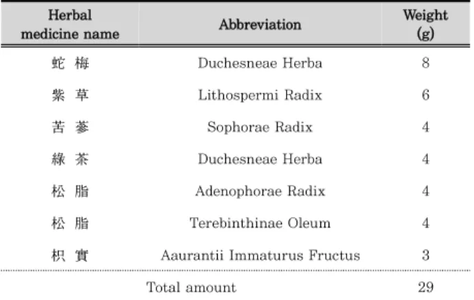
본 실험에 사용한 천연 소재 복합물의 구성 약재들은 ㈜옴 니허브에서 국산 약재를 구입하였고, 대전대학교에서 정선 후 사용하였고, 그 내용과 분량 (1첩)은 다음과 같다(Table 1).

Herbal

medicine name Abbreviation Weight

(g)

蛇 梅 Duchesneae Herba 8

紫 草 Lithospermi Radix 6

苦 蔘 Sophorae Radix 4

綠 茶 Duchesneae Herba 4

松 脂 Adenophorae Radix 4

松 脂 Terebinthinae Oleum 4

枳 實 Aaurantii Immaturus Fructus 3

Total amount 29

Table 1. The Composition of Natural Herb Mixture

2. 동물 및 사료

실험동물인 수컷 4주령의 NC/Nga 마우스 (16 ∼ 22g)는

㈜라온바이오에서 공급 받아 실험 당일까지 고형사료 (Altromin CO., Germany)와 물을 충분히 공급하고 온도 22

± 2℃, 습도 55 ± 15%, 12시간-12시간 (light-dark cycle)의 환경에서 2주간 적응시킨 후 실험에 사용하였다. 본 실험은 대전대학교 동물실험윤리 위원회의 승인 (동물사용 윤리위원회 승인 번호–DJUARB 2017-016)을 받아 동물윤리준칙에 의거 하여 실험하였다.

3. 시약 및 기기

사용된 시약은 2,4-Dinitrochlorobenzene (DNCB : Sigma Co., U.S.A.), Acetone (Duksan, Korea), Olive oil (Sigma Co., U.S.A.) Ethyl ether (Sigma Co., U.S.A.), Formaldehyde (Duksan, Korea), Mouse cytokine milliplex map immunoassay kit (Millipore Co., U.S.A.), Mouse IgE kit (Millipore Co., U.S.A.), Mouse/Rat FGF basic ELISA kit (R&D system, U.S.A.), Mouse VEGF ELISA kit (R&D system, U.S.A.) 등을 사용하였으며, 기기는 rotary vacuum evaporator(Büchi B-480 Co., Switzerland), freeze dryer (EYELA FDU-540 Co., Japan), centrifuge (Sigma Co., U.S.A.), Luminex (Millipore Co., U.S.A.), ELISA reader (Molecular Devices Co., U.S.A.), Light Microscope (Carl Zeiss, Co., Germany) 등을 사용하였다.

4. 시료 제조

천연 소재 복합물 2첩 (58 g)에 증류수 1,500 ㎖를 넣고 3 시간 동안 환류추출 후 여과액을 얻어 rotary vacuum evaporator에서 감압 농축하였다. 농축된 용액을 freeze dryer로 동결 건조하여 분말 13.2 g을 얻었으며, 얻어진 분말은 초저온 냉동고 (-80℃)에서 보관하여 사용하였다.

5. AD 병태 모델 제작 및 시료 처리

AD 병태 모델을 제작하기 위해 6주령이 된 NC/Nga 마우 스의 귀 하단부에서부터 꼬리 상단부까지 전체를 제모하고 24 시간 방치 후 1%의 DNCB 용액 (아세톤․올리브오일 = 3 : 1) 150 ㎕를 제모 부위에 도포하고, 3일 후 2차 도포하였다. 1차 도포 후 7일차부터 실험 종료 시까지 1주일에 3회 DNCB 용액 100 ㎕를 지속적으로 도포하였다.

실험 그룹은 AD를 유발시키지 않은 정상군 (이하, Normal),

AD 유발 후 증류수만을 도포하는 대조군 (이하, Control),

0.5% dexamethasone를 도포하는 양성대조군 (이하, Dexa)과

25% 농도로 천연 소재 복합물을 도포하는 실험군 (이하, NHM)

등 그룹 당 6마리씩 총 4개의 그룹으로 나누어 매일 오후 3시에

실험을 진행하였다.

(3)

6. 혈청 내 사이토카인 및 면역 글로불린 생성량 측정

실험 종료 후 ether로 마취한 상태에서 심장 천자법을 이용 하여 채혈한 다음 3,000 rpm에서 20분간 원심 분리하여 혈청을 분리하였다. 분리된 혈청을 가지고 IL-4, IL-5, IL-6, IL-13, TNF-α, IFN-γ, IgE 생성량을 Mouse cytokine milliplex map immunoassay kit과 Mouse immunoglobulin kit을 이용하여 Luminex로 측정하였다.

7. 혈액 내 면역세포 수 측정

실험 종료 후 ether로 마취한 상태에서 심장 천자법을 이용 하여 채혈한 다음 전혈 250 ㎕를 K2 EDTA tube에 담아 케 이피엔티 (KP&T, Korea)에 분석 의뢰하여 백혈구 (WBC), 호산구 (Eosinophil), 림프구 (Lymphocyte), 단핵구 (Monocyte) 수를 측정하였다.

8. 혈청 내 피부재생 인자 측정

실험 종료 후 ether로 마취한 상태에서 심장 천자법을 이용 하여 채혈한 다음 3,000 rpm에서 20분간 원심 분리하여 혈 청을 분리하였다. 분리된 혈청을 가지고 FGF 및 VEGF 생성 량을 Mouse/Rat FGF basic ELISA kit과 Mouse VEGF ELISA kit을 이용하여 ELISA reader로 측정하였다.

9. 조직학적 검사

배부의 피부를 떼어내어 각 실험군 별로 적출한 조직은 10%

중성 포르말린에 48시간 고정하여 고정이 완료된 조직들을 흐르는 수돗물에서 12시간 수세한 뒤 조직 내의 고정액을 완전 제거하였다. 조직의 탈수를 위해 60%에서부터 100% 알코올에 이르기까지 농도 상승 순으로 탈수하고, xylene에 투명과정을 거친 다음 파라핀 블럭을 제작하였다. 제작된 블럭은 박편절 단기 (microtome)를 이용해 3∼4 ㎛ 두께로 절편을 만들어 탈 파라핀 및 함수과정을 거친 다음 hematoxyline-eosin (H&E) 염색과 Masson-trichrome (M&T) 염색을 실시하여 광학현 미경상에서 관찰 및 사진 촬영 하였다.

10. 육안 및 관능 평가

AD 유발 후 실험 시작 9주령부터 실험 종료 13주령까지 NC/Nga 마우스의 임상적 육안 평가법으로 관능 평가를 실시 하였다. 평가 항목은 홍반 (Erythema), 가려움과 건조 피부 (Pruritus & Dry skin), 부종과 혈종 (Edema & Hematoma), 짓무름 (Excoriation), 태선화 (Lichenification) 등으로 5가 지이다. 각각의 항목은 없음 (0), 약함 (1), 중증도 (2), 심함 (3) 으로 채점하였다.

11. 통계처리

본 연구의 실험 결과는 평균값±표준 편차 (mean±S.D.)로 표시하였다. 각 처리군의 비교는 one-way analysis of variance (ANOVA) 방법을 이용하였고, Student’s t-test를 사용하여 통계적 유의성을 검증하였으며, 정상군과 비교한 대 조군의 결과는

###

p<0.001,

##

p<0.01,

#

p<0.05로 대조군과 비교한 실험군의 결과는

***

p<0.001,

**

p<0.01,

*

p<0.05로 표기하였다.

Ⅲ. 결 과

1. 혈청 내 사이토카인 및 IgE 생성량에 미치는 영향 혈청 내 사이토카인 및 IgE 생성량을 측정한 결과, 대조군은 정상군에 비해 모든 결과에서 유의성 있는 증가가 나타났다.

반면, NHM 도포군은 대조군과 비교하였을 때 사이토카인 IL-4 (56.24%,

**

p<0.01) 및 IL-5 (57.36%,

***

p<0.001), IL-6 (20.53%,

*

p<0.05), IL-13 (10.59%,

*

p<0.05), TNF-α (29.97%,

**

p<0.01), IFN-γ (13.52%,

*

p<0.05) 생성량과 IgE (30.15%,

***

p<0.001) 생성량을 유의성 있게 감소시켰 다(Table 2). 이와 같은 결과는 양성대조군인 Dexa 도포군의 IL-4 및 IL-5, IL-6, TNF-α, IgE 생성량에 대한 감소 효 능이 NHM 도포군에서도 나타났으며, Dexa 도포군에서 유의성 있는 감소 효능이 나타나지 않은 IL-13과 IFN-γ에서도 유 의성 있는 결과가 도출되었다. 특히, AD의 표적 인자인 IgE 생성량을 큰 폭으로 감소시키는 것이 확인되었다(Table 2).

Cytokine and IgE level (pg/㎖)

Groups

Normal Control Dexa NHM

IL-4 17.2±4.6 99.0±6.0### 35.1±3.4** 43.3±8.8**

IL-5 69.7±10.9 814.1±138.3### 362.6±8.1*** 347.2±28.4***

IL-6 121.5±4.1 1051.6±116.1### 863.1±691.7* 835.8±172.6*

IL-13 397.1±28.5 2210.6±237.9### 2137.7±496.2 1976.6±328.4*

TNF-α 351.5±24.2 1954.1±187.5### 1180.6±89.5** 1368.4±266.7**

IFN-γ 912.5±14.6 1678.8±158.3## 1501.6±165.8 1451.8±247.3*

IgE 181.6±31.3 2272.6±820.5### 1861.3±936.8** 1587.3±447.9***

Luminex assays were used to measure serum levels of cytokine and IgE. The results are mean±standard deviation (SD) for six mice per group (###p<0.001, ##p<0.01 compared to Normal; ***p<0.001, **p<0.01, *p<0.05 compared to Control).

Table 2. The effects of NHM on serum cytokine and IgE in a DNCB-induced mouse model of atopic dermatitis

(4)

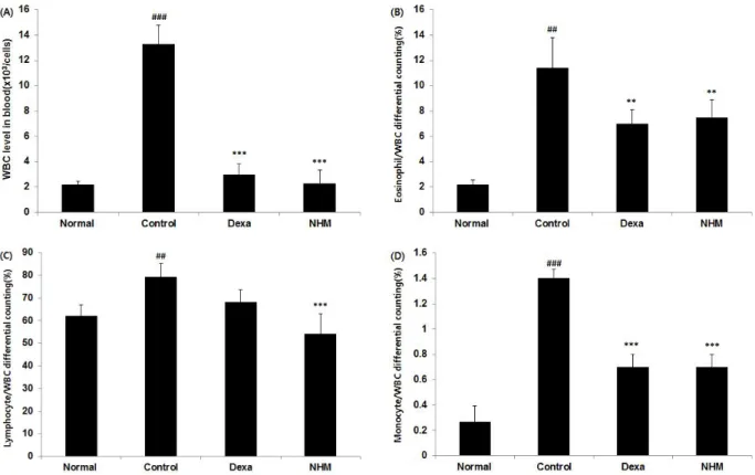
2. 혈액 내 면역세포 수에 미치는 영향

혈액 내 면역세포 수를 측정한 결과, 대조군은 정상군에 비해 모든 결과에서 유의성 있는 증가가 나타났다. 반면, NHM 도 포군은 대조군과 비교하였을 때 백혈구 수 (82.66%,

***

p<0.001) (Fig. 1A) 및 백혈구 내 호산구 (34.21%,

**

p<0.01)(Fig. 1B), 림프구 (31.78%,

***

p<0.001)(Fig. 1C), 단핵구 (50.00%,

***

p<0.001)(Fig. 1D) 수를 유의성 있게 감소시켰다(Fig. 1).

이와 같은 결과는 양성대조군인 Dexa 도포군의 백혈구 수 및 백혈구 내 호산구, 단핵구 수에 대한 감소 효능이 NHM 도포 군에서도 나타났으며, Dexa 도포군에서 유의성 있는 감소 효 능이 나타나지 않은 백혈구 내 림프구 수를 NHM 도포군에서 유의성 있게 감소시키는 것이 확인되었다(Fig. 2C).

Fig. 1. The effects of NHM on white blood cell (WBC) (A), eosinophil (B), lymphocyte (C), monocyte (D) in a DNCB-induced mouse model of atopic dermatitis. The results are mean±standard deviation (SD) for six mice per group (###p<0.001, ##p<0.01 compared to Normal; ***p<0.001,

**p<0.01 compared to Control).

3. 혈액 내 피부 재생 인자에 미치는 영향

혈액 내 피부 재생 인자를 측정한 결과, 대조군은 정상군에 비해 FGF 생성량은 유의성 있는 증가가 나타났으나, VEGF 생성량에서는 차이가 나타나지 않았다. 반면, NHM 도포군은 대조군과 비교하였을 때 FGF (88.71%,

***

p<0.001)(Fig. 2A)

및 VEGF (34.91%,

***

p<0.001)(Fig. 2B) 생성량를 유의성 있게 증가시켰다(Fig. 2). 이와 같은 결과는 양성대조군인 Dexa 도포군이 피부 재생 인자 중 FGF 생성량만을 유의성 있게 증가 시켰으나, NHM 도포군은 FGF 생성량과 VEGF 생성량 역시 유의성 있게 증가시키는 것이 확인되었다(Fig. 2B).

Fig. 2. The effects of NHM on fibroblast growth factor (FGF) (A), vescular endothelial growth factor (VEGF) (B) in a DNCB-induced mouse model of atopic dermatitis. ELISA assays were used to measure serum levels of FGF and VEGF. The results are mean± standard deviation (SD) for six mice per group (###p<0.001 compared to Normal; ***p<0.001 compared to Control).

(5)

4. 조직변화에 미치는 영향

등 조직 변화를 확인한 결과, H&E 염색(Fig. 3A)에서 대조 군은 정상군에 비하여 표피층(Epidermis, E-붉은 화살표)가 두꺼워지고 세포의 침윤(Inflammatory, I-붉은 사각형)으로 인한 세포의 밀도가 높게 나타났으며, M&T 염색(Fig. 3B)에 서는 진피층(Dermis, D-붉은 화살표)이 감소하고 콜라겐 (Collagen, C-노란 사각형, 담청색 염색)이 생성을 확인할 수 있었다. 반면, NHM 도포군은 H&E 염색에서 대조군에 비해 표피층이 두께가 줄어들고 세포 침윤이 적었으며, 신생 혈관 (Blood vessel, V-붉은 다이아몬드)과 피지선(Sebaceous gland, G-붉은 원형)이 확인되었고 M&T 염색에서도 대조군에 비해 진피층의 두께가 넓으며, 콜라겐 형성에 따른 섬유질이 광범위 하게 많은 것이 확인되었다(Fig. 3). 이와 같은 결과는 양성대조군인 Dexa 도포군과 NHM 도포군 모두 대조군에 비해 조직변화를 정상군에 가깝게 회복을 시키고 있음이 확인되었 으며, 차이가 있다면 Dexa 도포군은 H&E 염색 시 모낭(Hair follicle, F-노란 원형)이 관찰되었다(Fig. 3A).

Fig. 3. Histopathological effects of NHM on dorsal skin in a DNCB-induced mouse model of atopic dermatitis. Representative histological images of dorsal skin tissues were stained using hematoxylin and eosin (A) or Masson-trichrime (B) (magnification, 200×).
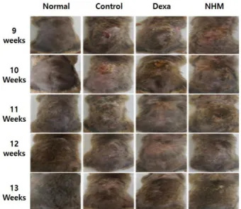
5. 육안 및 관능 평가

AD 유발 후 실험 시작 전인 9주령에서 대조군과 양성대조군, 실험군은 정상군에 비해 홍반, 태선화, 부종과 짓무름 등이 심한 것이 확인되었다(Fig. 4). 이후 시간이 지속될수록 양성 대조군인 Dexa 도포군과 NHM 도포군은 정상군에 비해 건조 함과 부종이 남아있기는 했으나 대조군에 비해 빠른 회복을 보여 실험 종료 주인 13주령에서는 정상군과 가까운 회복이 되었음을 확인되었다(Fig. 4). 이와 같은 결과는 피부염 지수 (Index score)에 반영되어 대조군에 비해 종료 주인 13주차에 약 3점 이상의 차이가 나타났다(Fig. 5). 특히, NHM 도포군은 양성대조군에 비해 피부염 지수에서 11주령 차에서 AD 증상을 빠르게 완화시키는 것으로 확인되었다.

Fig. 4. The effects of NHM on skin lesions typical in a DNCB- induced mouse model of atopic dermatitis. Images of skin lesions were taken in the 9 to 13 weeks.

Fig. 5. The effects of NHM on index score in a DNCB-induced mouse model of atopic dermatitis. The scoring represents a clinical index that was evaluated each week from 9 to 13 weeks. Clinical skin index of dermatitis was defined asthesum of the individual scores graded as 0(none), 1(mild), 2(moderate), 3(severe) foreach offivesignsand symptoms(Erythema, Pruritus & Dry skin, Edema &

Hematoma, Excoriation, Lichenification) ; Symptoms were evaluated by skin dryness, eruption and wound on the dorsal skin.

Ⅳ. 고 찰

기존 AD 환자들은 신속한 치료가 가능한 內治法과 서양의학

치료제를 선호함에 따라 활용 가치가 낮아져 사장되어 지던

外治法이 최근 들어 재조명 받고 있다. 이는 AD 치료제로 사용

되던 항히스타민, 스테로이드 등의 처리가 AD의 주된 증상인

가려움증을 억제하기엔 충분하지 못하고 잦은 부작용으로 인해

장기적으로 사용할 수 없는 문제점을 보완하고자 천연물을 활

용한 치료에 대한 필요성이 대두되고 한의학계에서도 안전하고

효과적인 AD 외용제 치료에 대한 연구가 활발히 진행된 결과로,

스프레이 및 입욕제, 크림 등 다양한 형태로 활용되고 있는

추세이다

24-26)

.

(6)

본 연구에서 사용한 천연 소재 복합물 (Natural herb mixture, NHM)은 문헌에서 AD와 유사한 병증의 피부 질환과 실험적으로 효능이 입증된 천연물 소재를 혼합한 복합물을 통해 항AD 효능과 더불어 AD로 인해 발생되는 2차적 피부 질환 개선에 대한 평가를 위해 NC/Nga 마우스에게 DNCB를 처리 하여 그 효능을 확인하였다.

AD의 다양한 원인 중 대표적인 Th1 세포와 Th2 세포의 불균형에 의한 면역학적 요인은 급성 피부 병변에서 IL-4, IL-5, IL-13 등의 Th2 사이토카인이 과발현 된다

27)

. 이처럼 Th2 사이토카인은 선천 면역을 저하시키고 알러지를 유발할 뿐만 아니라 직접적으로 피부 장벽 기능을 저하시킨다고 보고 되고 있다

28)

. 그 예로 Th2 사이토카인 중 IL-4는 피부 장벽 손상 후 회복을 저해시키며, 필라그린 (filaggrin)의 발현과 세라마이드 (ceramide) 합성을 억제하며

29,30)

, 각질층에서 세 라마이드는 IL-6에 의해 염증 반응을 더욱 심화시키고 TNF-α 에 의해 전신반응으로 유도되게 된다

27)

. 또한, 과도하게 생성된 Th2 사이토카인은 이소 타입 전환 (isotype- switching)을 통해 AD 병변에 대한 판단이 되는 면역글로불린 E (immunoglobulin E, IgE)로 전환되며

31)

, Th1/Th2를 분 비하는 IFN-γ 의 증가는 염증 부위에 대한 비만 세포의 침윤과 밀접하게 관련되어 있다

32)

. 본 연구 결과, NHM 도포군은 대 조군에 비해 Th2 사이토카인과 IgE 생성량을 유의성 있게 감 소시켰다. 특히, NHM 도포는 감소 효능이 양성대조군과 비 슷한 것으로 확인되었고 양성대조군에서 유의적 감소가 나타 나지 않은 IL-13과 IFN-γ 역시 유의적으로 감소시킴에 따라 AD 치료뿐만 아닌 Th2 사이토카인 상승으로 인해 발생하는 천식, 비염과 같은 알러지 질환에 활용될 수 있는 가능성을 시사하고 있다.

이와 같은 결과는 면역세포 수를 측정한 결과에 반영되어 인체 내 바이러스, 세균, 기생충 등의 침투 시 탐식작용, 항원 제시, 이물질 인식, 항체생산 등의 기능을 하는 백혈구 수를 유의성 있게 감소시켰다

33)

. 또한, 백혈구 중 제I형 알러지 질 환인 AD에 관여하는 호산구 (eosinophil)와 식균 작용 및 염 증성 사이토카인을 생성하는 대식세포 (macrophage), 획득 면역의 중심적 역할을 하는 림프구 (lymphocyte)의 유의적인 감소가 나타났다

34)

. 이는 백혈구에서 분비되는 단백활성 물질인 사이토카인 감소 결과와 부합되어 NHM 도포가 면역·알러지 질환인 AD에 효능이 있음이 검증되었다.

NHM 도포가 항AD 효능이 입증됨에 따라 AD의 다양한 임상 소견 중 긁는 행위로 인해 AD 발병 후 상처가 지속되고 회복 후에도 흉터로 이어지는 점에 착안하여 효과적이고 신속한 피 부재생에 대한 여부를 확인하고자 하였다.

상처 치유 과정에서 약 6일 가량 호중구 (neutrophil)와 대 식세포 등이 상처 주위로 몰리게 되는 염증 단계가 종료되어 증식단계로 이행되면 혈관 형성 (angiogenesis), 섬유화 (fibroplasia) 등 상처가 발생한 조직이 재생되고 섬유조직이 교원질 (collagen) 층으로 바뀌며 재형성 단계를 통해 종료된

35-37)

. 이처럼 상처 치유단계에서는 혈관 형성의 모든 단계에

작용하는 VEGF와 혈관 형성의 주된 촉진 인자로 알려진 FGF가 중요하다고 할 수 있다. 본 연구 결과, NHM 도포군은 피부재생인자인 FGF와 VEGF 생성을 대조군에 비해 유의성 있게 증가시켰다. 이와 같은 결과는 실험 종료 후 H&E와

M&T 염색을 통한 조직변화에 반영되어 NHM 도포군은 대조 군에 비해 신생 혈관의 형성과 염증 세포의 침윤이 적고 표피 및 진피층의 정상화와 더불어 교원질 형성이 활발하게 진행되 었고 이는 육안, 관능 평가 결과로 이어져 NHM 도포는 단순 AD 치료제가 아닌 AD로 인해 발생되는 2차적 손상에 대해서도 빠른 회복을 할 수 있음을 보여주고 있다.

이상의 결과를 종합해 볼 때, NHM 도포는 AD 동물 모델 에서 AD 원인과 특징이라 할 수 있는 사이토카인과 IgE, 면 역세포 수의 생성량 감소와 동시에 피부재생에 대한 대표적 인자를 증가시킴에 따라 면역반응과 염증을 통한 AD 치료제 뿐만 아니라 AD에 의한 2차 질환까지도 개선될 수 있음이 증 명되었다. 특히, 양성대조군에서 효능이 나타나지 않은 IL-13 및 IFN-γ , VEGF 생성량의 결과가 NHM 도포군에서 유의적 으로 효능이 나타남에 따라 NHM 도포가 기존 약물에 대해 보완할 수 있음을 보여주고 있다. 추후 본 연구결과를 바탕으로 임상 연구가 뒷받침 되고 피부 재생인자 중 transforming growth factor (TGF)-β , matrix metalloproteinases (MMPs) 등에 대한 연구 결과가 보강된다면 안전하고 효과적인 AD 환자 에게 다양하게 활용될 수 있는 외치제임을 시사하고 있다.

Ⅴ. 결 론

본 연구에서는 천연 소재 복합물 (NHM)이 아토피피부염 개선과 피부재생에 대한 효과를 객관적으로 검증하고자 아토 피피부염을 유발한 동물 모델을 통해 혈액학적, 조직학적, 육 안 및 관능평가 등을 실시한 결과 다음과 같은 결론을 얻었다.

1. NHM 도포군은 대조군에 비해 혈청 내 사이토카인 IL-4, IL-5, IL-6, IL-13, TNF-α , IFN-γ 등의 사이토카 인 및 IgE 생성량을 유의성 있게 감소시켰다.

2. NHM 도포군은 대조군에 비해 혈액 내 면역세포 중 백 혈구 및 백혈구 내 호산구, 대식세포, 림프구 수를 유의성 있게 감소시켰다.

3. NHM 도포군은 대조군에 비해 혈청 내 FGF 및 VEGF 생성량을 유의성 있게 증가시켰다.

4. NHM 도포군은 대조군에 비해 H&E 및 M&T 염색을 통해 신생혈관의 형성과 염증 세포의 침윤 적고 표피 및 진피층의 정상화와 더불어 교원질 형성이 활발하게 진행 되었다.

5. NHM 도포군은 대조군에 비해 육안 평가를 통해 홍반, 부종, 건조함 등의 피부 개선이 빠르게 진행되었으며, 관능 평가인 피부염 지수 역시 감소시켰다.

이상의 결과들로 보아 NHM 도포는 아토피피부염뿐만이

아닌 아토피피부염에 의한 2차적 손상까지 개선시킬 수 있음이

실험적으로 규명되었다. 보다 다양한 기전 확인을 통해 보완

할 점은 있으나 이 같은 유의적인 전임상 단계의 결과는 외치

제로써 폭넓게 활용될 수 있음을 제시해 주고 있다.

(7)

감사의 글

이 논문은 2017학년도 대전대학교 교내학술연구비 지원에 의해 연구되었음.

References

1. Morales-Suárez-Varela M, Gallardo-Juan A, García- Marcos L, Gimeno-Clemente N, López-Silvarrey- Varela Á, Miner-Canflanca I, Batlles-Garrido J, Blanco-Quiros A, Busquets-Monge RM, Domínguez- Aurrecoechea B, Arnedo-Pena A. The impact of atmospheric pollutants on the prevalence of atopic eczema in 6-7-year-old schoolchildren in Spain;

ISAAC Phase III. Iranian Journal of Allergy, Asthma and Immunology. 2013 ; 12(3) : 220.

2. Korean statistical information sevice. Statistic.

Available from : URL : http://kosis.kr/statisticsList/

statisticsList_01List.jsp?vwcd=MT_ZTITLE&parentI d=D#SubCont

3. Wú-qiān. yī-zōng-jīn-jiàn. China : Rénmín- wèishēng publisher. 1963. p 1890.

4. Lassus. Clinical comparison of alclometasone dipropionate cream 0.05% with hydrocortisone butyrate cream 0.1% in the treatment of atopic dermatitis in children. J Int Med Res. 1983 ; 11(5) : 315-9.

5. Callen J, Chamlin S, Eichenfield LF, Ellis C, Girardi M, Goldfarb M, Hanifin J, Lee P, Margolis D, Paller AS, Piacquadio D, A systematic review of the safety of topical therapies for atopic dermatitis. British Journal of Dermatology. 2007 ; 156(2) : 203-21.

6. Pariser D. Topical corticosteroids and topical calcineurin inhibitors in the treatment of atopic dermatitis: focus on percutaneous absorption. Am J Ther. 2009 ; 16(3) : 264-73.

7. Breuer K, Werfel T, Kapp A. Safety and efficacy of topical calcineurin inhibitors in the treatment of childhood atopic dermatitis. American journal of clinical dermatology. 2005 ; 6(2) : 65-77.

8. Frankel HC, Qureshi AA. Comparative effectiveness of topical calcineurin inhibitors in adult patients with atopic dermatitis. American journal of clinical dermatology. 2012 ; 13(2) : 113-23.

9. Tennis P, Gelfand JM, Rothman KJ. Evaluation of cancer risk related to atopic dermatitis and use of topical calcineurin inhibitors. British Journal of Dermatology. 2011 ; 165(3) : 465-73.

10. Lee DJ, Jeon IH, Kim HS, Cho IY, Jang SI.

Antioxidative and anti-inflammatory effect of ethanol extract from duchesnea chrysantha. Journal

of Physiology & Pathology in Korean Medicine.

2012 ; 26(1) : 59-66.

11. Lee DJ, Cho IY, Jang SI. Anti-allergy Effect of Ethanol Extract from Duchesnea chrysantha. Journal of Physiology & Pathology in Korean Medicine.

2012; 26(6) : 902-7.

12. Kang SH, Kim GJ. Effects of Lithospermum erythrorhizon on the cytokine gene expression in human keratinocytes. The Journal of Korean Medicine Ophthalmology and Otolaryngology and Dermatology. 2013 ; 26(1) : 50-62.

13. Choi SB, Bae GS, Jo IJ, Park KC, Seo SH, Kim DG, Shin JY, Gwak TS, Lee JH, Lee GS, Park SJ.

The anti-inflammatory effect of Lithospermum Erythrorhizon on lipopolysaccharide-induced inflammatory response in RAW 264.7 cells. The Korea Journal of Herbology. 2013 ; 28(2) : 67-73.

14. Kim SH, Jung HS, Lee JY, Kim DG, Cho BG. Effect of Lithospermum erythrorhizon on the atopic dermatitis. The Journal of Pediatrics of Korean Medicine. 2004 ; 18(1) : 63-75.

15. Lee JY, Park SS. The Effect of Allergic Inflamation by Sophora Flavescens Aiton Extract Ion Through Inhibition of the NFĸB, JNK and p38 Pathway. Journal of Sasang Constitutional Medicine.

2009 ; 21(1) : 139-49.

16. Jeong SI, Choi BM, Yun YG, Lee JW, Jang SI. A noble therapeutic approach of atopic dermatitis by development of Th2 chemokine inhibitors from natural products: inhibitory effect of Sophora flavescens extract in atopic dermatitis model mice, NC/Nga. Herbal Formula Science. 2009 ; 17(1) : 141-51.

17. Yu ML, Koo YS, Kim DH. Effects of Sophorae Radix (SFR) on Allergic Inflammation related Atopy Dermatitis. The Journal of Daejeon Oriental Medicine.

2006 ; 15(2) : 135-48.

18. Katiyar SK, Ahmad N, Mukhtar H. Green tea and skin. Archives of Dermatology. 2000 ; 136(8) : 989-94.

19. Katiyar SK. Skin photoprotection by green tea:

antioxidant and immunomodulatory effects. Current Drug Targets-Immune, Endocrine & Metabolic Disorders. 2003 ; 3(3) : 234-42.

20. Seo YA. Analgesic and anti-inflammatory activity of Resina Pini. Natural Product Sciences. 2007 ; 13(4) : 347-54.

21. Jung SA, Choi YY, Yang WM. Anti-atopic dermatitis effects of Rafinesque via regulation of immune response and nerve growth factor. Journal of Korean Medicine. 2016 ; 37(1) : 10-20.

22. Park JM, Chae JW. Effects of Aurantii Immaturus

(8)

Fructus (AI) on Atopic Dermatitis (AD) Induced by DNCB in Mice. The Journal of Pediatrics of Korean Medicine. 2015 ; 29(1) : 27-43.

23. Kim HK, Lee EJ, Kweon YT, Hwang KH, Joo HH, Song BK. The Study on the antiallergic Action of Poncirus trifoliata. The Journal of Korean Oriental Internal Medicine. 2000 ; 21(1) : 156-61.

24. Koblenzer CS. Itching and the Atopic Skin. J.

Allerrgy Clin. Immun. 1999 ; 104(3) : S109-13.

25. Mihara K, Kuratani K, Matusui T, Yokota K. Vital Role of the Itch-scratch Response in Development of Spontaneous Dermatitis in NC/Nga Mice. J.

Dermatol, 2004 ; 151(2) : 335-45.

26. Kim GW, Kim DH. Effects of Ato-tang on DNCB- induced atopic dermatitis in Mice. The Korea Journal of Herbology. 2016 ; 31(4) : 79-85.

27. Kim HJ, Shin JU, Lee KH. Atopic dermatitis and skin barrier dysfunction. Allergy asthma respir dis. 2013 ; 1(1) : 20-8.

28. Kubo A, Nagao K, Amagai M. Epidermal barrier dysfunction and cutaneous sensitization in atopic diseases. The Journal of clinical investigation. 2012

; 122(2) : 440-7.

29. Hatano Y, Terashi H, Arakawa S, Katagiri K.

Interleukin-4 suppresses the enhancement of ceramide synthesis and cutaneous permeability barrier functions induced by tumor necrosis factor-α and interferon-γ in human epidermis.

Journal of investigative dermatology. 2005 ; 124(4) : 786-92.

30. Kim BE, Leung DY, Boguniewicz M, Howell MD.

Loricrin and involucrin expression is down- regulated by Th2 cytokines through STAT-6.

Clinical Immunology. 2008 ; 126(3) : 332-7.

31. Ki HP, Jang SI, Yun YG. Ameliorative Effects of Hwangnyeonhaedok-tang on Atopic Dermatitis.

Herbal formula science. 2013 ; 21(1) : 80-90.

32. Abramovits W. Atopic dermatitis. J am acad dermatol. 2005 ; 53(1) : 86-93.

33. Sim BY, Kim SH, Kim DH. Anti-inflammatory effect of Baecksunpijibujabokhap-bang in Atopic dermatitis model mice. The Korea Journal of Herbology. 2014 ; 29(3) : 51-8.

34. Lee BW, Gim SB, Song HH, Ji JG, Bak JW, Kim DH. Study on the immune modulatory activity of seokjahaeki-tang using atopic dermatitis animal models. Korean J. Oriental physiology & pathology.

2012 ; 26(4) : 446-54.

35. Li J, Chen J, Kirsner R. Pathophysiology of acute wound healing. Clinics in dermatology. 2007 ; 25(1) : 9-18.

36. Kirsner RS, Eaglstein WH. The wound healing process.

Dermatologic clinics. 1993 ; 11(4) : 629-40.

37. Singer AJ, Clark RA. Cutaneous wound healing.

New England journal of medicine. 1999 ; 341(10) :

738-46.

수치

Table 1. The Composition of Natural Herb Mixture
Table 2. The effects of NHM on serum cytokine and IgE in a DNCB-induced mouse model of atopic dermatitis
Fig. 1. The effects of NHM on white blood cell (WBC) (A), eosinophil (B), lymphocyte (C), monocyte (D) in a DNCB-induced mouse model of atopic dermatitis
Fig.  3.  Histopathological  effects  of  NHM  on  dorsal  skin  in  a  DNCB-induced mouse model of atopic dermatitis

참조

관련 문서

Design Theories of Ship and Offshore Plant, Fall 2015, Myung-Il Roh 1.. Design Theories of Ship and Offshore

Thethesi sof&#34; &#34;by Gombri ch,oneofthegreatart hi stori ansi n 20th century,i soneofthosethathavebeen di sputed i n ourdays.Forthemysteri ousway i nwhi

IL-6의 발현은 control 그룹에서 가장 낮았으며, 주입하는 LPS 농도가 증가될수록 IL-6의 발현이 증가되는 경향을 보였다.. LPS 투여

Design Theories of Ship and Offshore Plant, Fall 2016, Myung-Il Roh 1.. Design Theories of Ship and Offshore

Design Theories of Ship and Offshore Plant, Fall 2017, Myung-Il Roh 1.. Design Theories of Ship and Offshore

Design Theories of Ship and Offshore Plant, Fall 2014, Myung-Il Roh 33 G 1 : New position of center of mass after the object on the deck moves.. to

Design Theories of Ship and Offshore Plant, Fall 2017, Myung-Il Roh 1.. Design Theories of Ship and Offshore

Statistical analyses were performed using SPSS software (version 12.0, SPSS Inc, Chicago, IL, USA). Measurements of the alveolar bone thickness from the root