Minimally Invasive (Laparoscopic or Robotic) Reduced Port (Single Port) Distal Pancreatectomy
11
0
0
전체 글
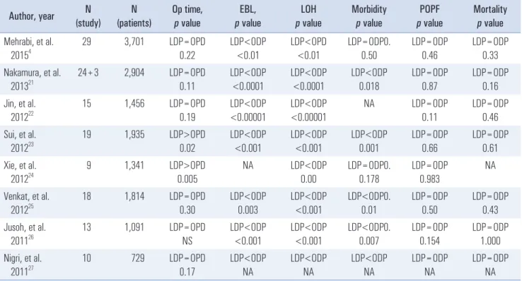
(2) 6. Chang Moo Kang. Table 1. Recently published meta-analysis comparing LDP with open DP N (study). N (patients). Op time, p value. EBL, p value. LOH p value. Morbidity p value. POPF p value. Mortality p value. 29. 3,701. LDP=OPD 0.22. LDP<ODP <0.01. LDP<OPD <0.01. LDP=ODP0. 0.50. LDP=ODP 0.46. LDP=ODP 0.33. 24+3. 2,904. LDP=OPD 0.11. LDP<ODP <0.0001. LDP<ODP <0.0001. LDP<ODP 0.018. LDP=ODP 0.87. LDP=ODP 0.16. Jin, et al. 201222. 15. 1,456. LDP=OPD 0.19. LDP<ODP <0.00001. LDP<ODP <0.00001. NA. LDP=ODP 0.11. LDP=ODP 0.46. Sui, et al. 201223. 19. 1,935. LDP>OPD 0.02. LDP<ODP <0.001. LDP<ODP <0.001. LDP<ODP 0.001. LDP=ODP 0.66. LDP=ODP 0.61. Xie, et al. 201224. 9. 1,341. LDP>OPD 0.005. NA. LDP<ODP 0.00. LDP=ODP0. 0.178. LDP=ODP 0.983. NA. Venkat, et al. 201225. 18. 1,814. LDP=OPD 0.30. LDP<ODP 0.003. LDP<ODP <0.001. LDP<ODP0. 0.01. LDP=ODP 0.50. LDP=ODP 0.43. Jusoh, et al. 201126. 13. 1,091. LDP=OPD NS. LDP<ODP <0.001. LDP<ODP <0.001. LDP<ODP0. 0.007. LDP=ODP 0.154. LDP=ODP 1.000. Nigri, et al. 201127. 10. 729. LDP=OPD 0.17. LDP<ODP NA. LDP<ODP NA. LDP<ODP NA. LDP=ODP NA. LDP=ODP NA. Author, year Mehrabi, et al. 20154 Nakamura, et al. 201321. LDP = laparoscopic distal pancreatectomy; ODP = open distal pancreatectomy; EBL = estimated blood loss; LOH = length of hospital stay; POPF = postoperative pancreatic fistula; NA = not available. estimated intraoperative blood loss. Above all, cosmetic effects from laparoscopic port incisions have not been evaluated and need to be considered when interpreting meta-analysis data (Table 1). It is still being debated whether randomized controlled studies are needed to provide scientific evidence for which surgical approach is superior.3,4 Recently, some expert surgeons have tried to reduce the number of trocars in conventional laparoscopic surgery to enhance LDP cosmetic and minimally invasive effects. It seems that reduced-port or single-port laparoscopic surgery is frequently performed for standard laparoscopic procedures including appendectomies, cholecystectomies, and colectomies.5-7 Barbaros et al.8 reported the first single-incision laparoscopic DP performed in a 59-year-old female to treat pancreatic metastasis from renal cell carcinoma. Since then, the number of cases treated with laparoscopic single port (LSP) or laparoscopic reduced port (LRP) DP procedures has increased (Fig. 1). In this review, we summarize the currently available literatures reporting laparoscopic single-port or reduced-port distal pancreatectomy, including current technical advances and future trajectories of these procedures.. Trend of publication LSP/LRP-DP. 30. 28. 25 22. 20 15. 13 13 11 9.33. 10. 7.3. 5 2. 1 1 1. 4. 3 1.5. 2.75 3. 3 1. 0 2010. 2011. 2012. 2013. 2014. 2015. 2016. Fig. 1. Chronological trends for publications reporting LSP/LRP-DP on PubMed and KoreaMed. Scientific reports on laparoscopic DP are gradually increasing. Of note, the number of patients per published report during one year is also increasing (yellow). Four recent publications present a comparative analysis with conventional LDP. Blue column = number of publications; Red column = number of patients; Yellow column = average patients per publication.. Currently Available Surgical Platforms and Short-term Outcomes When reviewing the publications reporting laparoscopic. Journal of Minimally Invasive Surgery Vol. 20. No. 1, 2017.
(3) 2. 2. 2. 1. 2. 2. 1. 2. 1. 1. 59. 40. 39. 32. 33. 53. 40. 46. 33. 32. 45. 44. 71. 60. Barbaros, et al. 20108. Chang, et al. 201228. Morales-Conde, et al. 201329. Kim, et al. 201530. Machado, et al. 201313. Misawa, et al. 201231. Srikanth, et al. 201332. Machado, et al. 201533. 2. 1. NA. 2. Gender (1:male 2:female). Authors, year. Age. NET. IPMN. NET. IPMN. NET. NET. NET. SCN. MCN. NET. SPN. NET. Pancreatic cyst. Pancreatic metastasis. Diagnosis. 0.9. 6. 3.5. 2. 1.2. 2. 3.5. 3.5. 6.5. 3.3. 6. 3.5. 3. SpDP. DPS. DPS. SpDP. SpDP. SpDP. DPS. SpDP. DPS. SpDP. SpDP. DPS. SpDP. DPS. 135. 340. 300. 110. 117. 174. NA. 225. 240. 174. 143. 140. 233. 330. Tumor Opname Optime size. 50. 200. 250. 50. 50. 50. NA. 240. 0. Minimal. 50. 35. 100. 100. EBL. Table 2. Review of published case series for laparoscopic single-port or reduced-port DP. 0. 0. 0. 0. 0. 0. NA. 0. 0. 0. 0. 0. 0. 0. 0. 1. 1. 0. 0. 0. 1. 0. 0. 1. 0. 0. 0. 1. TransfuPOPF sion. 1. 4. 3. 1. 2. 2. 5. 5. 7. 4. 7. 2. 3. 7. LOH. Gastric retraction suture Gastric retraction suture Gastric retraction suture Gastric retraction suture Gastric retraction suture Gastric retraction suture. GelPOINT®+additional Reverse Trendelenburg 5-mm port GelPOINT®+additional Reverse Trendelenburg 5-mm port GelPOINT®+additional Reverse Trendelenburg 5-mm port GelPOINT®+additional Reverse Trendelenburg 5-mm port GelPOINT®+additional Reverse Trendelenburg 5-mm port GelPOINT®+additional Reverse Trendelenburg 5-mm port. NA. NA. NA. Direct retraction with grasper. EndoGrab device. Prolene suture. Polyprepelene suture. Gatric retraction. Gastric retraction suture. Reverse Trendelenburg. Position. Single incision with multiports. SILS. SILS. GelPoint. Glove port+additional 5-mm port. SILS. SILS. SILS. System. Minimally Invasive (Laparoscopic or Robotic) Reduced Port (Single Port) Distal Pancreatectomy. 7. www.e-jmis.org.
(4) Authors, year. Gender (1:male 2:female). 2. 2. 1. 2. 1. 2. 2. 2. 1. 2. 1. 1. 2. Age. 37. 47. 57. 42. 39. 43. 29. 51. 28. 55. 20. 69. 40. Table 2. Continued 1. Journal of Minimally Invasive Surgery Vol. 20. No. 1, 2017. SCN. NET. SPN. IPMN. NET. MCN. NET. SCN. IPMN. NET. IPMN. MCN. NET. Diagnosis. 3.4. 1.3. 2.7. 3.2. 1.7. 4. 2.3. 3.3. 5.2. 4.5. 7. 3.7. 3. SpDP. SpDP. SpDP. SpDP. SpDP. SpDP. SpDP. SpDP. SpDP. SpDP. SpDP. SpDP. SpDP. 110. 170. 120. 149. 178. 189. 200. 198. 196. 210. 189. 120. 179. Tumor Opname Optime size. 50. 50. 50. 50. 50. 50. 100. 50. 50. 100. 100. 100. 50. EBL. 0. 0. 0. 0. 0. 0. 0. 0. 0. 0. 0. 0. 0. 0. 0. 0. 0. 0. 1. 0. 0. 1. 0. 0. 0. 0. TransfuPOPF sion. 3. 2. 1. 2. 1. 3. 1. 1. 2. 2. 2. 1. 2. LOH. Position Reverse Trendelenburg Reverse Trendelenburg Reverse Trendelenburg Reverse Trendelenburg Reverse Trendelenburg Reverse Trendelenburg Reverse Trendelenburg Reverse Trendelenburg Reverse Trendelenburg Reverse Trendelenburg Reverse Trendelenburg Reverse Trendelenburg Reverse Trendelenburg. System GelPOINT®+additional 5-mm port GelPOINT®+additional 5-mm port GelPOINT®+additional 5-mm port GelPOINT®+additional 5-mm port GelPOINT®+additional 5-mm port GelPOINT®+additional 5-mm port GelPOINT®+additional 5-mm port GelPOINT®+additional 5-mm port GelPOINT®+additional 5-mm port GelPOINT®+additional 5-mm port GelPOINT®+additional 5-mm port GelPOINT®+additional 5-mm port GelPOINT®+additional 5-mm port. Gastric retraction suture. Gastric retraction suture. Gastric retraction suture. Gastric retraction suture. Gastric retraction suture. Gastric retraction suture. Gastric retraction suture. Gastric retraction suture. Gastric retraction suture. Gastric retraction suture. Gastric retraction suture. Gastric retraction suture. Gastric retraction suture. Gatric retraction. 8 Chang Moo Kang.
(5) Zang, et al. 201534. Yao, et al. 201312. Authors, year. 2. 2. 2. 2. 2. 2. 2. 2. 2. 36. 42. 22. 34. 34. 39. 73. 27. 45. 1. 2. 20. 8. 2. 2. 49. 46. Gender (1:male 2:female). Age. Table 2. Continued 2. Nesidioblastosis. MCN. MCN. NET. MCN. Pancreatic cyst. MCN, SPN. Arterial Aneurysm. Pancreatic cyst. SCN. Fibromatosis. MCN. NET. Diagnosis. NA. 6.2. 4. 1.2. 3. 3.5. 3.5. 3.5. 4.5. 4.5. 3.5. 5. 2.8. SpDP. SpDP. DPS. SpDP. SpDP. SpDP. SpDP. DPS. DPS. DPS. SpDP. DPS. SpDP. 140. 95. 155. 170. 165. 115. 110. 170. 125. 150. 240. 300. 140. Tumor Opname Optime size. Minimal. 200. 200. 50. 100. 50. 30. 10. 100. 10. 500. 500. 50. EBL. 0. NA. NA. NA. NA. NA. NA. NA. NA. NA. NA. NA. 0. 0. 0. 0. 0. 0. 0. 0. 0. 0. 1. 0. 0. 0. TransfuPOPF sion. 6. 5. 7. 8. 10. 8. 6. 6. 8. 7. 9. 8. 5. LOH. Position. Single incision with multi-ports. Single incision with multi-ports. Single incision with multi-ports. Single incision with multi-ports. Single incision with multi-ports. Single incision with multi-ports. Single incision with multi-ports. Single incision with multi-ports. Single incision with multi-ports. Single incision with multi-ports. Single incision with multi-ports. Single incision with multi-ports. Supine, reverse Trendelenburg. Reverse Trendelenburg. Reverse Trendelenburg. Reverse Trendelenburg. Reverse Trendelenburg. Reverse Trendelenburg. Reverse Trendelenburg. Reverse Trendelenburg. Reverse Trendelenburg. Reverse Trendelenburg. Reverse Trendelenburg. Reverse Trendelenburg. GelPOINT®+additional Reverse Trendelenburg 5-mm port. System. Gastric retraction suture. Gastric encircling plastic tube. Gastric encircling plastic tube. Gastric encircling plastic tube. Gastric encircling plastic tube. Gastric encircling plastic tube. Gastric encircling plastic tube. Gastric encircling plastic tube. Gastric encircling plastic tube. Gastric encircling plastic tube. Gastric encircling plastic tube. Gastric encircling plastic tube. Gastric retraction suture. Gatric retraction. Minimally Invasive (Laparoscopic or Robotic) Reduced Port (Single Port) Distal Pancreatectomy. 9. www.e-jmis.org.
(6) Journal of Minimally Invasive Surgery Vol. 20. No. 1, 2017. Lee, et al. 201618. Kuroki, et al. 201435. Authors, year. 2. 1. 1. 2. 1. 1. 49. 36. 43. 70. 73. 53. 1. 64. 2. 2. 68. 41. 2. 60. 1. 2. 63. 71. 1. 79. Gender Age (1:male 2:female). Table 2. Continued 3. NET. NET. Squamoid cyst. mcn. mcn. ipmc. SPN. IPMN. NET. Pancreatic metastasis. NET. Chronic pancreatitis. Pancreatic metastasis. Diagnosis. 3.5. 0.9. 2. 2.1. 6. 1.2. 2.2. 2.3. NA. NA. NA. NA. NA. SpDP. SpDP. SpDP. SpDP. SpDP. SpDP. SpDP. SpDP. SpDP. SpDP. SpDP. SpDP. SpDP. 120. 253. 126. 185. 149. 137. 164. 144. 238. 235. 232. 271. 345. Tumor Opname Optime size. NA. NA. NA. NA. NA. NA. NA. NA. 200. 20. 70. 60. 20. EBL. 0. 0. 0. 0. 0. 0. 0. 0. 0. 0. 0. 0. 0. 0. 0. 0. 0. 0. 0. 0. 0. 0. 0. 0. 0 (bleeding). 0. TransfuPOPF sion. 6. 10. 7. 8. 8. 6. 7. 8. NA. NA. NA. NA. NA. LOH. Position. Glove port+/- additional Right semidecubitus 5-mm port. Glove port+/- additional Right semidecubitus 5-mm port. Glove port+/- additional Right semidecubitus 5-mm port. Glove port+/- additional Right semidecubitus 5-mm port. Glove port+/- additional Right semidecubitus 5-mm port. Glove port+/- additional Right semidecubitus 5-mm port. Glove port+/- additional Right semidecubitus 5-mm port. Glove port+/- additional Right semidecubitus 5-mm port. SILS+2-mm additional Reverse Trendelenburg port. SILS+2-mm additional Reverse Trendelenburg port. SILS+2-mm additional Reverse Trendelenburg port. SILS+2-mm additional Reverse Trendelenburg port. SILS+2-mm additional Reverse Trendelenburg port. System. Intraperitoneal retractor. Intraperitoneal retractor. Intraperitoneal retractor. Intraperitoneal retractor. Intraperitoneal retractor. Intraperitoneal retractor. Intraperitoneal retractor. Intraperitoneal retractor. “Stomach-hanging method”. “Stomach-hanging method”. “Stomach-hanging method”. “Stomach-hanging method”. "Stomach-hanging method". Gatric retraction. 10 Chang Moo Kang.
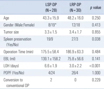
(7) 11. Intraperitoneal retractor Glove port+/- additional Right semidecubitus 5-mm port 6 0 NA. LRP-DP (N=30). p value. 43.3±15.9. 48.2±16.0. 0.250. 12/18. 0.413. 3.3±1.5. 3.4±1.7. 0.855. NET. Spleen preservation (Yes/No). 19/9. 27/3. 0.038. Operation Time (min). 175.5±58.4. 186.9±63.3. 0.484. EBL (ml). 139.1±158.2. 75.8±56.6. 0.141. 6.6±1.9. 3.0±2.2. 4/24. 26/4. 1.000. 2. 0. 0.229. 1.7. LSP-DP (N=28). 1. SpDP. 157. 0. Intraperitoneal retractor Glove port+/- additional Right semidecubitus 5-mm port 6 0 NA 2.5. SpDP. 175. 0. Intraperitoneal retractor Glove port+/- additional Right semidecubitus 5-mm port 6 0 NA 5.8. DPS. 127. 0. Intraperitoneal retractor Glove port+/- additional Right semidecubitus 5-mm port 7 0 NA 1.7. SpDP. 165. 0. Intraperitoneal retractor Glove port+/- additional Right semidecubitus 5-mm port 6 0 0 NA 178 SpDP 5. Table 3. Comparative analysis of laparoscopic single-port and reducedport DP. Gender (Male:Female) Tumor size. 46. squamoid cyst 1 31. SCM 2 69. MCM 2 64. 2. PSN. Age. 20. Gender Age (1:male 2:female). single port (LSP) or reduced port (LRP)-DP, a total of eight case reports and eight case series were identified in PubMed and KoreaMed. Among them, 12 publications described individual patient short-term perioperative outcomes, and a total of 58 patients were selected to evaluate perioperative outcomes after LSP/LRP-DP (Table 2). There were four retrospective9-12 comparative analyses between LSP/LRP-DP and conventional LDP. The most frequently used surgical system for LRP- DP was single-port with an additional 2-mm or 5-mm assist port. Pure LSP-DP was performed in only 14 patients (24.1%). The success rate for LSP/LRP-DP was very high. Conversion to multiport conventional laparoscopic DP was reported in just two patients (3.4%). Several methods for facilitating pancreas exposure were described. These included using sutures for gastric retraction, a plastic tube for gastric circling, and the use of an intraperitoneal retractor, or direct retraction with a laparoscopic grasper. Spleen-preserving DP is known to be a time and labor consuming procedure, and thus, an advanced laparoscopic technique is required for preserving the spleen during LDP. However, it is interesting to note that a spleenpreserving procedure was performed even in 46 patients (78%). A summary of perioperative outcomes showed that 20 patients were male, and 37 were female with a mean age of 45.9 ±16.0 years (gender information was missing in one report13). Most pathologic diagnoses were benign or borderline malignant tumors of the pancreas with a mean tumor diameter of 3.4±1.6 cm. Only four patients (6.9%) were found to have malignant tumors (intraductal papillary mucinous neoplasm with cancerous transformation (n=1) and pancreatic metastasis (n=3)). The mean operation time was 181.5±60.8 min, and. LOH (days) POPF (Yes/No). Authors, year. Table 2. Continued 4. Diagnosis. Tumor Opname Optime size. EBL. TransfuPOPF sion. LOH. System. Position. Gatric retraction. Minimally Invasive (Laparoscopic or Robotic) Reduced Port (Single Port) Distal Pancreatectomy. Conversion to conventional DP. 8/19*. <0.001. *Missing gender data in one report. www.e-jmis.org.
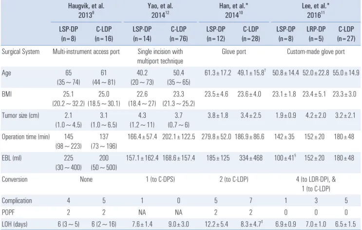
(8) 12. Chang Moo Kang. Comparative analysis between LSP/ RP-DP and conventional LDP. the mean estimated intraoperative blood loss was 99.9±109.9 ml. No patients required intraoperative transfusion. The mean hospital stay was 4.9±2.7 days. There was no surgery-related mortality. Comparative analysis between LSP-DP and LRPDP showed that the spleen preserving rate was much higher (p=0.038) and that the hospital stay was reduced (6.6±1.9 days, vs. 3.0±2.2 days, p<0.001) in LRP-DP (Table 3). This suggests that LRP-DP may be more reliable in selected DP cases requiring advanced surgical techniques. There were no significant differences between the two groups in terms of age, gender, tumor size, and postoperative pancreatic fistula formation. In LSP-DP, only two patients (7.1%) were found to convert to conventional laparoscopic DP, and four cases (14.3%) required an additional port (conversion to LRP-DP) for safe completion of the operation. After taking potential publication bias into account, current published data on LSP/LRP-DP carefully suggest that (1) both LSP-DP and LRP-DP are feasible and safe in select patients and that (2) LRP-DP seems to be more effective in spleenpreserving procedures and enhances postoperative recovery.. There are only four studies which compared the perioperative outcomes between LSP/LRP-DP and conventional DP, including the most recent report by Lee.11 Among them, two10,11 were reported by members of Korean Society of Endoscopic and Laparoscopic Surgery (KSELS).14 The perioperative outcomes investigated in each study are summarized in Table 4. Even though the conclusions derived from these studies were based on a limited number of the cases and retrospective study designs associated with unintended selection bias, all studies indicated that LSP/LRP-DP was comparable to conventional DP in patients who required DP for benign and borderline (low grade) malignant tumors in distal pancreas. The technical difficulty of the procedure and the resulting stress for the surgeon were not evaluated in these studies, which will continue to be the main obstacles to make LRP/ LSP-DP routine in clinical practice. Technical advances and more surgical experiences are needed to define the potential role of LSP/LRP-DP in minimally invasive pancreatic surgery.. Table 4. Review of comparative analyses between LSP-DP/ LRP-DP and conventional LDP (C-LDP) Haugvik, et al. 20139 LSP-DP (n=8) Surgical System Age BMI. Yao, et al. 201412. C-LDP (n=16). LSP-DP (n=14). Multi-instrument access port 65 (35~74). 61 (44~81). 25.1 25.0 (20.2~32.2) (18.5~30.1). Tumor size (cm). 2.1 (1.0~4.5). 3.1 (1.0~6.5). Operation time (min). 145 (98~223). 137 (73~196). EBL (ml). 225 (30~400). 200 (50~500). Conversion. Han, et al.* 201410. C-LDP (n=76). LSP-DP (n=12). Single incision with multiport technique 40.2 (20~73). 50.4 (35~65). 22.6 23.3 (18.4~27) (21.3~25.2) 4.3 (1.2~11). C-LDP (n=28). Glove port. 23.5±4.6. 23.6±4.0. 3.8±1.8. 3.4±2.5. 3.2±2.1. 166.4±57.4 202.1±122.5 279.8±52.0 186.9±86.6. 142±35. 152±20. 180±48. 157.1±162.4 168.6±157.4. 100±41§. 152±20. 180±48. 185±125. 1 (to C-DPS). 334±468. 2 (to C-LDP). 4 (to LDR-DP), & 1 (to C-LDP). 1. 0. 5. 7. POPF. 2. 2. NA. NA. 2. 2. 6 (2~16) ‡. 7.6±1.4 §. *Data from the members of KSELS. p=0.035, p=0.028, p=0.035. Journal of Minimally Invasive Surgery Vol. 20. No. 1, 2017. 23.1±1.8 23.4±5.1 23.3±3.0 4.2±2.0. 5. †. C-LDP (n=27). 61.3±17.2 49.1±15.8† 50.8±14.4 52.0±22.8 55.0±14.9. 4 6 (3~5). LRP-DP (n=5). Custom-made glove port. Complication LOH (days). LSP-DP (n=8). 1.9±0.9. None. 3.7 (0.7~6). Lee, et al.* 201611. 9.0±3.0. 12.2±5.4. ‡. 8.3±4.7. 1. 3. 5. 0. 0. 0. 6.9±0.9. 7.0±1.0. 6.5±1.5.
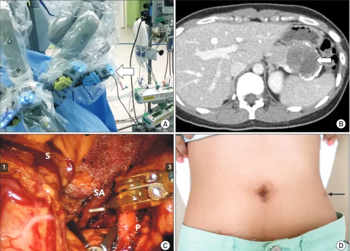
(9) 13. Minimally Invasive (Laparoscopic or Robotic) Reduced Port (Single Port) Distal Pancreatectomy. Potential Technique: Robotic Single Site plus ONE port Distal Pancreatectomy Despite the increasing number of laparoscopic DPs being performed and the advance of laparoscopic instruments, the fatigue and stress resulting from limited motion for instrument manipulation in the narrow surgical space (in current single port system) needs to be considered when performing LSP/ LRP-DP. Therefore, in order to improve intraoperative surgical quality, technical innovation is essential. In theory, robotic surgical systems can overcome the limitations of laparoscopic surgery.15,16 This technology may work even with LSP/RP-DP. A robotic single-site surgical system has been introduced to facilitate laparoscopic single-port surgery.17-19 Additionally the. stable, 3-D operation field can enhance a surgeon’s ergonomic environment. This enables surgeons to avoid the situation of right and left disorientation for triangular configuration during laparoscopic single-port surgery. It is thought that most intraoperative stress and fatigue results from the mechanics of the laparoscopic single-port surgical system. However, the robotic surgical system automatically calculates the movement of the surgeon’s console with the help of specially designed curved trocars and semi-flexible instruments, making it possible for the surgeon’s right and left hand to control the rightand left-sided screen instruments even if the instrument is attached to the left and right robotic arm, respectively.18,20 If an additional robotic arm is added through another trocar in the abdomen, a wrist-like motion of instrument can be produced in the robotic single-site surgical system allowing for a more. A. B. C. D. Fig. 2. Technical innovation of the robotic surgical system for LSP/RP DP. Robot setting for robotic single-site plus ONE port DP. Note a third robotic arm (white thick arrow) in the left lateral flank of the patient (three left-sided robotic arms) (A). Large pancreatic tumor with marginal calcification (white thin arrow) (B). The stomach was retracted with a single site robotic arm, and the splenic artery was effectively dissected using another robotic arm from the single-site robotic surgical system. A third robotic arm allowed for angulating wrist motion (C). Postoperative wound. Left lateral flank wound from the third robotic arm is away from the midline. The operative wound is hardly visible (black arrow) (D). S = stomach; SA = splenic artery; P = pancreas. www.e-jmis.org.
(10) 14 effective reduced-port surgery (Fig. 2A). Considering there is no wrist like-motion in pure robotic single site robotic surgical system, this technical advantages from additional port will be great helpful. In addition, preoperative surgical rehearsal is another advantage of robotic surgery. Surgical procedures can be simulated and techniques modified before applying them to patients, which allows for improved surgical quality and safety. Beginning in October 2015, we have been applying our robotic single-site plus ONE port DP technique in selected cases. A total of six cases, including a recent case of pancreatic enucleation, have already been performed safely using this new technique (unpublished). A case of robotic single-site plus ONE port DP case is briefly introduced in this review. A 24-year-old female patient was admitted to the hospital due to the incidental finding of a mass in the pancreatic tail (Fig. 2B). Based on the presumed diagnosis of a solid pseudopapillary pancreatic neoplasm, she underwent robotic single-site plus ONE port DP. Total operation time was 160 minutes, and the estimated intraoperative blood loss was less than 50 ml. When dissecting splenic vessels, angulating motion of surgical instrument through additional port made surgical procedure effective and easy (Fig. 2C). No POPF was noted. She was discharged on the seventh postoperative day. Postoperatively, the wound appeared to be healing well (Fig. 2D). This case suggests that the main obstacles of the LSP/LRP system, which include surgical stress and ineffective instrument manipulation, can be resolved by using a robotic surgical system. More experience is required to determine the exact role of the robotic single-site surgical system for performing LSP/LRP-DP.. Conclusion and Future Perspectives Despite the lack of randomized controlled studies, the accumulating number of LDP cases strongly suggests that LDP is a safe and effective surgical option for treating benign and borderline malignant tumors of the left pancreas. Currently, some efforts are being made to reduce the number of external wounds resulting from LSP/LRP-DP. LSP/LRP-DP is an emerging technique, and only a limited number of cases have been performed, however, the currently available published data show that LSP/LRP-DP is feasible, safe, and even comparable to conventional LDP. According to the literatures, a spleen-preserving procedure can be performed without increasing perioperative risk by this approach. It is difficult to estimate the limitations of instrumental movement and the surgeon’s intraoperative stress and fatigue during LSP/LRPDP. However, these technical limitations may be obstacles to the widespread use of LSP/LRP-DP. Further technical innovation and advances are required for reliable minimally invaJournal of Minimally Invasive Surgery Vol. 20. No. 1, 2017. Chang Moo Kang. sive LDP. It is expected that more reliable clinical data based on a larger number of patients will be published from expert laparoscopic surgeons in the near future. Minimally invasive surgeons will continue to work to reduce postoperative pain and number of external wound, increasing the quality of life associated with laparoscopic procedures.. References 1) Soper NJ, Brunt LM, Dunnegan DL, Meininger TA. Laparoscopic distal pancreatectomy in the porcine model. Surg Endosc 1994;8:57-60; discussion 60-51. 2) Cuschieri A, Jakimowicz JJ, van Spreeuwel J. Laparoscopic distal 70% pancreatectomy and splenectomy for chronic pancreatitis. Ann Surg 1996;223:280-285. 3) Kang CM. Should we randomize our patients in the name of the “scientific evidence”? Surgery 2015;158:1742-1743. 4) Mehrabi A, Hafezi M, Arvin J, et al. A systematic review and meta-analysis of laparoscopic versus open distal pancreatectomy for benign and malignant lesions of the pancreas: it’s time to randomize. Surgery 2015;157:45-55. 5) Xu AM, Huang L, Li TJ. Single-incision versus three-port laparoscopic appendectomy for acute appendicitis: systematic review and meta-analysis of randomized controlled trials. Surg Endosc 2015;29:822-843. 6) Yamazaki M, Yasuda H, Koda K. Single-incision laparoscopic cholecystectomy: a systematic review of methodology and outcomes. Surg Today 2015;45:537-548. 7) Carus T. Current advances in single-port laparoscopic surgery. Langenbecks Arch Surg 2013;398:925-929. 8) Barbaros U, Sumer A, Demirel T, et al. Single incision laparoscopic pancreas resection for pancreatic metastasis of renal cell carcinoma. JSLS 2010;14:566-570. 9) Haugvik SP, Rosok BI, Waage A, Mathisen O, Edwin B. Singleincision versus conventional laparoscopic distal pancreatectomy: a single-institution case-control study. Langenbecks Arch Surg 2013;398:1091-1096. 10) Han HJ, Yoon SY, Song TJ, et al. Single-port laparoscopic distal pancreatectomy: initial experience. J Laparoendosc Adv Surg Tech A 2014;24:858-863. 11) Lee H, Heo JS, Choi SH, Choi DW. Safety and Feasibility of Single Incision Laparoscopic Spleen Preserving Distal Pancreatectomy. J Minim Invasive Surg 2016;19:89-96. 12) Yao D, Wu S, Li Y, Chen Y, Yu X, Han J. Transumbilical singleincision laparoscopic distal pancreatectomy: preliminary experience and comparison to conventional multi-port laparoscopic surgery. BMC Surg 2014;14:105. 13) Machado MA, Surjan RC, Makdissi FF. First single-port laparoscopic pancreatectomy in Brazil. Arq Gastroenterol 2013;50:310312..
(11) Minimally Invasive (Laparoscopic or Robotic) Reduced Port (Single Port) Distal Pancreatectomy. 14) The Korean Society of Endoscopic & Laparoscopic Surgeons. The Korean Society of Endoscopic & Laparoscopic Surgeons [Internet]. Bundang: The Korean Society of Endoscopic & Laparoscopic Surgeons; c2008 [cited on Sep 20, 2016]. Available from: http:// www.ksels.or.kr/. 15) Ballantyne GH. Robotic surgery, telerobotic surgery, telepresence, and telementoring. Review of early clinical results. Surg Endosc 2002;16:1389-1402. 16) Hashizume M, Konishi K, Tsutsumi N, Yamaguchi S, Shimabukuro R. A new era of robotic surgery assisted by a computerenhanced surgical system. Surgery 2002;131:S330-333. 17) Qadan M, Curet MJ, Wren SM. The evolving application of singleport robotic surgery in general surgery. J Hepatobiliary Pancreat Sci 2014;21:26-33. 18) Lee SH, Jung MJ, Hwang HK, Kang CM, Lee WJ. The first experiences of robotic single-site cholecystectomy in Asia: a potential way to expand minimally-invasive single-site surgery? Yonsei Med J 2015;56:189-195. 19) Konstantinidis KM, Hirides P, Hirides S, Chrysocheris P, Georgiou M. Cholecystectomy using a novel Single-Site((R)) robotic platform: early experience from 45 consecutive cases. Surg Endosc 2012;26:2687-2694. 20) Morelli L, Guadagni S, Di Franco G, Palmeri M, Di Candio G, Mosca F. Da Vinci single site(c) surgical platform in clinical practice: a systematic review. Int J Med Robot 2016;12:724-734. 21) Nakamura M, Nakashima H. Laparoscopic distal pancreatectomy and pancreatoduodenectomy: is it worthwhile? A meta-analysis of laparoscopic pancreatectomy. J Hepatobiliary Pancreat Sci 2013;20:421-428. 22) Jin T, Altaf K, Xiong JJ, et al. A systematic review and metaanalysis of studies comparing laparoscopic and open distal pancreatectomy. HPB (Oxford) 2012;14:711-724. 23) Sui CJ, Li B, Yang JM, Wang SJ, Zhou YM. Laparoscopic versus open distal pancreatectomy: a meta-analysis. Asian J Surg 2012;35:1-8. 24) Xie K, Zhu YP, Xu XW, Chen K, Yan JF, Mou YP. Laparoscopic distal pancreatectomy is as safe and feasible as open procedure: a. 15. meta-analysis. World J Gastroenterol 2012;18:1959-1967. 25) Venkat R, Edil BH, Schulick RD, Lidor AO, Makary MA, Wolfgang CL. Laparoscopic distal pancreatectomy is associated with significantly less overall morbidity compared to the open technique: a systematic review and meta-analysis. Ann Surg 2012;255:1048-1059. 26) Jusoh AC, Ammori BJ. Laparoscopic versus open distal pancreatectomy: a systematic review of comparative studies. Surg Endosc 2012;26:904-913. 27) Nigri GR, Rosman AS, Petrucciani N, et al. Metaanalysis of trials comparing minimally invasive and open distal pancreatectomies. Surg Endosc 2011;25:1642-1651. 28) Chang SK, Lomanto D, Mayasari M. Single-port laparoscopic spleen preserving distal pancreatectomy. Minim Invasive Surg 2012;2012:197429. 29) Morales-Conde S, Rubio-Manzanares M, Alarcon I, Barranco A, Socas M. [Single port distal pancreatectomy without spleen preserving]. Cir Esp 2013;91:541-543. 30) Kim EY, You YK, Kim DG, et al. Dual-incision laparoscopic spleen-preserving distal pancreatectomy. Ann Surg Treat Res 2015;88:174-177. 31) Misawa T, Ito R, Futagawa Y, et al. Single-incision laparoscopic distal pancreatectomy with or without splenic preservation: how we do it. Asian J Endosc Surg 2012;5:195-199. 32) Srikanth G, Shetty N, Dubey D. Single incision laparoscopic distal pancreatectomy with splenectomy for neuroendocrine tumor of the tail of pancreas. J Minim Access Surg 2013;9:132-135. 33) Machado MA, Surjan RC, Makdissi FF. Laparoscopic Distal Pancreatectomy Using Single-Port Platform: Technique, Safety, and Feasibility in a Clinical Case Series. J Laparoendosc Adv Surg Tech A 2015;25:581-585. 34) Zhao G, Hu M, Liu R, et al. Single-port retroperitoneoscopic pancreatectomy: preliminary results from the first 3 patients. J Clin Gastroenterol 2014;48:559-562. 35) Kuroki T, Adachi T, Okamoto T, Kanematsu T. Single-incision laparoscopic distal pancreatectomy. Hepatogastroenterology 2011;58:1022-1024.. www.e-jmis.org.
(12)
수치


+5

관련 문서
limit the crack growth by increasing # of site of crazing or
Incheon is still advancing ceaselessly as demonstrated in its plentiful milestones including, the expansion of the Incheon Port and opening of the Incheon Metro subway
Patient had laparoscopic surgery on the adnexal tumor and excised tissue was removed through Douglas pouch incision by single surgeon.. Results: The mean age
The strategy of Russian new eastern policy and Chinese One Belt Load using ports in the east sea area, Korean new northern policy, Japanese west coast
At first, I arranged the current conditions and function of container terminal in the port of Busan through the existing literature study on the function
Results of the study, first, enhanced support services for route expansion to take advantage of the geographical advantage of the Busan port, second,
If owing to circumstances beyond the control of the Assured either the contract of carriage is terminated at a port or place other than the destination named therein or the
Secondly, the important factors to attract T/S cargo at Kwangyang Port are geographical location, promotion activity to introduce Port and expansion of
